Review
. 2024 Sep 30;7(12):e202302083.
doi: 10.26508/lsa.202302083. Print 2024 Dec.

Cellular stress and epigenetic regulation in adult stem cells

Affiliations
Review

Cellular stress and epigenetic regulation in adult stem cells

Joey Llewellyn et al. Life Sci Alliance. .

Abstract

Stem cells are a unique class of cells that possess the ability to differentiate and self-renew, enabling them to repair and replenish tissues. To protect and maintain the potential of stem cells, the cells and the environment surrounding these cells (stem cell niche) are highly responsive and tightly regulated. However, various stresses can affect the stem cells and their niches. These stresses are both systemic and cellular and can arise from intrinsic or extrinsic factors which would have strong implications on overall aging and certain disease states. Therefore, understanding the breadth of drivers, namely epigenetic alterations, involved in cellular stress is important for the development of interventions aimed at maintaining healthy stem cells and tissue homeostasis. In this review, we summarize published findings of epigenetic responses to replicative, oxidative, mechanical, and inflammatory stress on various types of adult stem cells.


Conflict of interest statement

The authors declare that they have no conflict of interest.

Figures

Figure 1.
Figure 1.. Stem cells and their niche.
(A) Hematopoietic stem cells (HSCs) are largely found residing in the bone marrow microenvironment. Cycling HSCs are found to localize near the sinusoid whereas those that are quiescent tend to localize closer to the endosteal (bone-lining) wall. Cells neighboring HSCs in the bone marrow (e.g., mesenchymal stem cells [MSCs] and arteriole endothelial cells) often hold roles in regulating HSC function and maintenance via the secretion of cytokines, growth factors, and other small molecules. (B) Mesenchymal/stromal stem cells (MSCs) are found to reside in several adult (adipose tissue, compact bone, bone marrow, dental pulp, liver) and embryonic (endometrium, placenta, umbilical cord) tissues. MSCs directly regulate cellular function and maintenance of nearby cells within their respective niches, including adult stem cells, through several secretory mechanisms. (C) The existence of neural stem cells (NSCs) has been established in rodents in the subgranular zone of the hippocampal dentate gyrus and the subventricular zone of the lateral ventricles. Subventricular zone NSCs produce (inhibitory) oligodendrocytes and interneurons, whereas subgranular zone NSCs generate (excitatory) granule neurons. The controversy regarding the existence and activity of NSCs in humans has not yet been fully resolved. (D) The niche residency of intestinal stem cells (ISCs) has been established largely within the crypt base (below the villi) along the intestinal epithelium. Their location has been long debated; however, their exact position along the crypt base has yet to be fully resolved. ISCs hold multipotent differentiation capacity, where ISCs adjacent to longer villi are biased towards producing enterocytes whereas those adjacent to shorter villi will largely produce secretory cells (e.g., goblet cells). (E) Muscle/satellite stem cells (MuSCs) are uniquely identified by their anatomical location between the basal lamina and myofiber of skeletal muscle tissue. Unlike other adult stem cell niches, the MuSC local microenvironment is anatomically dynamic during muscle regeneration and attributable to regulatory networks by MuSCs and other functionally diverse resident cells.
Figure 2.
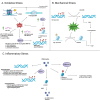
Figure 2.. A schematic illustration of epigenetic changes in HSCs because of various stressors.
(A) HSCs under IR-induced replicative stress displayed an accumulation of yH2AX foci without DDR activation accompanied by impaired S phase progression, senescence markers, and a decrease in DNA helicase components. Ott1 maintains HSC quiescence during replicative stress, where HSCs exhibit premature aging phenotypes. EZH2 is down-regulated in murine HSCs under severe replicative stress, leading to stem cell exhaustion. CBP is a transcriptional coactivator and histone acetyltransferase involved in several processes, such as differentiation, proliferation, and gene regulation. (B) PRC1 and PRC2 complexes are key epigenetic regulators in gene silencing via histone modifications. Autophagy regulates oxidative stress by clearing mitochondria, but its repression increases hypomethylation in genes that accelerate myeloid differentiation. (C) Inflammatory signals reduce expression of the histone demethylase UTX/KDM6A, impairing DNA repair and increasing oxidative stress. Global gene expression analyses have identified down-regulation of SWI/SNF-related genes, which are involved in chromatin remodeling and transcriptional silencing. Inflamed HSCs show increased expression of P-selectin (Selp), driven by heightened NF-κB pathway activity, which up-regulates inflammation-related genes and contributes to HSC functional decline. Chronic inflammation also induces a senescence-associated secretory phenotype in HSCs, thereby perpetuating inflammation and impairing HSC function.
Figure 3.
Figure 3.. A schematic illustration of epigenetic changes in MSCs because of various stressors.
(A) In MSCs, ROS-induced DNA damage induced prolonged DDR activation and increased yH2AX foci driving premature senescence. In addition, miR-210, miR-29a-3p, and miR-30c-5p confer resistance to oxidative stress in MSCs, although upstream regulatory mechanisms of these miRNAs require further investigation. In aging MSCs, oxidative stress inhibits osteogenic differentiation by increasing EZH2 levels, which elevate H3K27me3 at the Foxo1 promoter. (B) Dnmt3b was found to bind to the Shh promoter region and directly catalyze hypermethylation. HDAC1 negatively correlates with osteogenesis by deacetylating the Jag1 promoter in BMSCs, down-regulating JAG1 and the NOTCH signaling pathway, whereas mechanical stimulation reduces HDAC1 levels, rescuing JAG1-mediated osteogenesis. (C) Histone variant mH2A1.1 modifications play a significant role in epigenetic regulation via the TLR4 pathway, inducing SASP. Increased H3K9me3 in aged MSCs leads to a repressive chromatin state, cellular senescence, reduced self-renewal capacity, and elevated expression of aging-related genes such as Cdkn2a (p16) and Cdkn1a (p21), with KDM4B loss exacerbating these effects by raising H3K9me3 and HP1α levels, ultimately impairing MSC functionality and regenerative capacity. Overexpression of AHCY leads to genome hypermethylation, whereas CBX3/HP1γ contributes to increased levels of H3K9me3.
Figure 4.
Figure 4.. A schematic illustration of epigenetic changes in ISCs because of various stressors.
(A) Replicative stress has been linked to genome-wide hypermethylation to the DNA methylome in the intestinal epithelium. Simulations displayed that promoter regions marked with H3K27me3 were found to undergo DNA hypermethylation during DNA repair in response to replicative stress in irradiated ISCs. This was associated with an increase in DNMT activity recruited to open chromatin regions undergoing repair and containing low levels of DNA methylation. (B) ISCs under oxidative stress demonstrate age- and stress-induced DNA damage accumulation in ISCs using yH2AvD which suppresses ribosome biogenesis. (C) Signaling molecules such as Wnt, Bmp, and Notch, produced by Paneth cells, regulate ISC fate and function by allowing Atoh1 transcription factor to bind, enabling cells to revert to a primitive state. Epigenetic modifications such as the incorporation of histone variant H2A.Z mediated by Znhit1, are central to ISC plasticity, influencing key genes such as Lgr5 and TGF-β crucial for self-renewal and differentiation, with specific roles for MTG8, MTG16, and Id3 in modulating chromatin accessibility and maintaining ISC identity.
Figure 5.
Figure 5.. A schematic illustration of epigenetic changes in NSCs because of stress.
LPS adversely affects NSC viability and differentiation by inducing key epigenetic changes, particularly histone modifications, with prolonged LPS exposure increasing Jmjd2b and NF-κB p65 expression, leading to reduced H3K9me3 levels at gene promoters essential for NSC function and neurogenesis, thus profoundly altering the epigenetic landscape of NSCs.
Figure 6.
Figure 6.. A schematic illustration of epigenetic changes in MuSCs because of various stressors.
(A) Bmi1 is shown to maintain MuSC pools and myogenesis during muscle via repression of the ink4a locus after muscle injury and during oxidative stress. Bmi1 overexpression in mouse models and patients enhances MT1-mediated protection against oxidative stress, improving muscle strength and regeneration, likely through gene silencing mechanisms involving PRC1. (B) PRMT5 represses p21 expression via H3R8 dimethylation at regulatory sites upstream of the murine p21 gene. PRMT7 regulates p21 expression through DNMT3b-mediated hypermethylation at the p21 promoter, influencing H4R3me2 and H3K4me3 levels. (C) Inflammatory signals, especially CCR2 ligands, elevate CCR2 expression in myogenic progenitors, inhibiting their fusion into myofibers by activating MAPKp38δ/γ signaling and phosphorylating MyoD. Chronic inflammatory signals reduce Kmt5a activity, leading to a decline in H4K20me1 levels, disrupting MuSC quiescence by down-regulating Notch target genes. In addition, chronic inflammation disrupts p38α/β MAPK, adding repressive H3K27me3 marks on myogenic gene promoters such as Pax7.

References

    1. Evans MJ, Kaufman MH (1981) Establishment in culture of pluripotential cells from mouse embryos. Nature 292: 154–156. 10.1038/292154a0 - DOI - PubMed
    1. Martin GR (1981) Isolation of a pluripotent cell line from early mouse embryos cultured in medium conditioned by teratocarcinoma stem cells. Proc Natl Acad Sci U S A 78: 7634–7638. 10.1073/pnas.78.12.7634 - DOI - PMC - PubMed
    1. Takahashi K, Tanabe K, Ohnuki M, Narita M, Ichisaka T, Tomoda K, Yamanaka S (2007) Induction of pluripotent stem cells from adult human fibroblasts by defined factors. Cell 131: 861–872. 10.1016/j.cell.2007.11.019 - DOI - PubMed
    1. Takahashi K, Yamanaka S (2006) Induction of pluripotent stem cells from mouse embryonic and adult fibroblast cultures by defined factors. Cell 126: 663–676. 10.1016/j.cell.2006.07.024 - DOI - PubMed
    1. Clevers H (2015) What is an adult stem cell? Science 350: 1319–1320. 10.1126/science.aad7016 - DOI - PubMed

LinkOut - more resources