. 2024 Dec 10;74(1):128-140.
doi: 10.1136/gutjnl-2024-332902.

Nuclear translocation of plasma membrane protein ADCY7 potentiates T cell-mediated antitumour immunity in HCC

Affiliations

Nuclear translocation of plasma membrane protein ADCY7 potentiates T cell-mediated antitumour immunity in HCC

Jianan Chen et al. Gut. .

Abstract

Background: The potency of T cell-mediated responses is a determinant of immunotherapy effectiveness in treating malignancies; however, the clinical efficacy of T-cell therapies has been limited in hepatocellular carcinoma (HCC) owing to the extensive immunosuppressive microenvironment.

Objective: Here, we aimed to investigate the key genes contributing to immune escape in HCC and raise a new therapeutic strategy for remoulding the HCC microenvironment.

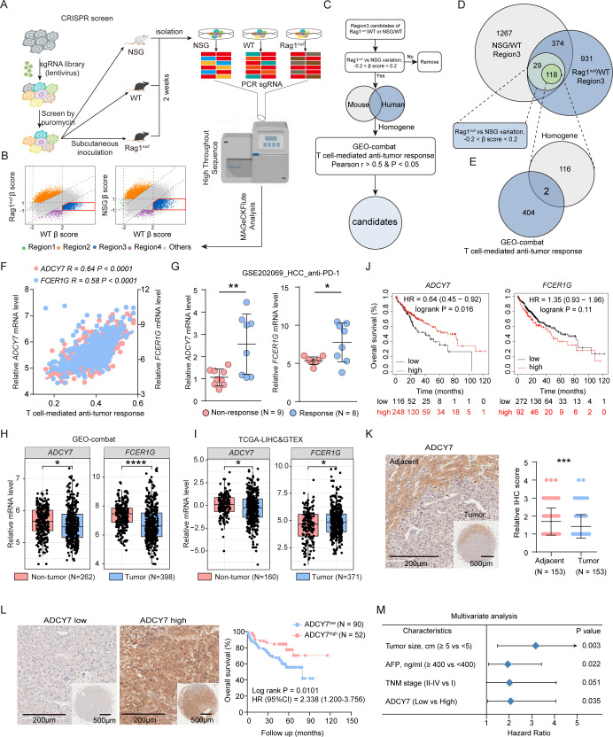
Design: The genome-wide in vivo clustered regularly interspaced short palindromic repeats (CRISPR) screen library was conducted to identify the key genes associated with immune tolerance. Single-cell RNA-seq (scRNA-seq), flow cytometry, HCC mouse models, chromatin immunoprecipitation and coimmunoprecipitation were used to explore the function and mechanism of adenylate cyclase 7 (ADCY7) in HCC immune surveillance.

Results: Here, a genome-wide in vivo CRISPR screen identified a novel immune modulator-ADCY7. The transmembrane protein ADCY7 undergoes subcellular translocation via caveolae-mediated endocytosis and then translocates to the nucleus with the help of leucine-rich repeat-containing protein 59 (LRRC59) and karyopherin subunit beta 1 (KPNB1). In the nucleus, it functions as a transcription cofactor of CCAAT/enhancer binding protein alpha (CEBPA) to induce CCL5 transcription, thereby increasing CD8+ T cell infiltration to restrain HCC progression. Furthermore, ADCY7 can be secreted as exosomes and enter neighbouring tumour cells to promote CCL5 induction. Exosomes with high ADCY7 levels promote intratumoural infiltration of CD8+ T cells and suppress HCC tumour growth.

Conclusion: We delineate the unconventional function and subcellular location of ADCY7, highlighting its pivotal role in T cell-mediated immunity in HCC and its potential as a promising treatment target.

Keywords: HEPATOCELLULAR CARCINOMA; IMMUNE RESPONSE.


Conflict of interest statement

Competing interests: None declared.

Figures

Figure 1
Figure 1. In vivo CRISPR/Cas9 screen identifies potential candidates as determinants of T cell-mediated antitumour immunity. (A, B) The schematic of the genome-wide in vivo CRISPR/Cas9 knockout library screen. (C) The detailed workflow of potential candidate selection. (D) 118 homologous genes between human and mouse species were obtained according to the selection criteria. (E) 2 genes (ADCY7 and FCER1G) strongly associated with T cell-mediated antitumour responses were remained among the above-mentioned 118 homogenes. (F) Pearson correlation of ADCY7 or FCER1G with T cell-mediated antitumour responses. (G) Expression of ADCY7 and FCER1G in HCC patients responding to ICIs in GSE202069 cohort. (H, I) The expression status of ADCY7 and FCER1G in HCC from GEO-combat (H) and TCGA-LIHC and GTEX (I) data sets. (J) Patients with low ADCY7 expression have worse prognosis using the online KM-Plotter tool based on TCGA database. (K) HCC TMA cohort shows a low ADCY7 protein expression in HCC tissues compared with adjacent non-tumour tissues. (L) Kaplan-Meier analysis demonstrates that low ADCY7protein expression correlated with worse prognosis. (M) Multivariate analysis shows ADCY7, tumour size and AFP level are independent risk factor for HCC. Error bars in (G, K) indicate the mean±SD. The p value and the R value in (F) were calculated using Pearson’s correlation analysis. P values in (G–I) were calculated by two-tailed Student’s t-test. P values in (J, L) were calculated by log-rank test. P value in (K) was calculated by Mann-Whitney U test. P value in (M) was calculated by Cox regression analysis. *p<0.05, **p<0.01, ***p<0.001, ****p<0.0001. HCC, hepatocellular carcinoma; TMA, tissue microarray.
Figure 2
Figure 2. ADCY7 re-expression positively correlates CD8+ T cell-mediated antitumour immunity in HCC. (A) Western blot confirms the stable re-expression of ADCY7 in Hepa1-6 cells. (B, C) The images of tumour xenografts (B) and the tumour growth curves (C) of C57BL/6J mice; n=9 mice for NC group and n=10 mice for ADCY7 re-expression group. (D, E) Flow cytometry results exhibit a higher infiltration of CD3+ T, CD8+ T cells after ADCY7 re-expression, but not CD4+ T and NK cells (n=4). (F, G) Similar results are observed in IHC staining of CD3+ T, CD8+ T, CD4+ T and NK cells (n=5). (H, I) IHC staining of ADCY7, CD3+ T cells and CD8+ T cells in HCC TMA cohort confirms the higher infiltration in patients with high ADCY7 expression. (J) Western blot confirms the stable re-expression of ADCY7 in HCC cells. (K, L) T cell migration assay on the coincubation of HCC cells with healthy donor CD8+ T cells (n=3). Error bars in (C, E, G, I, K, L) indicate the mean±SD. P value in (C) was calculated by two-way analysis of variance. P values in (E, G, I, K, L) were calculated by two-tailed Student’s t-test. *p<0.05, **p<0.01, ***p<0.001, ****p<0.0001. HCC, hepatocellular carcinoma; NS, not significant; TMA, tissue microarray.
Figure 3
Figure 3. ADCY7 hampers HCC progression in a CCL5-dependent manner. (A, B) Enrichment analysis of KEGG (A) and GSEA (B) showed that ADCY7 is significantly associated with the chemokine signalling pathway based on GEO-combat data set. (C) The human chemokine protein array was used to determine chemokine levels in supernatants of HepG2 cells. (D, E) qPCR (D) and western blotting (E) show that CCL5 is upregulated in the ADCY7 re-expression group. (F) T cell migration assay showed CCL5 knockdown drastically suppressed CD8+ T cells migration induced by ADCY7 re-expression (n=3). (G, H) The images (G) and tumour growth curves (H) of C57BL/6J mice inoculated with negative control (NC) cells, ADCY7 re-expression cells, ADCY7 re-expression and shCCL5 cells and shCCL5 cells, respectively; n=8 mice for ADCY7 re-expression group and n=6 for other groups. (I) The general images of orthotopic tumours derived from C57BL/6J mice. (J) The longest diameters of orthotopic tumours (n=6). (K, L) Flow cytometry results exhibit a higher infiltration of CD3+ T, CD8+ T and IFN-γ+CD8+ T cells with ADCY7 re-expression but reduced after CCL5 knockdown (n=4). (M, N) Similar results are observed in IHC staining of CD8+ T and CD4+ T cells (n=5). Error bars in (D, F, H, J, L, N) indicate the mean±SD. P values in (D, F, J, L, N) were calculated by two-tailed Student’s t-test. P values in (H) were calculated by two-way analysis of variance. *p<0.05, **p<0.01, ***p<0.001, ****p<0.0001. HCC, hepatocellular carcinoma; NS, not significant.
Figure 4
Figure 4. Nuclear ADCY7 upregulates CCL5 expression by binding to its promoter sequence. (A, B) Subcellular distribution of ADCY7 in ADCY7 re-expressing HCC cells by western blot (A) and STED (B) analysis. LMNB1 as a nuclear internal reference and GAPDH as the cytoplasm reference, indicating the purity of the extracted nuclear and cytoplasm proteins. (C, D) ChIP-qPCR indicated ADCY7 could bind to CCL5 gene promoter sequence (n=3). (E) Prediction of putative transcription factors of CCL5 via different kinds of public databases. ‘All’ indicates the full 2000 bp sequence of CCL5 promoter; ‘P6’ indicates the −1100 to −900 sequence. (F, G) CEBPA silence more significantly reduced the CCL5 mRNA expression than MYB in HCCLM3 cells (n=3). The mRNA expression of ADCY7 and CCL5 in NC and ADCY7 groups are from the same wells in figure 4F,G. (H) Co-IP assay showed the interaction of ADCY7 with CEBPA in HCC cells. (I) Western blot showed no significant difference of CEBPA expression in indicated HCC cells. (J) ChIP-qPCR showed stronger binding capacity of CEBPA to CCL5 promoter in ADCY7 re-expression cells than that in control cells (n=3). (K) ChIP-qPCR suggested after CEBPA silence, ADCY7 failed to enhance CCL5 transcription (n=3). (L) Western blot analysis of ADCY7, CEBPA and CCL5 levels after CEBPA silence. (M) Schematic of ADCY7 domain structure. (N) The subcellular distribution of ADCY7 truncations identified by cytoplasmic and nuclear western blot. (O) qPCR analysis of CCL5 mRNA expression in HepG2 cells transfected with empty vector and ADCY7 truncations (n=5). (P) The interactions between ADCY7 fragments and CEBPA were evaluated via Co-IP in HepG2 cells. Error bars in (D, F, G, J, K, O) indicate the mean±SD, and p values were calculated by two-tailed Student’s t-test. **p<0.01, ***p<0.001, ****p<0.0001. ChIP, chromatin immunoprecipitation; Co-IP, coimmunoprecipitation; EV, extracellular vesicle; HCC, hepatocellular carcinoma; NS, not significant; WT, wild-type.
Figure 5
Figure 5. ADCY7 accesses the nuclei under the help of caveolae-mediated endocytosis and LRRC59 and KPNB1. (A, B) Co-IP (A) and STED (B) assays showed the nuclear interaction and colocalisation of ADCY7 with CAV-1 in HCC cells. (C) The distribution of ADCY7 in subcellular organisation after Filipin III treatment (15 µM used for 12 hours). (D) Schematic model of ADCY7 nuclear translocation. (E–G) STED (E, F) and Co-IP (G) assays showed the nuclear colocalisation and interaction of ADCY7 with LRRC59 and KPNB1 in HCC cells. (H, I) Co-IP assay indicated ADCY7 was unable to interact with KPNB1 after LRRC59 silence. (J) Western blot analysis showed reduced ADCY7 expression in the nucleus after LRRC59 silence. (K–M) Western blot analysis showed total ADCY7 and cytoplasmic or nuclear ADCY7 levels by importazole (IPZ) treatment in indicated HCC cells (0, 6 or 8 µM used for 48 hours). Co-IP, coimmunoprecipitation; HCC, hepatocellular carcinoma.
Figure 6
Figure 6. Exosomal ADCY7 retards HCC progression. (A, B) Identification of exosomes by TEM (A) and western blot (B). (C) Identification of ADCY7 exists in exosomes by LC-MS/MS in HCCLM3 (ADCY7) cells. (D) Confocal microscopy analysis showed exosomal ADCY7 is able to enter into HCCLM3 cells. (E) Identification of exosomes from hepa1-6 ADCY7-reexpression cells by TEM and western blot. (F) Treatment schedule of exosomes derived from control and ADCY7 re-expression Hepa1-6 cells in the C57BL/6J mice bearing HCC tumours. (G, H) The images of tumour xenografts (G) and tumour growth curves (H) of C57BL/6J mice treated with exosomes derived from control and ADCY7 re-expression Hepa1-6 cells. n=9 mice for NC-exo group and n=7 for ADCY7-exo group. (I, J) FCM revealed higher percentage of CD8+ T cells (I) and IFN-γ+CD8+ T cells (J) in ADCY7-exo groups (n=4). (K) mIHC analysis displays the staining of CCL5 and ADCY7 in NC-exo and ADCY7-exo groups. (L) A schematic model showing that ADCY7 penetrates the nucleus directly bounding to CCL5 gene promoter in order to draw CD8+ T cells into HCC. On the other hand, ADCY7 is packaged by exosomes to enhance CCL5 levels by entering into HCC cells with low ADCY7 expression. Error bars in (H, I, J) indicate the mean±SD. P value in (H) was calculated by two-way analysis of variance. P values in (I, J) were calculated by two-tailed Student’s t-test. *p<0.05, **p<0.01, ****p<0.0001. HCC, hepatocellular carcinoma; TEM, transmission electron microscopy.

References

    1. Sung H, Ferlay J, Siegel RL, et al. Global Cancer Statistics 2020: GLOBOCAN Estimates of Incidence and Mortality Worldwide for 36 Cancers in 185 Countries. CA Cancer J Clin . 2021;71:209–49. doi: 10.3322/caac.21660. - DOI - PubMed
    1. Duan H, Liu Y, Gao Z, et al. Recent advances in drug delivery systems for targeting cancer stem cells. Acta Pharm Sin B. 2021;11:55–70. doi: 10.1016/j.apsb.2020.09.016. - DOI - PMC - PubMed
    1. Yadav D, Kwak M, Chauhan PS, et al. Cancer immunotherapy by immune checkpoint blockade and its advanced application using bio-nanomaterials. Semin Cancer Biol. 2022;86:909–22. doi: 10.1016/j.semcancer.2022.02.016. - DOI - PubMed
    1. Wang S, Yang Y, Ma P, et al. CAR-macrophage: An extensive immune enhancer to fight cancer. EBioMedicine. 2022;76:103873. doi: 10.1016/j.ebiom.2022.103873. - DOI - PMC - PubMed
    1. Dhir M, Melin AA, Douaiher J, et al. A Review and Update of Treatment Options and Controversies in the Management of Hepatocellular Carcinoma. Ann Surg. 2016;263:1112–25. doi: 10.1097/SLA.0000000000001556. - DOI - PubMed

LinkOut - more resources