. 2024 Oct 22;11(10):ENEURO.0130-24.2024.
doi: 10.1523/ENEURO.0130-24.2024. Print 2024 Oct.

Adjacent Neuronal Fascicle Guides Motoneuron 24 Dendritic Branching and Axonal Routing Decisions through Dscam1 Signaling

Affiliations

Adjacent Neuronal Fascicle Guides Motoneuron 24 Dendritic Branching and Axonal Routing Decisions through Dscam1 Signaling

Kathy Clara Bui et al. eNeuro. .

Abstract

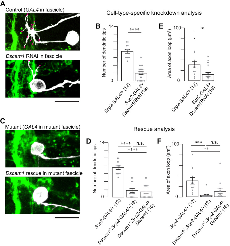
The formation and precise positioning of axons and dendrites are crucial for the development of neural circuits. Although juxtacrine signaling via cell-cell contact is known to influence these processes, the specific structures and mechanisms regulating neuronal process positioning within the central nervous system (CNS) remain to be fully identified. Our study investigates motoneuron 24 (MN24) in the Drosophila embryonic CNS, which is characterized by a complex yet stereotyped axon projection pattern, known as "axonal routing." In this motoneuron, the primary dendritic branches project laterally toward the midline, specifically emerging at the sites where axons turn. We observed that Scp2-positive neurons contribute to the lateral fascicle structure in the ventral nerve cord (VNC) near MN24 dendrites. Notably, the knockout of the Down syndrome cell adhesion molecule (Dscam1) results in the loss of dendrites and disruption of proper axonal routing in MN24, while not affecting the formation of the fascicle structure. Through cell-type specific knockdown and rescue experiments of Dscam1, we have determined that the interaction between MN24 and Scp2-positive fascicle, mediated by Dscam1, promotes the development of both dendrites and axonal routing. Our findings demonstrate that the holistic configuration of neuronal structures, such as axons and dendrites, within single motoneurons can be governed by local contact with the adjacent neuron fascicle, a novel reference structure for neural circuitry wiring.

Keywords: Axon; CNS; Drosophila; Dscam; dendrite; motoneuorn.


Conflict of interest statement

The authors declare no competing financial interests.

Figures

Figure 1.
Figure 1.
Neuronal fascicle spatially aligns with the position of MN24 dendrite formation. A, Top panel shows a schematic of MN24 (black) within the ventral nerve cord of an embryo at 15:00 AEL—all subsequent images are taken at 15:00 AEL unless otherwise specified. The axon stereotypically projects out of the soma, anteriorly along the edge of the longitudinal connective (LC), and away from the midline to target muscle 24 (M24). The bottom panel shows a representative fluorescence image of a lipophilic dye-labeled MN24. At 15:00 AEL, MN24 form their dendritic processes (magenta dots) at stereotyped positions on the axon routing. For all subsequent images, anterior is to the top, and medial is to the left. AC, anterior commissure. PC, posterior commissure. Scale bar, 10 µm. B, Representative fluorescence images of FasII-positive longitudinal fascicles within the ventral nerve cord. The stereotyped most lateral FasII-positive fascicle structure (arrowhead) provides a frame of reference to characterize the mediolateral position of MN24 dendrites. Gray dashed line depicts the midline. C, Distribution plots of the mediolateral positions of MN24 dendritic branches (white; n = 22 neurons) and FasII-positive lateral fascicle (dark gray; n = 77 hemisegments), where 0 µm indicates the distance from the CNS midline. We indicate the location of the measured area for each MN24 axonal loop in Extended Data Figure 1-1.
Figure 2.
Figure 2.
Dscam1 is required for MN24 neurite development. A, Representative fluorescence images of MN24 within wild-type (top panel) and Dscam1−/− mutant (bottom panel) backgrounds. B, C, Comparison of mean primary dendritic branch numbers (B) and axon routing areas. C, Within wild-type and Dscam1−/− mutant backgrounds; using Mann–Whitney U test. For all graphs, the sample size of neurons is denoted by the number in the parentheses of each genotype unless otherwise specified. For all subsequent statistical analyses, symbols indicate the following: ****p < 0.0001; ***p < 0.001; **p < 0.01; *p < 0.05; ns, not significant. D, Immunofluorescence staining of FasII at 15:00 AEL shows the visual comparison between axon terminals in wild-type (top panel) and Dscam1−/− mutant (bottom panel) backgrounds. Representative image displaying FasII staining in the wild-type and mutant backgrounds exhibits innervation by the SNa nerve branch (open circle). However, in the Dscam1−/− mutant background, the SNa sub-branches and the ISNb nerve have some mild targeting defects (magenta and green dots, respectively). E, Quantification of SNa innervation defects within wild-type (n = 76 hemisegments) and Dscam1−/− mutant (n = 67 hemisegments) backgrounds. Data is represented as a percentage—number of hemisegments with innervation defects over the total number of hemisegments observed. SNa innervation defects were characterized as mild (light gray) or severe (dark gray) when the SNa sub-branch had targeting defects or the SNa branch did not exit the nerve cord, respectively. F, Representative fluorescence images of FasII-positive axon tracts within wild-type (top panel) and Dscam1−/− mutant (bottom panel) backgrounds. G, Quantification of lateral fascicle defects within wild-type (n = 77 hemisegments) and Dscam1−/− mutant (n = 66 hemisegments) backgrounds. Data is represented as a percentage—length of lateral fascicle defects over total lateral fascicle length. Lateral fascicle defects were characterized as mild or severe when the lateral fascicle was thinning or contained a break, respectively. Scale bars, 10 µm in A, D, F.
Figure 3.
Figure 3.
MN24 axon routing supervenes proper cell body localization. AD, Representative fluorescence images of individually labeled MN24 cell bodies within the embryonic CNS in control (A, B) and Dscam1−/− mutant (C, D) backgrounds at different developmental stages (9:00 and 12:00 AEL, top and bottom panels, respectively). FLP recombinase-based stochastic labeling was used to genetically label MN24 within the hh-GAL4 pattern by expressing a membrane-bound epitope tag (FLAG). Dots represent the position of MN24 soma. Scale bar, 10 µm.
Figure 4.
Figure 4.
Dscam1 plays a cell-autonomous role for MN24 neurite development. A, Representative fluorescence images of MN24 in wild-type background expressing hh-GAL4 driver (top panel) and Dscam1 RNAi expressed under the control of the hh-GAL4 driver (bottom panel). B, E, Comparison of mean primary dendritic branch numbers (B) and axon routing areas (E) of MN24 in wild-type background expressing hh-GAL4 driver and Dscam1 RNAi expressed under the control of the hh-GAL4 driver; using Mann–Whitney U test. C, Representative fluorescence images of MN24 in Dscam1−/− mutant background expressing hh-GAL4 driver (top panel) and Dscam1−/− mutant background resupplied Dscam1 expressed under the control of the hh-GAL4 driver (bottom panel). D, F, Comparison of mean primary dendritic branch numbers (D) and axon routing areas (F) of MN24 in wild-type background expressing hh-GAL4 driver, Dscam1−/− mutant background expressing hh-GAL4 driver, and Dscam1−/− mutant background resupplied Dscam1 expressed under the control of the hh-GAL4 driver; using Kruskal–Wallis test followed by Dunn’s multiple-comparisons test. Scale bars, 10 µm in A, C.
Figure 5.
Figure 5.
Scp2-GAL4 driver allows labeling of lateral fascicle. A, Representative low-magnification images of a 15:00 AEL embryo labeled by membrane-bound GFP under the control of the Scp2-GAL4 driver. The expression pattern includes longitudinal fascicles but does not label neurons within the SNa nerve tract or afferent sensory axons. B, C, Representative high-magnification images in neuronal fascicles GFP-labeled under the control of Scp2-GAL4 driver (green) or immunostained with anti-FasII antibody (magenta). At 15:00 AEL, Scp2-positive fascicles include the medial and lateral fascicles (arrowheads) and exclude the intermedial fascicle (B). At 9:00 AEL, the Scp2-positive expression pattern labels an early fascicle that will later defasciculate in development (C). Scale bar, 40 µm in A and 10 µm in B, C.
Figure 6.
Figure 6.
Scp2-positive lateral fascicle provides noncell-autonomous Dscam1 for MN24 neurite development. A, Representative fluorescence images of MN24 in wild-type background expressing Scp2-GAL4 driver (top panel) and Dscam1 RNAi expressed under the control of the Scp2-GAL4 driver (bottom panel). B, E, Comparison of mean primary dendritic branch numbers (B) and axon routing areas (E) of MN24 in wild-type background expressing Scp2-GAL4 driver and Dscam1 RNAi expressed under the control of the Scp2-GAL4 driver, using Mann–Whitney U test. C, Representative fluorescence images of MN24 in Dscam1−/− mutant background expressing Scp2-GAL4 driver (top panel) and Dscam1−/− mutant background resupplied Dscam1 expressed under the control of the Scp2-GAL4 driver (bottom panel). D, F, Comparison of mean primary dendritic branch numbers (D) and axon routing areas (F) of MN24 in wild-type background expressing Scp2-GAL4 driver, Dscam1−/− mutant background expressing Scp2-GAL4 driver, and Dscam1−/− mutant background resupplied Dscam1 expressed under the control of the Scp2-GAL4 driver; using Kruskal–Wallis test followed by Dunn’s multiple-comparisons test. Scale bars, 10 µm in A, C. We show lateral fascicles in Dscam1 knockdown and knockout in Extended Data Figure 6-1 and MN24 soma position in Dscam1 knockout in Extended Data Figure 6-2.
Figure 7.
Figure 7.
Dscam1 in both Scp2-positive lateral fascicle and MN24 is sufficient to restore MN24 dendritogenesis and axon routing. A, Representative fluorescence images of MN24 within wild-type background (top panel), Dscam1−/− mutant background with combined Scp2- and MN24-specific expression of membrane-bound GFP (middle panel), and Dscam1−/− mutant background with combined Scp2- and MN24-specific resupply of Dscam1 (bottom panel). Scale bar, 10 µm. B, C, Comparison of mean primary dendritic branch numbers (B) and axon routing area (C) among MN24 in wild-type background, Dscam1−/− mutant background with Scp2- and MN24-specific expression of GFP membrane-bound, and Dscam1−/− mutant background with combined Scp2- and MN24-specific resupply of Dscam1, using Kruskal–Wallis test followed by Dunn’s multiple-comparisons test.
Figure 8.
Figure 8.
Proposed model for fascicle-mediated MN24 morphogenesis. A, Schematic illustrating the proposed model of how the fascicle structure mediates MN24 dendrite outgrowth and soma localization. B, Electron microscopy (EM) reconstruction of a single MN23/24 in first instar larva. Prominent morphological structures such as dendritic outgrowth and axon routing are retained in larval MN24. The backbone is indicated by gray. Blue dots indicate synaptic sites. Scale bar, 20 µm.

Update of

Similar articles

Cited by

References

    1. Alavi M, Song M, King GL, Gillis T, Propst R, Lamanuzzi M, Bousum A, Miller A, Allen R, Kidd T (2016) Dscam1 forms a complex with robo1 and the N-terminal fragment of slit to promote the growth of longitudinal axons. PLoS Biol 14:e1002560. 10.1371/journal.pbio.1002560 - DOI - PMC - PubMed
    1. Andrews GL, et al. (2008) Dscam guides embryonic axons by netrin-dependent and -independent functions. Development 135:3839–3848. 10.1242/dev.023739 - DOI - PMC - PubMed
    1. Avraham O, Hadas Y, Vald L, Hong S, Song MR, Klar A (2010) Motor and dorsal root ganglion axons serve as choice points for the ipsilateral turning of dI3 axons. J Neurosci 30:15546–15557. 10.1523/JNEUROSCI.2380-10.2010 - DOI - PMC - PubMed
    1. Avraham O, Hadas Y, Vald L, Zisman S, Schejter A, Visel A, Klar A (2009) Transcriptional control of axonal guidance and sorting in dorsal interneurons by the Lim-HD proteins Lhx9 and Lhx1. Neural Dev 4:21. 10.1186/1749-8104-4-21 - DOI - PMC - PubMed
    1. Brose K, Bland KS, Wang KH, Arnott D, Henzel W, Goodman CS, Tessier-Lavigne M, Kidd T (1999) Slit proteins bind Robo receptors and have an evolutionarily conserved role in repulsive axon guidance. Cell 96:795–806. 10.1016/S0092-8674(00)80590-5 - DOI - PubMed

Substances

LinkOut - more resources