

KIFC1 depends on TRIM37-mediated ubiquitination of PLK4 to promote centrosome amplification in endometrial cancer
- PMID: 39349439
- PMCID: PMC11442630
- DOI: 10.1038/s41420-024-02190-1
KIFC1 depends on TRIM37-mediated ubiquitination of PLK4 to promote centrosome amplification in endometrial cancer
Abstract
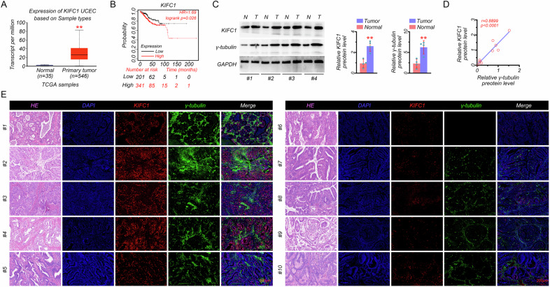
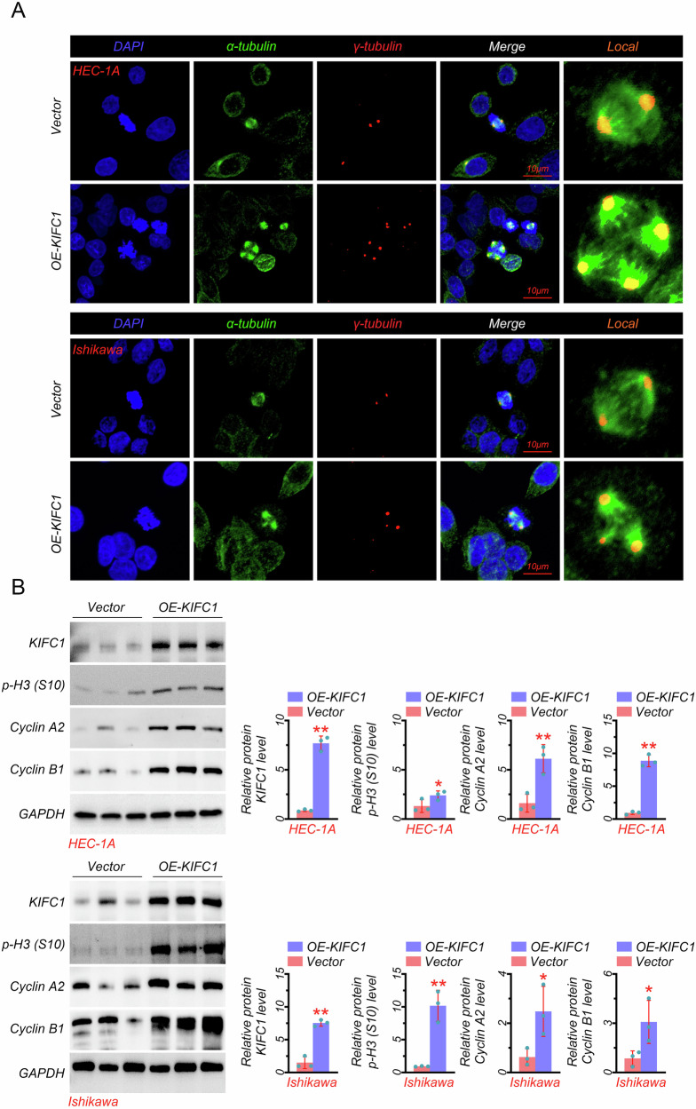
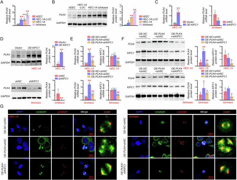
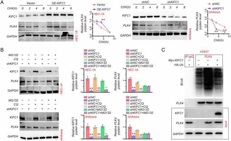
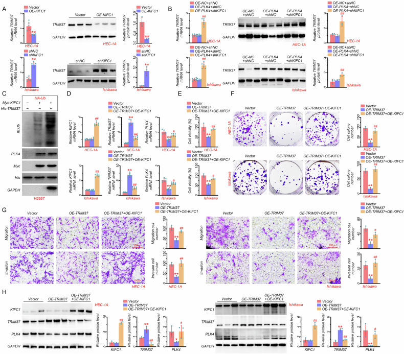
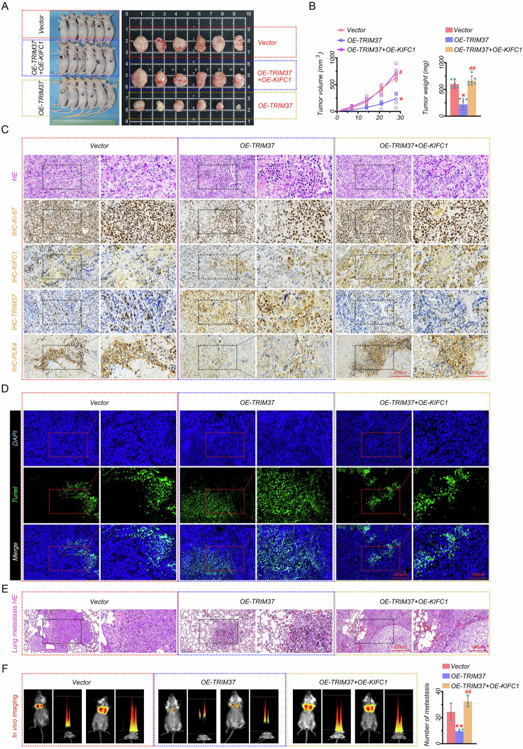
Endometrial cancer (EC), as one of the most common cancers, severely threatens female reproductive health. Our previous study has shown that Kinesin family member C1 (KIFC1) played crucial roles in the progression of EC. In addition, abnormal centrosome amplification, which was reported to be partially regulated by KIFC1, usually occurred in different cancers. However, whether KIFC1 promoted EC through centrosome amplification and the potential mechanism remain to be revealed. The present study demonstrated that overexpressed KIFC1, which exhibited a worse prognosis, had a positive correlation with an increased number of centrosomes in human EC samples. In addition, KIFC1 overexpression in EC cells prompted centrosome amplification, chromosomal instability, and cell cycle progression. Moreover, we demonstrated that KIFC1 inhibited E3 ubiquitin-protein ligase TRIM37 to maintain the stability of PLK4 by reducing its ubiquitination degradation, and finally promoting centrosome amplification and EC progression in vitro. Finally, the contributing role of KIFC1 and the inhibitory effect of TRIM37 on EC development and metastasis was verified in a nude mouse xenograft model. Our study elucidated that KIFC1 depends on TRIM37-mediated reduced ubiquitination degradation of PLK4 to promote centrosome amplification and EC progression, thus providing a potential prognostic marker and promising therapeutic target for EC in the future.
© 2024. The Author(s).
Conflict of interest statement
The authors declare no competing interests.
Figures

Similar articles
-
TRIM37 controls cancer-specific vulnerability to PLK4 inhibition.Nature. 2020 Sep;585(7825):440-446. doi: 10.1038/s41586-020-2710-1. Epub 2020 Sep 9. Nature. 2020. PMID: 32908304 Free PMC article.
-
KIFCI, a novel putative prognostic biomarker for ovarian adenocarcinomas: delineating protein interaction networks and signaling circuitries.J Ovarian Res. 2014 May 12;7:53. doi: 10.1186/1757-2215-7-53. eCollection 2014. J Ovarian Res. 2014. PMID: 25028599 Free PMC article.
-
Kinesin Family Member C1 (KIFC1) Accelerates Proliferation and Invasion of Endometrial Cancer Cells Through Modulating the PI3K/AKT Signaling Pathway.Technol Cancer Res Treat. 2020 Jan-Dec;19:1533033820964217. doi: 10.1177/1533033820964217. Technol Cancer Res Treat. 2020. PMID: 33034273 Free PMC article.
-
Computational benchmarking of putative KIFC1 inhibitors.Med Res Rev. 2023 Mar;43(2):293-318. doi: 10.1002/med.21926. Epub 2022 Sep 14. Med Res Rev. 2023. PMID: 36104980 Review.
-
TRIM37: a critical orchestrator of centrosome function.Cell Cycle. 2021 Dec;20(23):2443-2451. doi: 10.1080/15384101.2021.1988289. Epub 2021 Oct 21. Cell Cycle. 2021. PMID: 34672905 Free PMC article. Review.
References
-
- Hevir-Kene N, Rižner TL. The endometrial cancer cell lines Ishikawa and HEC-1A, and the control cell line HIEEC, differ in expression of estrogen biosynthetic and metabolic genes, and in androstenedione and estrone-sulfate metabolism. Chem Biol Interact. 2015;234:309–19. - PubMed
-
- Burke WM, Orr J, Leitao M, Salom E, Gehrig P, Olawaiye AB, et al. Endometrial cancer: a review and current management strategies: part I. Gynecol Oncol. 2014;134:385–92. - PubMed
-
- Parvin A, Wei BH, Hao SL, Yang WX, Tan FQ. KIFC1 overexpression promotes prostate cancer cell survival and proliferation by clustering of amplified centrosomes via interaction with Centrin 2. Biocell. 2021;45:1369–91.
LinkOut - more resources
Full Text Sources
