. 2024 Sep 30;14(1):22593.
doi: 10.1038/s41598-024-73358-z.

The IgLON family of cell adhesion molecules expressed in developing neural circuits ensure the proper functioning of the sensory system in mice

Affiliations

The IgLON family of cell adhesion molecules expressed in developing neural circuits ensure the proper functioning of the sensory system in mice

Katyayani Singh et al. Sci Rep. .

Abstract

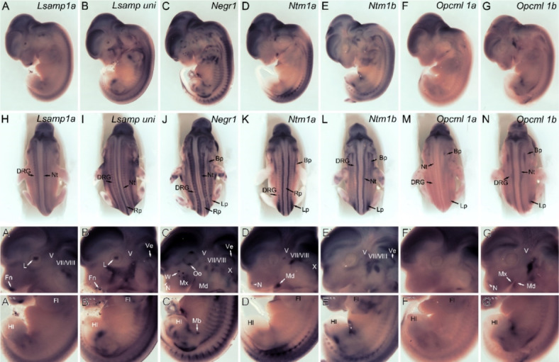
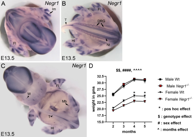
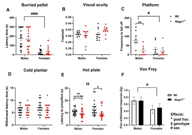
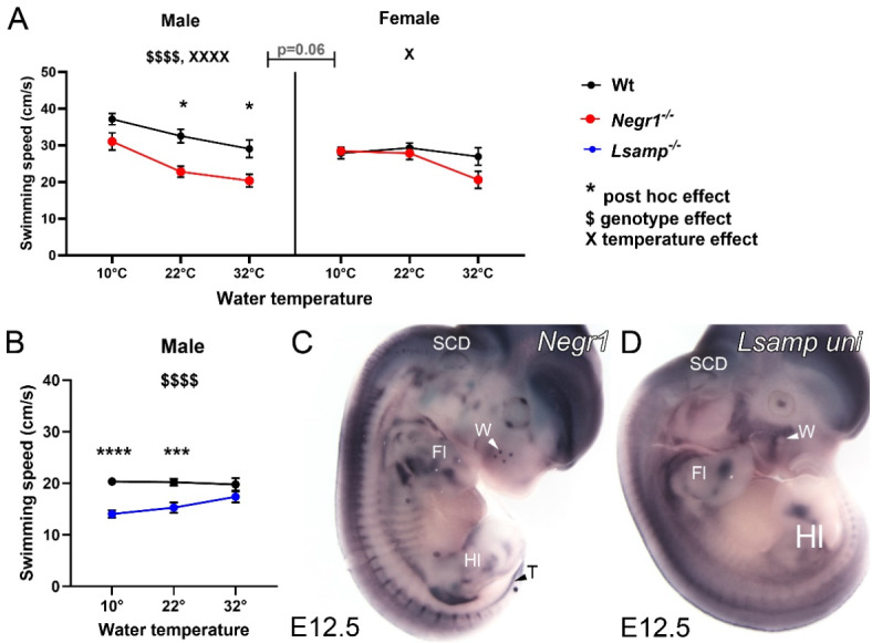
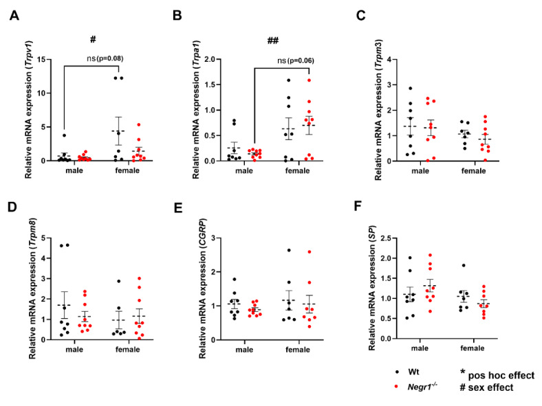
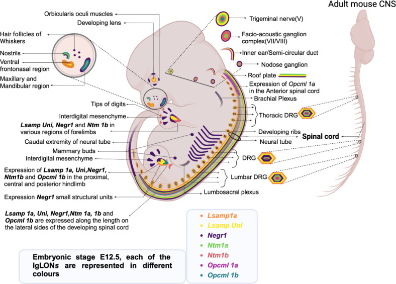
Deletions and malfunctions of the IgLON family of cell adhesion molecules are associated with anatomical, behavioral, and metabolic manifestations of neuropsychiatric disorders. We have previously shown that IgLON genes are expressed in sensory nuclei/pathways and that IgLON proteins modulate sensory processing. Here, we examined the expression of IgLON alternative promoter-specific isoforms during embryonic development and studied the sensory consequences of the anatomical changes when one of the IgLON genes, Negr1, is knocked out. At the embryonal age of E12.5 and E13.5, various IgLONs were distributed differentially and dynamically in the developing sensory areas within the central and peripheral nervous system, as well as in limbs and mammary glands. Sensory tests showed that Negr1 deficiency causes differences in vestibular function and temperature sensitivity in the knockout mice. Sex-specific differences were noted across olfaction, vestibular functioning, temperature regulation, and mechanical sensitivity. Our findings highlight the involvement of IgLON molecules during sensory circuit formation and suggest Negr1's critical role in somatosensory processing.

Keywords: DRG; IgLON; LSAMP; NEGR1; PNS; Sensory processing.


Conflict of interest statement

The authors declare no competing interests.

Figures

Fig. 1
Fig. 1
At embryonic age, E12.5 IgLONs are expressed in mice’s sensory precursor cells and spinal cord. Expression of Lsamp 1a, Lsamp uni, Negr1, Ntm 1a, Ntm 1b, Opcml 1a, and Opcml 1b, detected by whole-mount mRNA in situ hybridization. (AG) lateral and (HN) dorsal views of whole embryos. (A′–G′ and A″–G″) higher magnification images of the corresponding expression from the head and posterior body/hindlimb regions, respectively. Bp brachial plexus, DRG dorsal root ganglia, Fl forelimb, Fn frontonasal region, Hl hindlimb, Ve inner ear, L lens, Lp lumbosacral plexus, Mb mammary bud, Md mandibular, Mx maxilla, N nostril, Nt neural tube, Oo orbicularis occuli, Rp roof plate, W whiskers follicle, V trigeminal ganglion, VII–VIII facio-acoustic ganglion, X nodose ganglion.
Fig. 2
Fig. 2
Distribution of IgLON family alternative promoters at embryonal age E13.5 in mice. Expression of Lsamp 1a, Lsamp uni, Negr1, Ntm 1a, Ntm 1b, Opcml 1a, and Opcml 1b mRNA was detected by whole-mount in situ hybridization wild-type embryos. (AG) lateral and (HN) dorsal views of whole embryos. (A′–G′ and A″–G″) higher magnification images of corresponding mRNA probe expression from head and limb regions, respectively. DRG dorsal root ganglia, Fl forelimb, Hl hindlimb, Im interdigital mesenchyme, L lens, Mb mammary bud, N nostril, Nt neural tube, Ol olfactory lobe, Oo orbicularis occuli, Rp roof plate, W whiskers follicle.
Fig. 3
Fig. 3
Negr1 mRNA is expressed in developing sensory organs and body weight dynamics of adult Negr1−/− and wild-type male and female mice cohort used in the study. (AC) Whole-mount in situ hybridization for Negr1 in wild-type (Wt) embryos at E13.5 revealed strong expression in interdigital mesenchyme of developing forelimbs and hindlimbs, dorsal root ganglia, spinal cord, and tail region. (A) top, (B) bottom, (C) front view of whole-mount embryo. (D) Body weight dynamics of Wt and Negr1−/− male and female mice cohort used for the sensory testing. Two-way ANOVA and Three-way ANOVA with Bonferroni’s post hoc test were used to detect significant effects of genotype, sex, and monthly age on body weight from 2 to 5 months of mice (n = 20 mice in each group). For all plots, data show means ± SEM. Significance is denoted as follows: * for significant post hoc comparisons, $ for genotype, # for sex and ^ for months effects, *p < 0.05, $$p < 0.01, ####p < 0.0001. DRG dorsal root ganglia, FL forelimb, HL hindlimb, T the caudal extremity of the neural tube in the tail region.
Fig. 4
Fig. 4
Deletion of Negr1 resulted in impaired sensory function in mice (A) olfactory function test by buried food pellet test, (B) visual acuity test using optometer (C) equilibrium/balance at heights test done by using the elevated platform for frequency to fall off (D) cold sensitivity is measured by cold plantar test and (E) thermal allodynia is measured by hot plate test (F) mechanical sensitivity by 2 g Von Frey filament. For all plots, data show means ± SEM. Differences between the genotypes were tested by t-test or Mann–Whitney test. Two-way ANOVA, with post hoc Bonferroni comparisons, was used to check sex effects within the genotype between groups. Significance is denoted as follows: and * for significant comparisons between genotypes; *p < 0.05, **p < 0.01, ***p < 0.001 and #p < 0.05, ####p < 0.0001 for sex effects. Sample size was n = 20 in each group, i.e., each group containing male Wt (n = 20) and Negr1−/− (n = 20) mice, in female Wt (n = 20) and Negr1−/− (n = 20) mice, except n = 8–10 in each group for visual acuity and platform test.
Fig. 5
Fig. 5
Effect of water temperature on swimming speed in Negr1−/− and Lsamp−/− mice; and representative illustration showing complementary activity of Negr1 and Lsamp uni mRNA probes in developing sensory precursors. (A) The difference in swimming speed of Negr1−/− males, Negr1−/− females with Wt mice and (B) in Lsamp−/− males at different water temperatures (10 °C, 22 °C, 32 °C) (C) Complementary expression of Negr1 and Lsamp uni probes were observed in developing whiskers and in limb precursors at E12.5 embryo. For the plot, data show means ± SEM. Two-way ANOVA was used with post hoc Bonferroni comparisons to check genotype and water temperature effects within the genotype between groups. Three-way ANOVA was conducted only with Negr1−/− male and female data sets to check sex differences among all the groups. Significance is denoted as follows: $ for post hoc comparisons between genotypes; $$$$ p < 0.0001, X for temperature difference, X p < 0.05, XXXX p < 0.0001 temperature effects and *for post hoc from Two-way ANOVA *p < 0.05, ***p < 0.001, ****p < 0.0001. The sample size was n = 16–18 in each group. FL forelimb, HL hindlimb, SCD semi-circular duct, T the caudal extremity of the neural tube, W whisker follicle.
Fig. 6
Fig. 6
Relative mRNA expression levels of transient receptor potential channels and neuropeptides in DRG of Wt and Negr1−/− mice from both males and females. (A) Trpv1, (B) Trpa1, (C) Trpm3, (D) Trpm8, (E) CGRP (Calcitonin gene-related peptide), and (F) SP (Substance P). For all plots, data show means ± SEM. Differences between the genotypes were tested by t-test or Mann–Whitney test. Two-way ANOVA, with post hoc Bonferroni comparisons, was used to check sex and interaction effects within the genotype between groups. Significance is denoted as follows: and * for post hoc significant comparisons between genotypes; *p < 0.05, and #p < 0.05, ##p < 0.01 for sex effects. The sample size was n = 10 in each group, i.e., each group containing male and female of Wt and Negr1−/− mice; outliers were identified using the ROUT test.
Fig. 7
Fig. 7
Schematic illustration of IgLON molecule transcripts expressions at embryonic age E121.5 in developing sensory and peripheral structures in mice. (Image is created using Biorender).

Similar articles

References

    1. Catala, M. & Kubis, N. Gross anatomy and development of the peripheral nervous system. Handb. Clin. Neurol.115, 29–41. 10.1016/B978-0-444-52902-2.00003-5 (2013). - PubMed
    1. Baum, M. J. & Cherry, J. A. Processing by the main olfactory system of chemosignals that facilitate mammalian reproduction. Horm. Behav.68, 53–64. 10.1016/j.yhbeh.2014.06.003 (2015). - PubMed
    1. Burke, K. & Kobrina, A. Rodentia sensory systems. In Encyclopedia of Animal Cognition and Behavior (eds Vonk, J. & Shackelford, T. K.) (Springer, 2022). 10.1007/978-3-319-55065-7_765.
    1. Francis-West, P. H., Ladher, R. K. & Schoenwolf, G. C. Development of the sensory organs. Sci. Prog.85, 151–173. 10.3184/003685002783238852 (2002). - PMC - PubMed
    1. Collado, M. S. et al. The postnatal accumulation of junctional E-cadherin is inversely correlated with the capacity for supporting cells to convert directly into sensory hair cells in mammalian balance organs. J. Neurosci.31, 11855–11866. 10.1523/JNEUROSCI.2525-11.2011 (2011). - PMC - PubMed

Substances

LinkOut - more resources