. 2024 Sep 30;7(1):1222.
doi: 10.1038/s42003-024-06917-9.

STAT1 as a tool for non-invasive monitoring of NK cell activation in cancer

Affiliations

STAT1 as a tool for non-invasive monitoring of NK cell activation in cancer

Jin Young Min et al. Commun Biol. .

Abstract

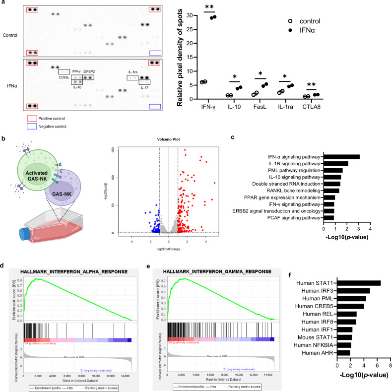
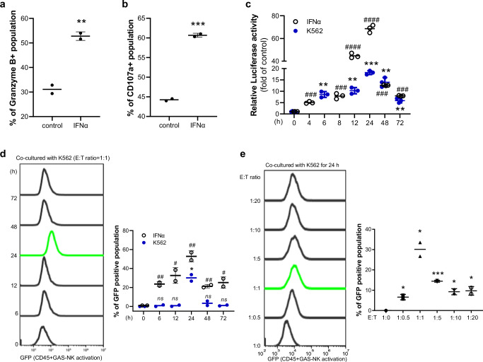
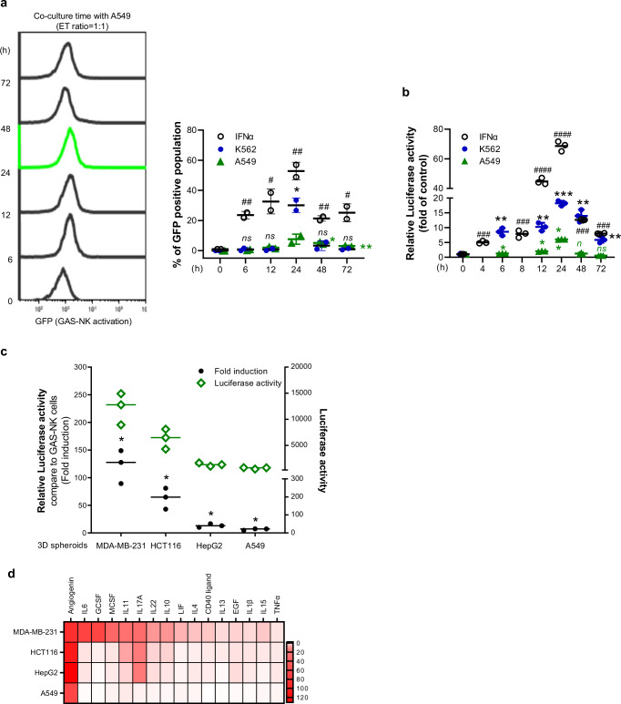
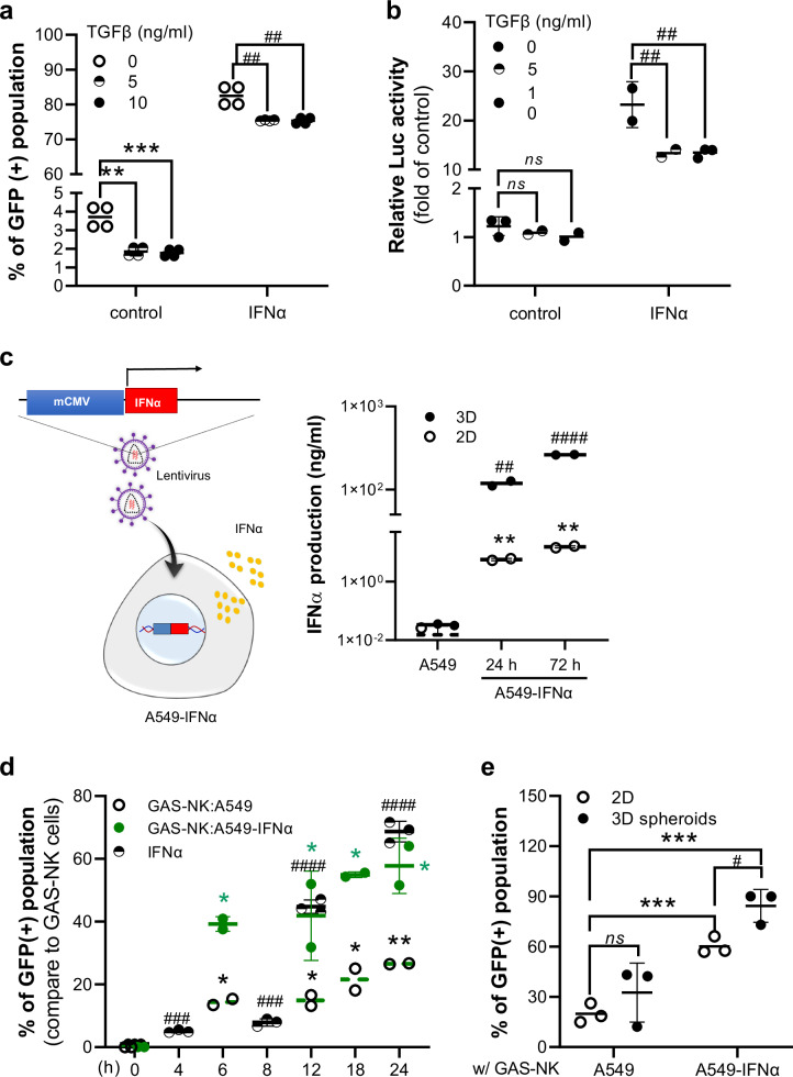
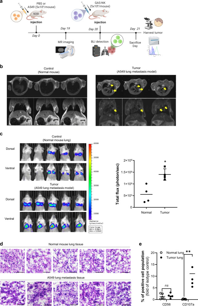
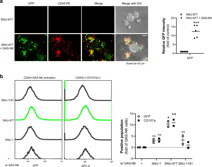
Natural killer (NK) cells play a crucial role in immunotherapy for cancer due to their natural ability to target and destroy cancer cells. However, current methods to visualize NK cells' activity against tumors in live organisms are limited. We introduce an imaging method that non-invasively tracks NK cell activation by cancer cells through the STAT1 protein. To achieve this, we modified NK cells to include a specific genetic sequence that binds to STAT1 when activated. These engineered NK cells (GAS-NK) demonstrate their functionality through various biological tests and analysis. Observations of changes in cancer environments and patient-derived cancer organoid models further confirm the effectiveness of this approach. Our method provides a way to monitor NK cell activity, which could improve the prediction and effectiveness of NK cell-based cancer therapies, contributing to advances in cancer treatment.


Conflict of interest statement

The authors declare no competing interests.

Figures

Fig. 1
Fig. 1. Visualization of IFNα-activated NK cell monitoring in vitro and in vivo.
a Illustrative diagram depicting the STAT1 activation mechanism in GAS-NK cells (created with BioRender.com). b Graph representing relative luciferase activity in GAS-NK cells over different treatment durations with IFNα (5000 units/mL). (n = 3, samples for all time points). c Quantitative analysis highlighting the increase in GFP-positive GAS-NK cell populations in response to IFNα exposure. (n = 3). d Immunoblot assay results illustrating the phosphorylation and total protein expression levels of STAT1 in GAS-NK cells, comparing IFNα-treated and untreated conditions. e Comparative in vivo imaging of control versus IFNα-primed GAS-NK cells (5 × 106/mouse), demonstrating significant differences in luminescent signaling. (n = 3 for both GAS-NK and primed GAS-NK). Statistical significance between groups was determined using paired Student’s t-tests, with significance levels denoted as follows: **P < 0.01; ***P < 0.001; ****P < 0.0001; ns, not significant.
Fig. 2
Fig. 2. Comparative analysis of GAS-NK cells with and without IFNα treatment.
a Human cytokine array analysis contrasting the secretion profiles of various cytokines by GAS-NK cells treated with IFNα for 24 hours versus untreated cells. The array images depict cytokine profiles for both conditions, with the upper image showing untreated GAS-NK cells and the lower one showing IFNα-treated cells. The accompanying bar graph quantifies cytokine levels, highlighting differences in the secretion of cytokines such as IFNγ, IL-10, Fas ligand, IL-1ra, and CTLA8 between the groups. (n  =  2; significance levels are denoted as follows: *P < 0.05; **P < 0.01). b A volcano plot highlights differentially expressed genes in GAS-NK cells following 24-hour IFNα treatment compared to untreated cells, with significant upregulation (red dots) and downregulation (blue dots) indicated. c A clustered bar chart presents Gene Ontology (GO) analysis of differentially expressed genes (DEGs), focusing on various signaling pathways. GSEA enrichment plots display gene clusters enriched in IFNα-treated GAS-NK cells over 24 hours, emphasizing (d) response to IFNα and (e) response to IFNγ. f Clustered bar chart analysis identifies transcription factors, with human STAT1 showing the highest significance.
Fig. 3
Fig. 3. Assessment of enhanced cytotoxicity and selective imaging in IFNα-stimulated GAS-NK cells co-cultured with K562 cells.
Increased expression of cytotoxic markers granzyme B (a) and CD107a (b) in IFNα-treated GAS-NK cells (5,000 U/mL) compared to controls (n = 2, control and IFNα). c, d Time-course analysis of luciferase activity (n = 3; samples for all time points) and GFP levels (n = 2; control and IFNα) in IFNα-treated and K562 co-cultured GAS-NK cells, showing significant variations (IFNα treatment: #; K562 co-culture: *). e Quantification of GFP-positive GAS-NK cells at different E:T ratios (1:0, 1:0.5, 1:1, 1:5, 1:10, 1:20) in co-culture with K562 cells (n = 2, control and IFNα). Statistical significance between groups was determined using paired Student’s t-tests, with significance levels denoted as follows: ** or ##, P < 0.01; *** or ###, P < 0.001; **** or ####, P < 0.0001; ns, not significant.
Fig. 4
Fig. 4. Impact of GAS-NK cell activation in co-culture with solid cancer cells.
a Histogram illustrating GFP intensity changes over time in GAS-NK cells co-cultured with A549 cells at an E:T ratio of 1:1. The accompanying quantitative graph combines these results with those from IFNα treatment and K562 cell co-culture (Fig. 3d), highlighting significant variations (A549 co-culture: *; IFNα treatment: #; K562 co-culture: *, blue color). (n = 2, samples for all time points). b Luciferase activity in GAS-NK cells co-cultured with A549 at an E:T ratio of 1:1, compared with results from IFNα treatment and K562 cell co-culture (Fig. 3c) (A549 co-culture: *; IFNα treatment: #; K562 co-culture: *, blue color). (n = 3, samples for all time points). c Line graph comparing luciferase activity of GAS-NK cells co-cultured with various solid tumor cell 3D spheroids (MDA-MB-231, HCT116, HepG2, A549) to GAS-NK cells alone. (n = 3, all other samples). d Tile plot indicating cytokine production levels in culture supernatants from four solid cancer cell lines, with color intensity changing from white to red indicating higher cytokine levels. Statistical significance between groups was determined using paired Student’s t-tests, with significance levels denoted as follows: * or #, P < 0.05; ** or ##, P < 0.01; *** or ###, P < 0.001; **** or ####, P < 0.0001; ns, not significant.
Fig. 5
Fig. 5. Analyzing GAS-NK cell response to tumor microenvironment cytokines.
a Response of GAS-NK cells to TGFβ (5 or 10 ng/ml) treatment, illustrated by changes in GFP intensity. A quantitative graph compares responses to IFNα and TGFβ treatments. Statistical significance is indicated in comparison to control and IFNα-treated cells (control: *; IFNα treatment: #). (n = 2, all other samples). b Luciferase activity of GAS-NK cells under the same treatment conditions, with statistical analysis against control and IFNα treatments (control: *; IFNα treatment: #; n = 3, control and IFNα). c Schematic of the A549-IFNα cell line and a comparative analysis of IFNα secretion levels in 2D and 3D cultures of A549 and A549-IFNα cells (statistical significance indicated for 2D vs. 3D comparisons; n = 3, control; * and IFNα; #). d Percentage of GFP-positive GAS-NK cells over time when co-cultured with A549 or A549-IFNα cells (E:T ratio = 1:1), integrating results from previous figures (IFNα treatment: *; A549: #; A549-IFNα: *, green color; n = 2 or 3, samples for all time points). e Graph quantifying the GFP( + ) GAS-NK cell population when co-cultured with 2D or 3D spheroids of A549 and A549-IFNα cells for 24 hours (n = 3, samples for all time points). (***P < 0.001; ns, not significant).
Fig. 6
Fig. 6. Tumor site-specific activation monitoring of GAS-NK cells in an A549 lung metastasis model.
a Time-lapse schematic of the experimental procedure involving A549 and GAS-NK cell injections, MRI imaging, BLI detection, and tumor tissue analysis in SCID mice (created with BioRender.com). b MRI images confirming lung metastasis model formation 19 days post-A549 tail vein injection, with tumors indicated by yellow arrows. c BLI images of control (top) and tumor groups (bottom) following tail vein injection of GAS-NK cells into normal and A549 lung metastasis SCID mice. Each image represents an independent experiment, with luminescence quantified as total flux photons/sec from targeted body areas. Statistical significance is indicated (*P < 0.05). (n = 5, control and tumor). d H&E stained lung tissue from normal and tumor-bearing mice, 24 hours post-GAS-NK cell injection. e Comparison of the percentages of CD56 and CD107a positive NK cell populations in lung tissues from normal and lung metastasis mice, with statistical significance noted (**P < 0.01; ns, not significant). (n = 3, control and tumor). Statistical significance between groups was determined using paired Student’s t-tests, with significance levels denoted as follows: *P < 0.05; **P < 0.01; ns, not significant.
Fig. 7
Fig. 7. Assessing GAS-NK cell activation with patient-derived cancer organoids.
a Fluorescence images of GFP and CD45-PE (red) post co-culture of tumor organoids (SNU-977) with CD45-PE stained GAS-NK cells (left panel). The accompanying graph quantifies GFP intensity in CD45-positive GAS-NK cells (right panel), with statistical significance indicated (**P < 0.001). (n = 5, SNU-977 and SNU-977 + GAS-NK). b Flow cytometry results of GFP and CD107a(+) populations in GAS-NK cells after 24-hour co-culture with three types of cancer organoids (SNU-1, SNU-977, SNU-1181) at an E:T ratio of 1:1. Bar charts depict fold changes in GFP and CD107a(+) populations in CD45( + )-GAS-NK cells (n = 2, all other samples). Statistical significance between groups was determined using paired Student’s t-tests, with significance levels denoted as follows: *P < 0.05; **P < 0.01; ***P < 0.001; ns, not significant.

Similar articles

Cited by

References

    1. Vivier, E., Tomasello, E., Baratin, M., Walzer, T. & Ugolini, S. Functions of natural killer cells. Nat. Immun.9, 503–510 (2008). - PubMed
    1. Krey, K., Babnis, A. W. & Pichlmair, A. System-Based Approaches to Delineate the Antiviral Innate Immune Landscape. Viruses12, 1196 (2020). - PMC - PubMed
    1. Islam, R., Pupovac, A., Evtimov, V. & Boyd, N. Sandrin MS. Enhancing a Natural Killer: Modification of NK Cells for Cancer Immunotherapy. Cells10, 1058 (2021). - PMC - PubMed
    1. Judge, S. J., Murphy, W. J. & Canter, R. J. Characterizing the Dysfunctional NK Cell: Assessing the Clinical Relevance of Exhaustion, Anergy, and Senescence. Frontiers in Cellular and Infection Microbiology10, 49 (2020). - PMC - PubMed
    1. Gangadaran, P. & Ahn, B. C. Molecular Imaging: A Useful Tool for the Development of Natural Killer Cell-Based Immunotherapies. Frontiers in Immunology8, 1090 (2017). - PMC - PubMed

Substances