. 2024 Sep 30;7(1):222.
doi: 10.1038/s42004-024-01304-1.

The molecular picture of the local environment in a stable model coacervate

Affiliations

The molecular picture of the local environment in a stable model coacervate

Atanu Baksi et al. Commun Chem. .

Abstract

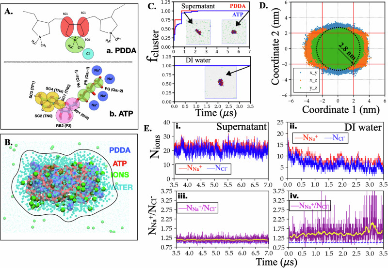
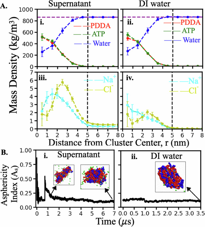
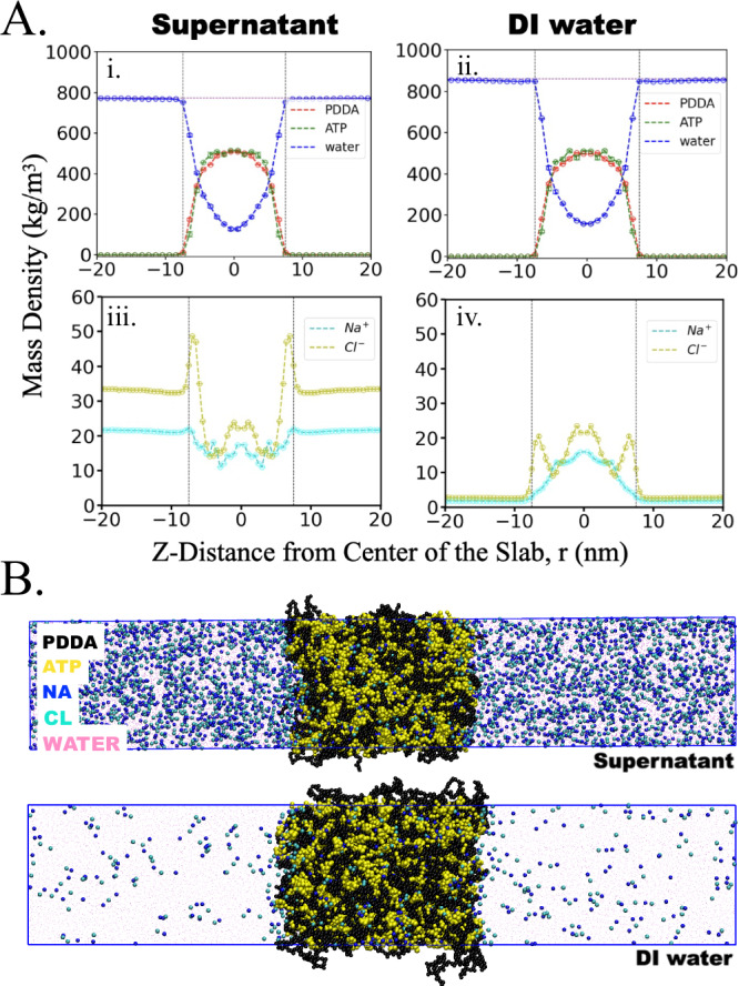
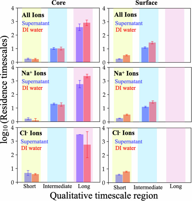
Complex coacervates play essential roles in various biological processes and applications. Although substantial progress has been made in understanding the molecular interactions driving complex coacervation, the mechanisms stabilizing coacervates against coalescence remain experimentally challenging and not fully elucidated. We recently showed that polydiallyldimethylammonium chloride (PDDA) and adenosine triphosphate (ATP) coacervates stabilize upon their transfer to deionized (DI) water. Here, we perform molecular dynamics simulations of PDDA-ATP coacervates in supernatant and DI water, to understand the ion dynamics and structure within stable coacervates. We found that transferring the coacervates to DI water results in an immediate ejection of a significant fraction of small ions (Na+ and Cl-) from the surface of the coacervates to DI water. We also observed a notable reduction in the mobility of these counterions in coacervates when in DI water, both in the cluster-forming and slab simulations, together with a lowered displacement of PDDA and ATP. These results suggest that the initial ejection of the ions from the coacervates in DI water may induce an interfacial skin layer formation, inhibiting further mobility of ions in the skin layer.


Conflict of interest statement

The authors declare no competing interests.

Figures

Fig. 1
Fig. 1. System definition, coacervate formation, and ionic nature of the coacervates.
A Schematic representation of atomistic-to-coarse-grained (CG) mapping of PDDA monomer, ATP molecule implemented in this work. B A snapshot of PDDA-ATP coacervate with associated ions and water beads just before transferring it to DI water. Counterions and water molecules enclosed in the black curved boundary (a curved surface in 3D) are at a distance smaller than 1 nm to any PDDA or ATP beads. Counterions and water outside this surface are discarded while transferring. C Time evolution of the fraction of PDDA and ATP molecules in the largest cluster (fcluster) in the supernatant (top) and DI water (bottom) for a selected simulation set. The same for all 8 sets are shown in Supplementary Figs. 18 and 19, respectively, for supernatant and DI water. We consider the cluster fully formed after fcluster reaches a stable value of 1.0. The snapshots in the insets are the simulation box at indicated simulation times where PDDA, ATP, and small ions beads are colored red, blue, and green, respectively. D Projection of the clustered PDDA and ATP beads' (coacervates) positions on three principal planes of the cluster for a selected set. The same for all 8 sets is shown in Supplementary Fig. 20. This projection informs about the coacervate size and shape (e.g., asphericity). We also used these projections to label the “surface” and “core” of coacervates. The overlapping rectangular area is used to identify a core represented by a sphere with a radius of rcore = 2.8 nm. In between rcore = 2.8 nm and rsurface = 5 nm is considered as surface. Here, coordinate 1 and 2 are the pairs of principal axes orthogonal to the projected axis. Three colored regions are projections of PDDA and ATP beads on three principal planes. E Time evolution of the number of Na+ and Cl ions within the coacervate in “supernatant” after the coacervate forms (i) and “DI water” (ii) for a selected set. Time evolution of the ratio of the number of Na+ to Cl ions within the coacervate in “supernatant” (iii) and “DI water” (iv) for a selected set. The same for all 8 sets are shown in Supplementary Figs. 1 and 2, respectively, for supernatant and DI water. Yellow lines in the lower panel indicate a running average (which is higher than 1) both in supernatant and DI water.
Fig. 2
Fig. 2. Density profiles and asphericity of the coacervates.
A Mass density profiles of PDDA, ATP, water, and ions in supernatant (left panels) and DI water (right panels). Error bars are the standard deviation of sample means. The density profiles are averaged over eight independent simulations. The vertical line at 5 nm is the guiding line indicating the maximum extent of the cluster. The horizontal line at 862 kg/m3 refers to equilibrated MARTINI 2.0 water density at 298 K. The molecular weight of each PDDA monomer in its fully dissociated state is 126.21 g/mol and that of an ATP molecule at the protonation state of −4e is 503.2 g/mol. The molecular weight of water, Na+ and Cl ions are 18.01, 22.99, and 35.45 g/mol, respectively. B Time evolution of asphericity index of the largest cluster in (i) supernatant (ii) DI water for a selected run. The same for all eight sets are shown in Supplementary Figs. 5 and 6 for supernatant and DI water, respectively. The relative (relative to mean) standard deviation in the last 2 microseconds simulation data averaged over all (8) sets is found to be 10.8% and 7.6% in supernatant and DI water, respectively. Attached snapshots in the inset are the clusters at indicated simulation times where PDDA, ATP, and ion beads are colored in red, blue, and green, respectively.
Fig. 3
Fig. 3. Density profiles and snapshots of slab simulations.
A Density profiles of (i) PDDA, ATP, and water in the supernatant, (ii) PDDA, ATP, and water in DI water, (iii) Na+, and Cl in the supernatant, and (iv) Na+, and Cl in DI water. B Snapshot of the PDDA-ATP slab with counterions and water beads (transparent pink) in the supernatant (top) and DI water (bottom).
Fig. 4
Fig. 4. Signature of rapid ejection of ions from coacervates upon transferring them to DI water.
The ions located within a spherical region of radius rcore = 2.8 nm are labeled as core ions whereas the ions located in the spherical region between rcore = 2.8 nm and rsurface = 5 nm labeled as surface ions at t = 0 ns. Results are shown for one simulation set. Results for other sets are shown in the Supplementary Fig. 20). Ion ejection from the surface (A) and from the core (B). The top panels in A and B show the time evolution of the label change of the ions initially labeled as “surface” and the time evolution of the label change of the ions initially labeled as “core”, respectively, after coacervates transferred to DI water. The initial 100 ns of the simulation data is zoomed in on the bottom panels of A and B to show the initial rapid ejection both from the surface (A) and from the core (B). C Schematic diagram of the ejection of initially-core and initially-surface ions.
Fig. 5
Fig. 5. Comparison of characteristic timescales (log10(τ)) describing ion residence correlation decays in “core” (left panel) and “surface” region (right panel).
The top panels report the timescales associated with overall ion dynamics whereas the middle and bottom panels report the same separately for Na+ ions and Cl ions, respectively. Each plot compares the dynamics of ions in the supernatant and DI water system.

Similar articles

Cited by

  • Coacervation in systems chemistry.
    Leon L, Santiago GM, Spruijt E. Leon L, et al. Commun Chem. 2024 Nov 22;7(1):275. doi: 10.1038/s42004-024-01358-1. Commun Chem. 2024. PMID: 39578544 Free PMC article.

References

    1. Bungenberg de Jong, H. & Kruyt, H. Coacervation (partial miscibility in colloid systems). Proc. K. Ned. Akad. Wet32, 849–856 (1929).
    1. Wagner, R. Some comments and questions äbout the germinal vesicular germinativa. Müllers Arch. Anat. Physiol. Sci. Med.268, 373–377 (1835).
    1. Valentin, G. Repertorium für anatomie und physiologie (Veit, 1837).
    1. Schwann, T. & Hünseler, F. Microscopic Investigations ön the Correspondence in the Structure and Growth of Animals and Plants 176 (W. Engelmann, 1910).
    1. Frankel, E. A., Bevilacqua, P. C. & Keating, C. D. Polyamine/nucleotide coacervates provide strong compartmentalization of mg2+, nucleotides, and RNA. Langmuir32, 2041–2049 (2016). - PubMed

LinkOut - more resources