. 2024 Oct;56(10):2246-2259.
doi: 10.1038/s12276-024-01322-y. Epub 2024 Oct 1.

CBL-b E3 ligase-mediated neddylation and activation of PARP-1 induce vascular calcification

Affiliations

CBL-b E3 ligase-mediated neddylation and activation of PARP-1 induce vascular calcification

Duk-Hwa Kwon et al. Exp Mol Med. 2024 Oct.

Abstract

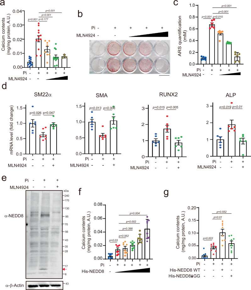
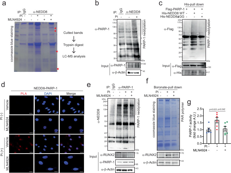
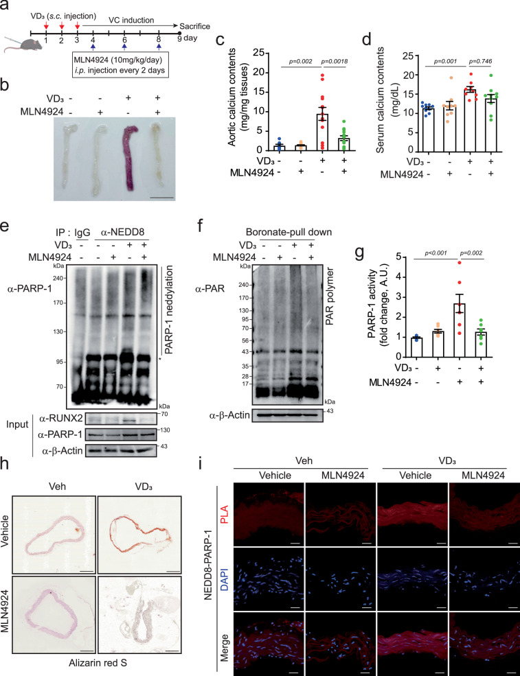
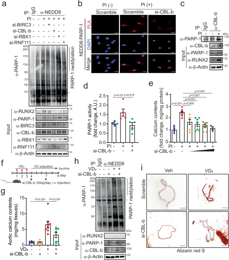
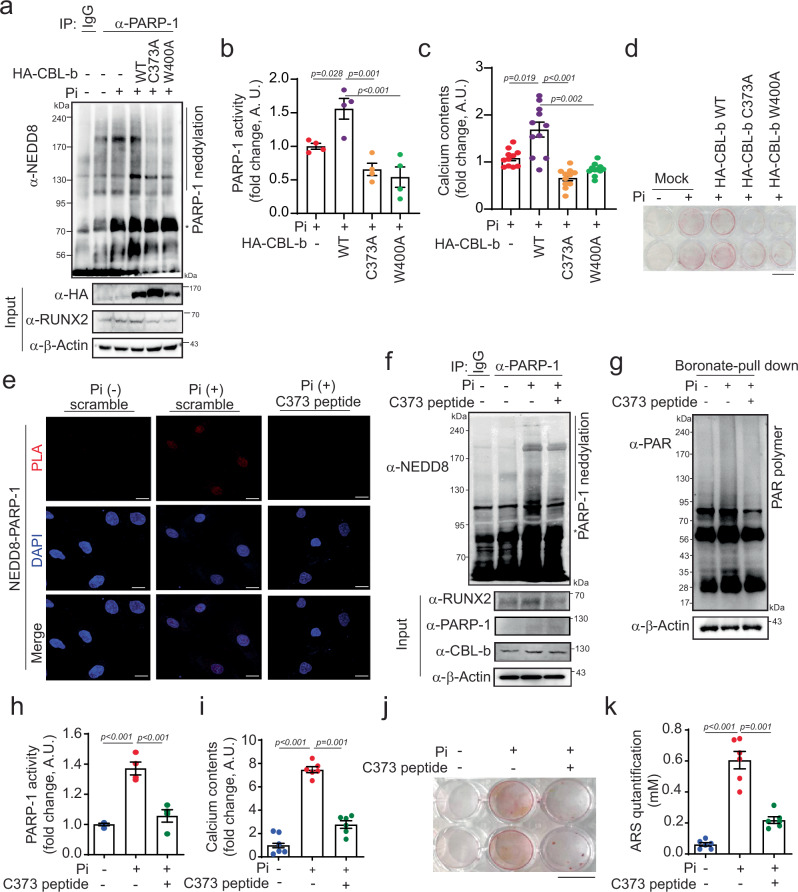
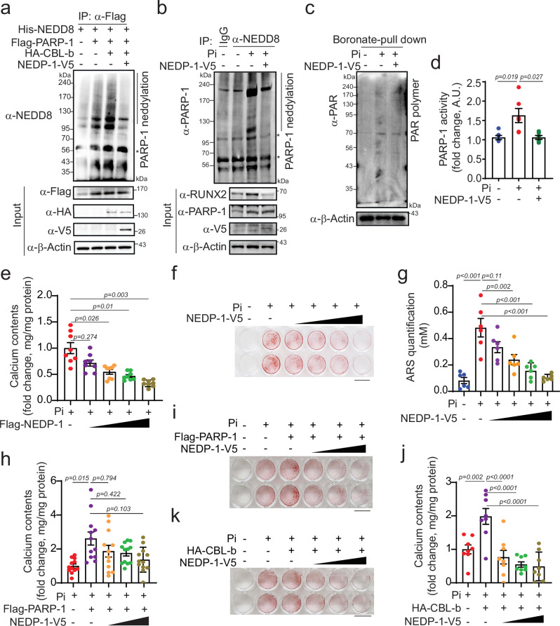
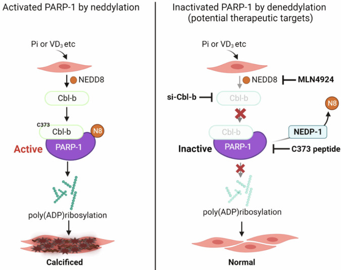
Vascular calcification (VC) refers to the accumulation of mineral deposits on the walls of arteries and veins, and it is closely associated with increased mortality in cardiovascular disease patients, particularly among high-risk patients with diabetes and chronic kidney disease (CKD). Neuronal precursor cell-expressed developmentally downregulated protein 8 (NEDD8) is a ubiquitin-like protein that plays a pivotal role in various cellular functions, primarily through its conjugation to target proteins and subsequent relay of biological signals. However, the role of NEDDylation in VC has not been investigated. In our study, we observed that MLN4924, an inhibitor of the NEDD8-activating E1 enzyme, effectively impedes the progression of VC. LC‒MS/MS analysis revealed that poly(ADP‒ribose) polymerase 1 (PARP-1) is subjected to NEDD8 conjugation, leading to an increase in PARP-1 activity during VC. We subsequently revealed that PARP-1 NEDDylation is mediated by the E3 ligase CBL proto-oncogene B (CBL-b) and is reversed by NEDD8-specific protease 1 (NEDP-1) during VC. Furthermore, the CBL-b C373 peptide effectively mitigated the inactive form of the E3 ligase activity of CBL-b, ultimately preventing VC. These findings provide compelling evidence that the NEDD8-dependent activation of PARP-1 represents a novel mechanism underlying vascular calcification and suggests a promising new therapeutic target for VC.


Conflict of interest statement

The authors declare no competing interests.

Figures

Fig. 1
Fig. 1. MLN4924 inhibits Pi-induced vascular calcification.
a Treatment with MLN4924 (0.001 µM, 0.01 µM, 0.1 µM, or 1 µM) inhibited Pi (4 mM)-induced calcium deposition in A10 cells. n = 4–12 per group, independent experiments. b A10 cells were treated with Pi and MLN4924 under the aforementioned conditions. Representative alizarin red S-stained VSMCs showing that mineralization was blocked by MLN4924 in Pi-treated A10 cells. Scale bar, 10 mm. c Quantification of alizarin red S staining. n = 6 per group, independent experiments. d mRNA levels of smooth muscle marker genes (SM22α and SMA) and osteogenic-related marker genes (RUNX2 and ALP) were measured in A10 cells treated with Pi and MLN4924. n = 6 per group, independent experiments. e NEDD8 immunoblotting was performed after A10 cells were treated with Pi and MLN4924. The red arrow indicates free NEDD8. f, g The amount of accumulated calcium was measured. Overexpression of NEDD8 potentiated Pi-induced calcium deposition in A10 cells. n = 8 per group, independent experiments (f). Inhibition of NEDD8 conjugation with NEDD8-ΔGG blunted Pi-induced calcium deposition. n = 8 per group, independent experiments (g). The data are shown as the means ± SEMs. Statistical significance was tested via ANOVA with Tukey’s HSD test and Dunnett’s T3 test.
Fig. 2
Fig. 2. NEDD8 is conjugated to PARP-1 in VC.
a A gel stained with Coomassie blue for mass spectrometry analysis. Cell extracts from A10 cells treated with Pi (4 mM) or MLN4924 (1 μM) were immunopurified with an anti-NEDD8 antibody and subjected to SDS‒PAGE. The bands indicated by arrows were then digested with trypsin and analyzed by mass spectrometry. b Cell lysates from Pi-treated A10 cells were immunoprecipitated with anti-NEDD8 and immobilized with anti-PARP-1. PARP-1 neddylation increased in Pi-induced VC. c Whole-cell lysates from 293 T cells transfected with the indicated constructs were subjected to Ni-NTA pull-down. PARP-1 neddylation was blunted in NEDD8-ΔGG compared with that in NEDD8 WT. d A PLA was performed. NEDD8-conjugated PARP-1 is dissociated by MLN4924 (1 μM). Scale bar=20 μM. e Immunoprecipitation with anti-PARP-1. Pi-induced PARP-1 neddylation is attenuated by MLN4924 (1 μM) in A10 cells. PARP-1 activity was measured by detecting the poly(ADP)-ribosylation (PAR) polymer (f) and conducting a PARP-1 enzyme activity assay (g). Pi-induced PARP-1 activity was blunted by MLN4924 treatment in A10 cells. n = 6 per group, independent experiments. Note: “*” indicates a nonspecific band. A.U., arbitrary units. The data are presented as the means ± SEMs. Statistical significance was tested via ANOVA with Tukey’s HSD test.
Fig. 3
Fig. 3. MLN4924 ameliorates VD3-induced VC.
a Treatment timeline for MLN4924 (10 mg/kg/day) with VD3 (5 × 105 IU/kg/day) in mice. b Whole aorta with alizarin red S staining. MLN4924 mitigated VD3-induced calcification. Scale bar, 10 mm. Measurement of calcium levels in the aorta (c) and serum (d) after MLN4924 treatment in VD3-injected mice. n = 9–11 per group, independent experiments. Treatment with MLN4924 inhibited calcium accumulation in the aorta but not in the serum of VD3-induced VC patients. e PARP-1 neddylation increased after injection of VD3, but was blocked by MLN4924. f Boronate bead-pull down assays were performed. PARP-1 activity, along with PAR polymer activity, was blunted by MLN4924. g Measurement of PARP-1 enzyme activity. h Alizarin red S staining of cross-sections of the aorta. Scale bar, 200 μΜ. i A PLA with anti-NEDD8 and anti-PARP-1 was performed on cross-sections. Scale bar, 20 μΜ, “*” indicates a nonspecific band. The data are presented as the means ± SEMs. Statistical significance was tested via ANOVA with the Bonferroni correction.
Fig. 4
Fig. 4. The E3 ligase CBL-b mediates PARP-1 neddylation in Pi-induced VC.
a Immunoblot analysis of immunoprecipitates from Pi-treated A10 cells following the depletion of BIRC3, CBL-b, RBX1 and RNF111 with an anti-NEDD8 antibody. b A PLA was performed with anti-NEDD8 and anti-PARP-1 in A10 cells transfected with siCBL-b and Pi. Scale bar, 20 µM. c PARP-1 activity was analyzed in Pi-treated A10 cells with CBL-b knockdown. n = 5 per group, independent experiments. d Endogenous PARP-1 interacted with CBL-b in A10 cells treated with Pi. e Measurement of the calcium content. The knockdown of CBL-b inhibited Pi-induced calcium accumulation in a dose-dependent manner. n = 4–7 per group, independent experiments. f Experimental procedure timeline. The mice were treated with si-CBL-b or scramble via tail vein injection twice during the VC induction period following VD3 injection (5 × 105 IU/kg/day). g Calcium assay. VD3-induced calcium accumulation was inhibited by CBL-b knockdown in the aorta. h Immunoprecipitation with an anti-NEDD8 antibody was performed. VD3-induced PARP-1 neddylation was blunted by CBL-b knockdown. i Vascular calcification in the aorta was determined by alizarin red S staining. Representative images are shown. Scale bar, 200 μΜ. “*” indicates a nonspecific band. The data are presented as the means ± SEMs. Statistical significance was tested via ANOVA with Tukey’s HSD test and the Bonferroni correction.
Fig. 5
Fig. 5. CBL-b E3 ligase activity at C373 promotes PARP-1 neddylation in Pi-induced VC.
a PARP-1 neddylation in Pi-treated A10 cells is dependent on CBL-b E3 ligase activity. b PARP-1 activity was determined via an anti-PARP-1 antibody. Mutants with inactive CBL-b E3 ligase activity showed decreased PARP-1 activity in Pi-treated A10 cells. n = 4 per group, independent experiments. c Measurement of the calcium content. Pi increased calcium accumulation, but this effect was blocked by inactive CBL-b E3 ligase mutants (C373A and W400A). n = 11 per group, independent experiments. d Alizarin red S staining was performed. Scale bar, 10 mm. e PLA assay. NEDD8 was associated with PARP-1 under Pi conditions, but was dissociated by the CBL-b C373 peptide. Scale bar, 20 µM. f Immunoblot analysis was performed via immunoprecipitation with anti-PARP-1. g A boronate pull-down assay was performed to detect the PAR polymer. h PARP-1 activity was analyzed. Pi-induced PARP-1 activity was attenuated by CBL-b C373. n = 4 per group, independent experiments. i Calcium assay. CBL-b C373 inhibited Pi-induced calcium deposition in A10 cells. n = 6 per group, independent experiments. j Calcification was determined by alizarin red S staining. Scale bar, 10 mm. k Quantification of alizarin red S staining was performed. n = 6 per group, independent experiments. “*” indicates a nonspecific band. The data are presented as the means ± SEMs. Statistical significance was tested via ANOVA with Tukey’s HSD test and Dunnett’s T3 test.
Fig. 6
Fig. 6. Administration of the CBL-b C373 peptide mitigated VD3-induced VC.
a Experimental procedure for administering the C373 peptide (1 mg/kg/day) via intraperitoneal injection to mice with VD3-induced VC. b Alizarin red S staining of the entire aorta in the mice. Scale bar, 10 mm. Aortic calcium (c) and serum calcium (d) levels were measured. Administration of the C373 peptide reduced VD3-induced calcium accumulation. n = 6 per group, independent experiments. e Immunoprecipitation with an anti-NEDD8 antibody was used to detect PARP-1 neddylation. PARP-1 neddylation was diminished by administration of the C373 peptide to the aortas of VD3-induced VC mice. f PARP-1 activity was detected by the PAR polymer with a boronate pull-down assay. The C373 peptide blunted VD3-induced PARP-1 activity. g Histology: alizarin red S staining (left panel scale bar, 200 μΜ) and PLA with anti-NEDD8 and anti-PARP-1 (right panel, scale bar, 20 μΜ) were performed on cross-sections. VD3 increased vascular calcification, but was inhibited by the C373 peptide. The administration of the CBL-b C373 peptide led to the dissociation of NEDD8 from PARP-1. “*” indicates a nonspecific band. The data are presented as the means ± SEMs. Statistical significance was tested via ANOVA with Tukey’s HSD test and Dunnett’s T3 test.
Fig. 7
Fig. 7. PARP-1 deneddylation by NEDP-1 attenuates VC.
a Cell lysates extracted from 293T cells transfected with the indicated plasmid DNAs were subjected to immunoprecipitation with anti-Flag. Overexpression of NEDP-1 resulted in the denaturation of PARP-1. b Transfection of NEDP-1 into A10 cells followed by treatment with Pi blocked Pi-induced PARP-1 neddylation. c Overexpression of NEDP-1 and Pi treatment in A10 cells led to decreased expression of the PAR polymer. d PARP-1 activity was measured under the same conditions. n = 5 per group. e Measurement of the calcium content. n = 8 per group, independent experiments. f Cells were stained with alizarin red S. A representative whole-well image is shown. Scale bar, 10 mm. g The extent of alizarin red S staining was quantified. n = 6 per group. h Pi-treated A10 cells were cotransfected with Flag-PARP-1 and NEDP-1-V5. Measurement of the calcium content. PARP-1 potentiated Pi-induced calcium deposition, which was blocked by the overexpression of NEDP-1. n = 12 per group, independent experiments. i Alizarin red S staining was performed under the same conditions. Scale bar, 10 mm. j A10 cells were cotransfected with HA-CBL-b and NEDP-1-V5. CBL-b also potentiated Pi-induced calcium deposition, which was blocked by NEDP-1 overexpression. n = 8 per group, independent experiments. k Alizarin red S staining under the same conditions. Scale bar, 10 mm. “*” indicates a nonspecific band. The data are presented as the means ± SEMs. Statistical significance was tested via ANOVA with Tukey’s HSD test and Dunnett’s T3 test.
Fig. 8
Fig. 8. Illustration of how PARP-1 activity, facilitated by NEDD8-conjugated PARP-1, induces vascular calcification.
A diverse range of stimuli, such as inorganic phosphate (Pi) or VD3, drive NEDD8 conjugation with PARP-1, which is mediated by CBL-b, and this process is reversed by NEDP-1. Notably, neddylation of PARP-1 leads to an increase in PARP-1 activity, which contributes to the progression of vascular calcification (VC). However, the NEDD8-activating E1 enzyme inhibitor MLN4924 effectively impedes the progression of VC. Additionally, a C373 peptide derived from CBL-b shows promise in preventing VC by mitigating the E3 ligase activity of the inactive form of CBL-b. Therefore, we propose that targeting the NEDD8-dependent activation of PARP-1 could be a potentially effective therapeutic approach for VC. Created with BioRender.com.

References

    1. Leopold, J. A. Vascular calcification: mechanisms of vascular smooth muscle cell calcification. Trends Cardiovasc. Med.25, 267–274 (2015). - PMC - PubMed
    1. Nicoll, R. & Henein, M. Arterial calcification: a new perspective? Int. J. Cardiol.228, 11–22 (2017). - PubMed
    1. Durham, A. L., Speer, M. Y., Scatena, M., Giachelli, C. M. & Shanahan, C. M. Role of smooth muscle cells in vascular calcification: implications in atherosclerosis and arterial stiffness. Cardiovasc. Res.114, 590–600 (2018). - PMC - PubMed
    1. Wu, M., Rementer, C. & Giachelli, C. M. Vascular calcification: an update on mechanisms and challenges in treatment. Calcif. Tissue Int.93, 365–373 (2013). - PMC - PubMed
    1. Hochstrasser, M. Origin and function of ubiquitin-like proteins. Nature458, 422–429 (2009). - PMC - PubMed

MeSH terms

LinkOut - more resources