. 2024 Sep 30;10(1):92.
doi: 10.1038/s41522-024-00560-2.

Microbiome-derived antimicrobial peptides show therapeutic activity against the critically important priority pathogen, Acinetobacter baumannii

Affiliations

Microbiome-derived antimicrobial peptides show therapeutic activity against the critically important priority pathogen, Acinetobacter baumannii

P J Alexander et al. NPJ Biofilms Microbiomes. .

Abstract

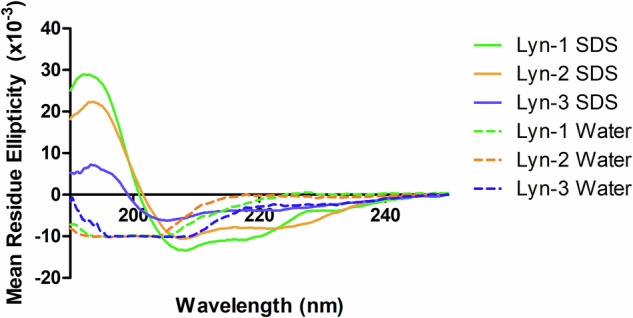
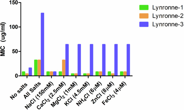
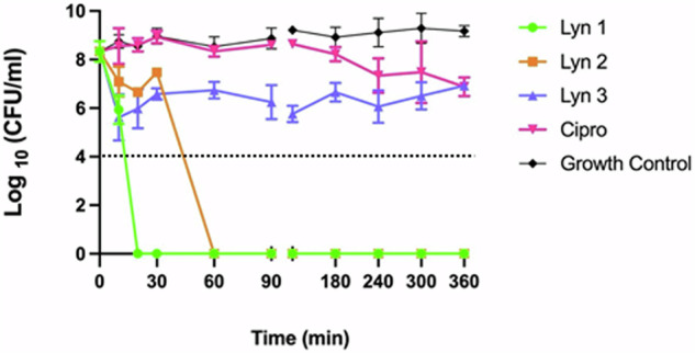
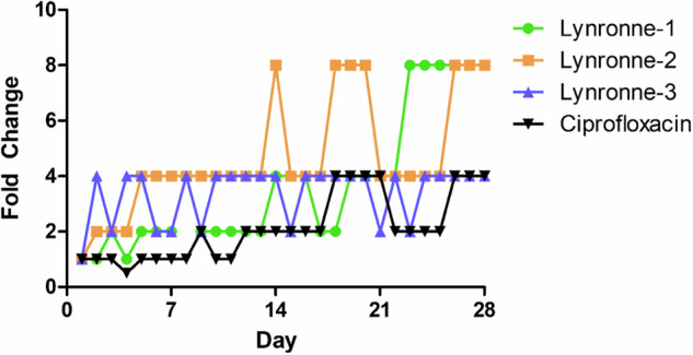
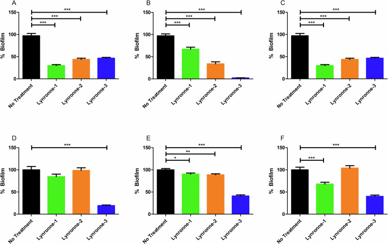
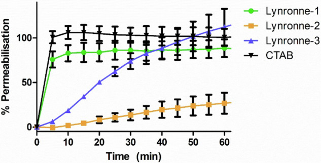
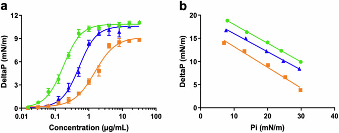
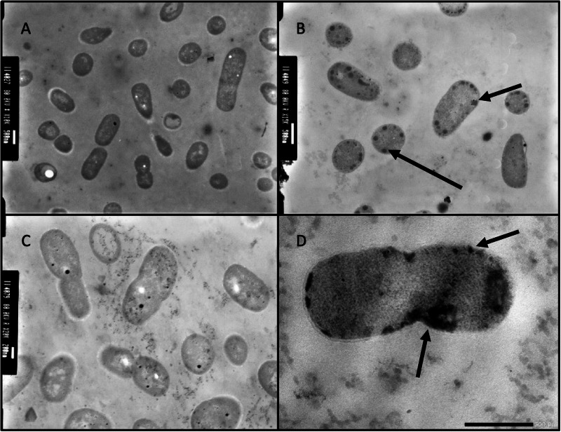
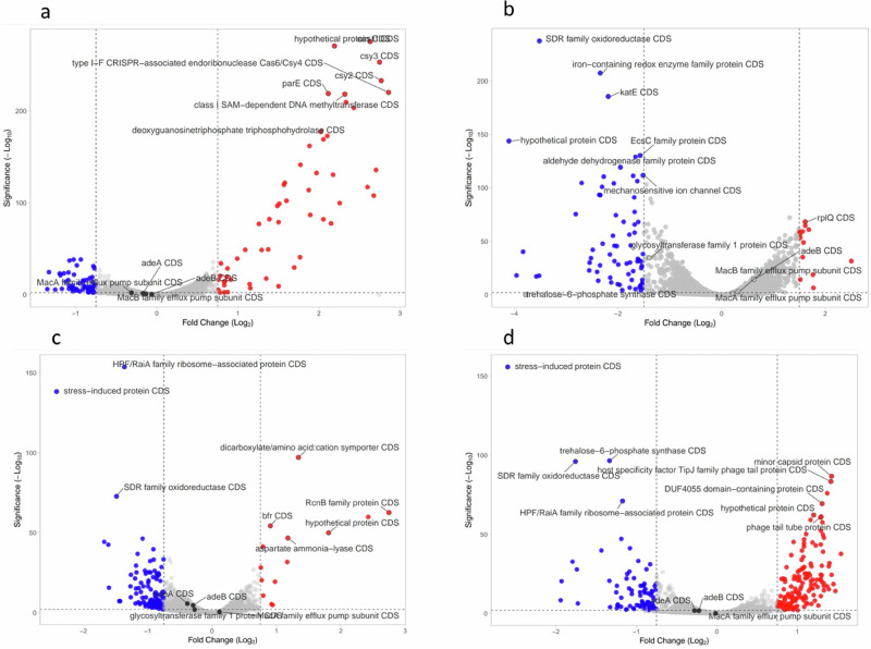
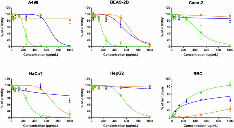
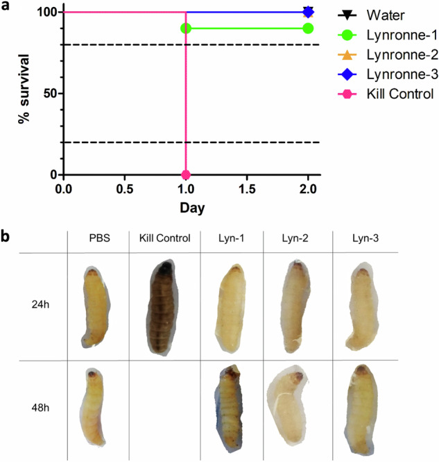
Acinetobacter baumannii is designated by the World Health Organisation as a critical priority pathogen. Previously we discovered antimicrobial peptides (AMPs), namely Lynronne-1, -2 and -3, with efficacy against bacterial pathogens, such as Staphylococcus aureus and Pseudomonas aeruginosa. Here we assessed Lynronne-1, -2 and -3 structure by circular dichroism and efficacy against clinical strains of A. baumannii. All Lynronne AMPs demonstrated alpha-helical secondary structures and had antimicrobial activity towards all tested strains of A. baumannii (Minimum Inhibitory Concentrations 2-128 μg/ml), whilst also having anti-biofilm activity. Lynronne-2 and -3 demonstrated additive effects with amoxicillin and erythromycin, and synergy with gentamicin. The AMPs demonstrated little toxicity towards mammalian cell lines or Galleria mellonella. Fluorescence-based assay data demonstrated that Lynronne-1 and -3 had higher membrane-destabilising action against A. baumannii in comparison with Lynronne-2, which was corroborated by transcriptomic analysis. For the first time, we demonstrate the therapeutic activity of Lynronne AMPs against A. baumannii.


Conflict of interest statement

The authors declare no competing interests.

Figures

Fig. 1
Fig. 1. Mean residue ellipticity of the Lynronne antimicrobial peptides in sterile water and 30 mM SDS, collected by far-UV circular dichroism (CD) spectrophotometry.
All AMPs were at 20 μg/ml, and the CD spectrum was collected between 185 and 250 nm. Dotted lines indicate results from Lyn-1, -2, -3 in water, and solid lines indicate results from Lyn-1, -2, -3 in 30 mM SDS. All readings were taken 5 times, and the average result taken for processing.
Fig. 2
Fig. 2. Antimicrobial activity of Lynronne AMPs against Acinetobacter baumannii DSM 30007 in presence of physiologic salts.
Salts were added into cation-adjusted Mueller Hinton broth, and stated concentrations were identified from previous studies. MICs were performed in triplicate. ‘No salts’ is cation-adjusted MH already containing Ca2+ with no additional CaCl2 (2.5 mM).
Fig. 3
Fig. 3. Time-dependent Lynronne AMPs-mediated activity against Acinetobacter baumannii DSM 30007.
4× MIC concentrations were used for all antimicrobials, and sterile PBS was used for the growth control. Dotted line indicates detection limit. Plate counts (CFU/ml) were taken at the time intervals of 0, 10, 20, 30, 60 and 1440 min. Broth cultures at a CFU/ml of 109 were incubated at 37 °C, 180 rpm for the duration of the assay. All treatments were tested in triplicate, and plate counts were taken in duplicate. Error bars denote standard deviation from the mean.
Fig. 4
Fig. 4. Resistance development during serial passage of Acinetobacter baumannii DSM 30007 over 28 days in the presence of sub-inhibitory concentrations of antimicrobials.
Wells containing the highest concentration of antimicrobial with growth observed from the most recent overnight MIC test were selected as the starting culture for serial passage. This figure is a representative of the mean change in MIC by 3 biological replicates. Ciprofloxacin was also included as a comparator, and blanks containing sterile MH broth used as a negative control. Fold change is indicative of doubling changes as compared to the initial MIC observed on day 1.
Fig. 5
Fig. 5. Antibiofilm activity of Lynronne antimicrobial peptides.
Effects of the Lynronne antimicrobial peptides on the growth and adhesion of Acinetobacter baumannii biofilms (AC) and the effects on established biofilms (DF). Graphs (A, D); A. baumannii DSM 30007. Graphs (B, E); A. baumannii DSM 102929. Graphs (C, F); A. baumannii clinical S26063. Biofilms were grown in cation-adjusted MH broth for 48 h at 37 °C in 96 well plates. Biofilm mass was determined using crystal violet staining and resolubilisation in acetic acid before OD600 readings were taken. Positive controls were established with the inclusion of sterile water, and negative controls were established using sterile cation-adjusted MH broth. Readings were taken with 12 technical replicates, with 3 biological replicates for each assay. Statistically significant differences between treatments and the positive control were determined using 1-way ANOVAs with Dunnett’s post-test. Error bars denote standard deviation.
Fig. 6
Fig. 6. Membrane permeabilisation of Lynronne-1, -2 and -3 over time based on propidium iodide fluorescence at 4× MIC.
Increased fluorescence indicates damage/pore formation in the bacterial cell membrane. 100% permeabilisation rate was established using average fluorescence by CTAB control once plateau had been achieved. Readings of excitation/emission at 540 nm and 590 nm were taken in triplicate, and the mean calculated for each time point. Error bars signify SEM as calculated using GraphPad Prism 5. Positive control established by cetrimonium bromide (CTAB) at 300 μM.
Fig. 7
Fig. 7. Evaluation of the insertion of the Lynronne AMPs into lipids of Acinetobacter baumannii.
Green lines: Lynronne-1; orange lines: Lynronne 2; Blue lines; Lynronne 3. The insertion of the Lynronne AMPs into total lipids extract of A. baumannii 30007 was measured using the Langmuir film balance (KIBRON apparatus). a Evaluation of the dose-dependent insertion of the Lynronne AMPs into monolayer of A. baumannii lipids. Lipid monolayers were obtained by spreading lipids extracted from A. baumannii 30007 at the water-air interface until reaching an initial surface pressure of 30 ± 0.5 mN/m. Increasing concentrations of Lynronne AMPs were then injected into the water sub-phase and their insertions were measured as the maximal variation of the surface pressure (DeltaP) usually reached within 20–30 min. DeltaP are expressed in mN/m (means ± S.D., n = 3). b Determination of the critical pressure of insertion. The lipid insertion of the Lynronne AMPs (at 1 µg/mL) was measured using lipid monolayers set-up at different initial surface pressures. Graph was used to calculate the critical pressure of insertion corresponding to the theoretical initial pressure at which no insertion can occurs. The critical pressure of insertion was graphically determined as the intercept of the linear slope with the X-axis when the DeltaP is equal to zero.
Fig. 8
Fig. 8. Transmission electron micrographs of Acinetobacter baumannii DSM 30007 cells after exposure to Lynronne-1, -2 and -3.
A untreated cells. B Lynronne-1. C Lynronne-2. D Lynronne-3. All peptides were at 4× MIC concentration, and cells were exposed for 60 min at 37 °C. Scale bars for image (A, B, D) are 500 nm, and image (C) is 200 nm. Black arrows in images (B, D) indicate vacuole aggregation at the bacterial cell membrane, indicating cell damage/response to AMP membrane exposure. These are representative images for each treatment condition.
Fig. 9
Fig. 9. Volcano plot representation of transcriptome change of A. baumannii DSM 30,007 cells after exposure to the Lynronne AMPs and ciprofloxacin.
a Ciprofloxacin. b Lynronne-1. c Lynronne-2. d Lynronne-3. Genes highlighted in blue were significantly downregulated (−log10 significance of >2, Log2 fold change of >0.75 in either direction). Cell cultures were challenged for 60 min at 1× MIC for all antimicrobials. All treatments were conducted in triplicate, and gene expression counts calculated using Geneious Prime (version 2022.2.2). Gene expression counts were compared for each treatment against the control using DESeq2 using Rstudio, and volcano plots created using VolcaNoseR version 2.0.
Fig. 10
Fig. 10. Evaluation of the toxicity of the Lynronne AMPs on human cell lines.
The toxicity of the Lynronne AMPs was measured against human cell lines from various organs. Human cells were exposed to increasing concentrations of the Lynronne AMPs. For cytotoxicity determination, A498 (kidney), BEAS-2B (lung), Caco-2 (intestine), HaCaT (skin), and HepG2 (liver) cells, cells were exposed for 48 h before measurement of the cell viability using resazurin assay. Results are expressed as percentage of cell viability using untreated cells as negative controls giving 100% viability. For haemolysis determination, human red blood cells (RBC) were exposed for 1 h before measurement of haemoglobin release. Results are expressed as percentage of haemolysis, using Triton X-100 (at 0.1%) as the positive control giving 100% haemolysis. Results are expressed as means ± S.D. (n3). Green lines: Lynronne-1; orange lines: Lynronne 2; Blue lines; Lynronne 3.
Fig. 11
Fig. 11. Toxicity determination of the Lynronne AMPs via Galleria mellonella injection model at 8x MIC.
a Survival curves of the Lynronne AMPs across 48 h after injection. Kill controls were established with 107 CFU/ml of Acinetobacter baumannii DSM 30007. b Table showing representative larval status at 24 h and 48 h after incubation at 37 °C. Status of larvae was determined using a combination of melanisation (as seen visibly in the 24 h Kill Control treatment), motility signs and responsiveness to physical stimuli. All treatments contained 10 replicates. >80% survival indicates low/negligible toxicity, between 20 and 80% indicates partial toxicity, and <20% survival indicates high toxicity.

Similar articles

Cited by

References

    1. van der Kolk, J. H., Endimiani, A., Graubner, C., Gerber, V. & Perreten, V. Acinetobacter in veterinary medicine, with an emphasis on Acinetobacter baumannii. J. Glob. Antimicrob. Resist.16, 59–71 (2019). - PubMed
    1. Heidari, H. et al. Molecular analysis of drug-resistant Acinetobacter baumannii isolates by ERIC-PCR. Meta Gene17, 132–135 (2018).
    1. Nocera, F. P., Attili, A. R. & De Martino, L. Acinetobacter baumannii: its clinical significance in human and veterinary medicine. Pathogens10,10.3390/pathogens10020127 (2021). - PMC - PubMed
    1. Nishimizu S., Shiota S., Oshiumi T., Takakura T. & Miyazaki E. A case of community-onset Acinetobacter pneumonia in a healthy person. IDCases. 24, e01133 (2021). - PMC - PubMed
    1. Liao, Y. et al Case report: acinetobacter baumannii septic arthritis in an immunocompetent infant. Front. med.10, 1135178 (2023). - PMC - PubMed

MeSH terms

LinkOut - more resources