. 2024 Sep 30;14(1):22608.
doi: 10.1038/s41598-024-73607-1.

LncRNA MIAT suppresses inflammation in LPS-induced J774A.1 macrophages by promoting autophagy through miR-30a-5p/SOCS1 axi

Affiliations

LncRNA MIAT suppresses inflammation in LPS-induced J774A.1 macrophages by promoting autophagy through miR-30a-5p/SOCS1 axi

Linqian Sun et al. Sci Rep. .

Abstract

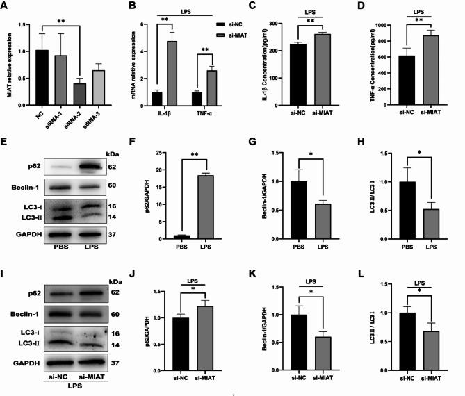
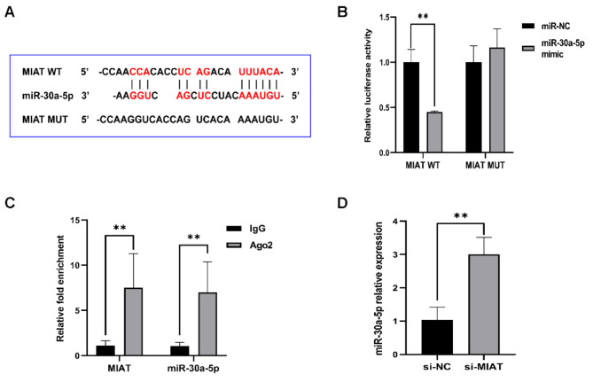
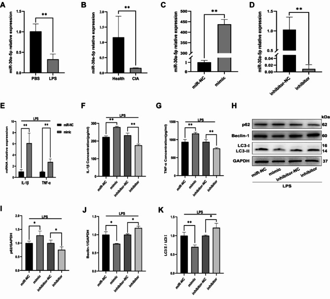
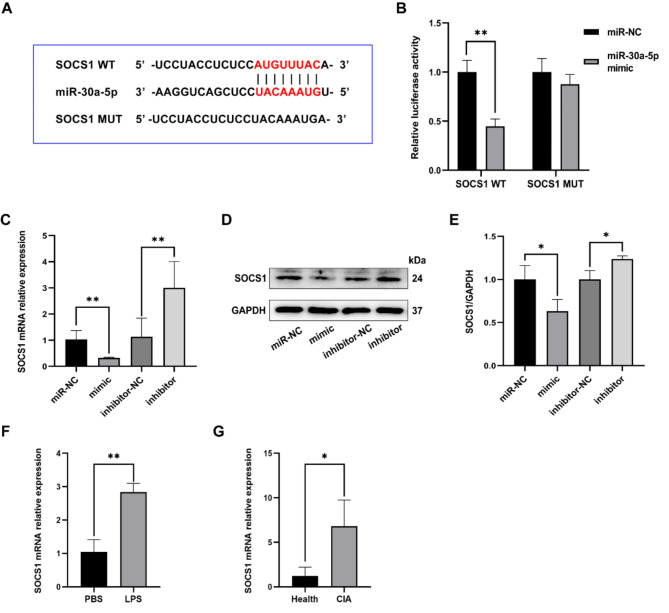
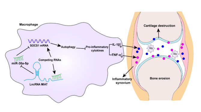
Accumulated data implicate that long noncoding RNA (lncRNA) plays a pivotal role in rheumatoid arthritis (RA), potentially serving as a competitive endogenous RNA (ceRNA) for microRNAs (miRNAs). The lncRNA myocardial infarction-associated transcript (MIAT) has been demonstrated to regulate inflammation. However, the role of MIAT in the inflammation of RA remains inadequately explored. This study aims to elucidate MIAT's role in the inflammation of lipopolysaccharide (LPS)-induced macrophages and to uncover the underlying molecular mechanisms. We observed heightened MIAT expression in LPS-induced J774A.1 cells and collagen-induced arthritis mouse models, in contrast to the expression pattern of miR-30a-5p. Silencing MIAT resulted in increased expression of the inflammatory cytokines IL-1β and TNF-α. Simultaneously, MIAT interference significantly impeded macrophage autophagy, evidenced by decreased expression of autophagy-related markers LC3-II and Beclin-1, alongside increased levels of p62 in LPS-induced J774A.1 cells. Notably, MIAT functioned as a ceRNA, sponging miR-30a-5p and exerting a negative regulatory influence on its expression. SOCS1 emerged as a target of miR-30a-5p, modulated by MIAT. Mechanistically, inhibiting miR-30a-5p reversed the impact of MIAT deficiency in promoting LPS-induced inflammation, while SOCS1 knockdown countered the cytokine inhibitory effect induced by silencing miR-30a-5p. In summary, this study indicates that lncRNA MIAT suppresses inflammation in LPS-induced J774A.1 macrophages by stimulating autophagy through the miR-30a-5p/SOCS1 axis. This suggests that MIAT holds promise as a potential therapeutic target for RA inflammation.

Keywords: Autophagy; Inflammation; LncRNA MIAT; Rheumatoid arthritis; miR-30a-5p/SOCS1 axis.


Conflict of interest statement

The authors declare no competing interests.

Figures

Fig. 1
Fig. 1
LncRNA MIAT expression is higher in CIA and LPS-induced J774A.1 macrophages. (A) The timeline for constructing CIA mouse models. (B) Photographs of healthy and CIA mouse paws (n = 6 for each group). (C) Arthritis severity scores in healthy and CIA mice (n = 6 for each group). (D) Representative micrographs of H&E staining of knee joint tissues from healthy and CIA mice (scale bar = 60 μm; n = 6 for each group). (E,F) IL-1β and TNF-α concentrations in serum from healthy and CIA mice (n = 6 for each group). (G) MIAT relative expression measured by RT-qPCR in healthy and CIA mice (n = 3 for each group). (HK) Expression of IL-1β and TNF-α measured by RT-qPCR or ELISA in normal J774A.1 cells and LPS-induced J774A.1 cells. (L) Relative expression of MIAT measured by RT-qPCR in normal J774A.1 cells and LPS-induced J774A.1 cells. Data obtained from more than three repeated experiments are presented as mean ± SD, **P < 0.01.
Fig. 2
Fig. 2
Knockdown of LncRNA MIAT induces inflammatory response and inhibits autophagy in LPS-induced J774A.1 cells. J774A.1 cells were transfected with si-MIAT or si-NC before LPS stimulation. (A) Relative expression of MIAT transfected with si-MIAT measured by RT-qPCR. (B) mRNA relative expression of TNF-α and IL-1β measured by RT-qPCR after knocking down MIAT. (C,D) IL-1β and TNF-ɑ concentration of cell supernatants after knocking down MIAT. (EH) Protein expression of LC3, p62, Beclin-1, and GAPDH after LPS stimulation evaluated by western blot analysis. (IL) Protein expression of LC3, p62, Beclin-1, and GAPDH after knocking down MIAT detected by western blot analysis. Data represent 3 biological replicates and are expressed as mean ± SD. *P < 0.05, **P < 0.01.
Fig. 3
Fig. 3
LncRNA MIAT functions as a molecular sponge for miR-30a-5p. (A) The predicted binding sequence between miR-30a-5p and MIAT. (B) Interaction between MIAT and miR-30a-5p confirmed by the dual-luciferase reporter assay. (C) RIP assay demonstrating binding relationships between MIAT and miR-30a-5p. (D) RT-qPCR assay examining miR-30a-5p expression. Data represent 3 biological replicates and are expressed as mean ± SD. **P < 0.01.
Fig. 4
Fig. 4
MiR-30a-5p modulates inflammatory response and autophagy in LPS-induced J774A.1 cells. J774A.1 cells were transfected with miR-30a-5p mimic or inhibitor before LPS stimulation. (A,B) MiR-30a-5p expression in LPS-induced J774A.1 cells and CIA mice assessed by RT-qPCR. (C,D) RT-qPCR of miR-30a-5p expression in J774A.1 cells transfected with miR-30a-5p mimic or inhibitor. (EG) TNF-α and IL-1β expression measured by RT-qPCR or ELISA. (HK) Protein expression of LC3, p62, Beclin-1, and GAPDH detected by western blot analysis. Data from more than three repeated experiments are presented as mean ± SD. *P < 0.05, **P < 0.01.
Fig. 5
Fig. 5
MiR-30a-5p directly interacts with SOCS1. (A) Predicted binding sequence between miR-30a-5p and SOCS1. (B) Binding relationship between miR-30a-5p and SOCS1 confirmed by dual-luciferase reporter assays with SOCS1 WT and SOCS1 MUT. (C) mRNA relative expression of SOCS1 measured by RT-qPCR. (D,E) Expression of SOCS1 detected by western blot analysis. (F,G) SOCS1 expression in LPS-induced J774A.1 cells and CIA mice assessed by RT-qPCR. Data from more than three repeated experiments are presented as mean ± SD. *P < 0.05, **P < 0.01.
Fig. 6
Fig. 6
LncRNA MIAT functions as a ceRNA for miR-30a-5p to regulate SOCS1 expression in J774A.1 cells. (A) SOCS1 mRNA levels measured by RT-qPCR in J774A.1 cells transfected with si-SOCS1. (B,C) SOCS1 protein expression detected by western blot analysis. (DF) TNF-α and IL-1β levels measured by RT-qPCR or ELISA. (G,H) SOCS1 expression detected by western blot analysis in each group. (IL) TNF-α and IL-1β levels measured by RT-qPCR or ELISA in each group. (MP) Protein expression of LC3, p62, Beclin-1, and GAPDH detected by western blot analysis. Data from more than three repeated experiments are presented as mean ± SD. *P < 0.05, **P < 0.01.
Fig. 7
Fig. 7
Potential mechanisms of MIAT inhibits inflammation in LPS-induced J774A.1 macrophages.

Similar articles

Cited by

References

    1. Smith, M. H. & Berman, J. R. What Is Rheumatoid Arthritis? JAMA327, 1194 (2022). - PubMed
    1. Kinne, R. W., Stuhlmüller, B. & Burmester, G. R. Cells of the synovium in rheumatoid arthritis. Macrophages. Arthritis Res. Ther.9, 224 (2007). - PMC - PubMed
    1. Athanasou, N. A. Synovial macrophages. Ann. Rheum. Dis.54, 392–394 (1995). - PMC - PubMed
    1. Ardura, J. A. et al. Targeting macrophages: friends or foes in Disease? Front. Pharmacol.10, 1255 (2019). - PMC - PubMed
    1. Ali, S. A., Peffers, M. J., Ormseth, M. J., Jurisica, I. & Kapoor, M. The non-coding RNA interactome in joint health and disease. Nat. Rev. Rheumatol.17, 692–705 (2021). - PubMed

MeSH terms

LinkOut - more resources