. 2024 Sep 30;24(1):337.
doi: 10.1186/s12876-024-03413-6.

OncomiR-181a promotes carcinogenesis by repressing the extracellular matrix proteoglycan decorin in hepatocellular carcinoma

Affiliations

OncomiR-181a promotes carcinogenesis by repressing the extracellular matrix proteoglycan decorin in hepatocellular carcinoma

Reem Amr Assal et al. BMC Gastroenterol. .

Abstract

Background: Proteoglycans are important tumor microenvironment extracellular matrix components. The regulation of key proteoglycans, such as decorin (DCN), by miRNAs has drawn attention since they have surfaced as novel therapeutic targets in cancer. Accordingly, this study aimed at identifying the impact of miR-181a in liver cancer and its regulatory role on the extracellular matrix proteoglycan, DCN, and hence on downstream oncogenes and tumor suppressor genes.

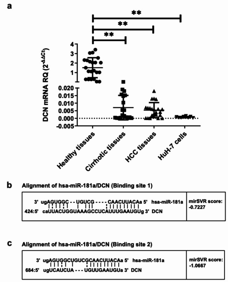
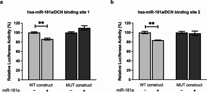
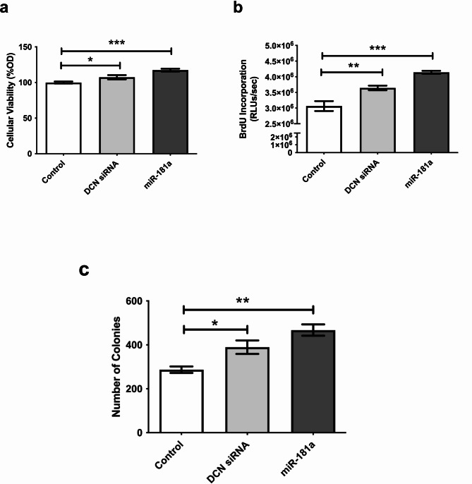
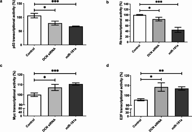
Results: DCN was under-expressed in 22 cirrhotic and HCC liver tissues compared to that in 11 healthy tissues of liver transplantation donors. Conversely, miR-181a was over-expressed in HCC liver tissues compared to that in healthy liver tissues. In silico analysis predicted that DCN 3'UTR harbors two high-score oncomiR-181a binding regions. This was validated by pmiRGLO luciferase reporter assay. Ectopic miR-181a expression into HuH-7 cells repressed the transcript and protein levels of DCN as assessed fluorometrically and by western blotting. DCN siRNAs showed similar results to miR-181a, where they both enhanced the cellular viability, proliferation, and clonogenicity. They also increased Myc and E2F and decreased p53 and Rb signaling as assessed using reporter vectors harboring p53, Rb, Myc, and E2F response elements. Our findings demonstrated that miR-181a directly downregulated the expression of its direct downstream target DCN, which in turn affected downstream targets related to cellular proliferation and apoptosis.

Conclusion: To our knowledge, this is the first study to unveil the direct targeting of DCN by oncomiR-181a. We also highlighted that miR-181a affects targets related to cellular proliferation in HCC which may be partly mediated through inhibition of DCN transcription. Thus, miR-181a could be a promising biomarker for the early detection and monitoring of liver cancer progression. This would pave the way for the future targeting of the oncomiR-181a as a therapeutic approach in liver cancer, where miR-181a-based therapy approach could be potentially combined with chemotherapy and immunotherapy for the management of liver cancer.

Keywords: Decorin (DCN); Hepatocellular carcinoma (HCC); Liver cancer; microRNA-181a (miR-181a).


Conflict of interest statement

The authors declare no competing interests.

Figures

Fig. 1
Fig. 1
Expression profiling of DCN in liver tissues and predicted targeting of DCN 3’UTR by miR-181a. (a) The expression of DCN was investigated in healthy, cirrhotic, and HCC liver biopsies in addition to HuH-7 cells. Each tissue sample was carried out in duplicates and repeated three times. (b, c) Alignment of the two high-score binding sites on DCN transcript with miR-181a seed sequence, as predicted by microRNA.org. Vertical lines indicate complementarity between the binding region of the mRNA and the seed sequence of the miRNA, while the dots indicate mismatches or GU wobbles. Also shown are the mirSVR scores, which factor in multiple features of the predicted miRNA: mRNA duplex, including (1) duplex features which includes base pairing at the seed region and 3’end of the miRNA; (2) sequence features which include A/U composition near the target sites and secondary structure accessibility; and (3) global features such as length of the UTR, relative position of the target site in the UTR and conservation score. The MirSVR downregulation scores correlate linearly with the extent of downregulation. Asterisks indicates statistically significant differences, where **p < 0.01
Fig. 2
Fig. 2
Impact of miR-181a on DCN mRNA and protein in HuH-7 cells. (a) HuH-7 cells were transfected with miR-181a mimics and the efficiency of miR-181a delivery was confirmed on the mRNA level using RTqPCR. (b) Relative expression of DCN was determined using RTqPCR in HuH-7 cells transfected with miR-181a mimics or specific siRNAs against DCN as a positive control. (c) Relative fluorescence intensity of DCN protein was quantified using a multilabel counter fluorometer after fluorescent labeling of transfected HuH-7 cells, and readings were plotted as % mean fluorescence intensity (MFI). The experiment was carried out in quadruplets and repeated three times. (d) Relative band intensity was quantified using ImageQuant software after western blotting to determine the DCN protein levels in transfected HuH-7 cells. The experiment was carried out in duplicates and repeated three times. (e) Representative image of the western blot results. The loading controls were run on the same gel. Then, the blot was cropped during the experiment as a different set of secondary antibodies was used for DCN and GAPDH. Thus, the figure represents two grouped blots from different parts of the same blot (the three upper bands represent the blot that was incubated with DCN antibody, while three lower bands represent the blot that was incubated with GAPDH antibody). The dashed line delineates the cropping site. The original uncropped blots of DCN and GAPDH are presented in Additional file 1 (Supplementary Figure S2). Asterisks indicates statistically significant differences, where *p < 0.05, ** p < 0.01, and ***p < 0.001
Fig. 3
Fig. 3
miR-181a targeting of DCN. Huh-7 cells were co-transfected with miR-181a mimics and either the wildtype (WT) or mutant (MUT) construct for each of the two predicted binding regions on DCN 3’UTR. Luciferase reporter assay was used to validate the independent and site-specific binding of miR-181a to (a) the first and (b) the second predicted binding regions on DCN 3’UTR. Asterisks indicates statistically significant differences, where ** p < 0.01
Fig. 4
Fig. 4
Impact of miR-181a on viability, proliferation, and clonogenicity of HuH-7 cells. HuH-7 cells were transfected with miR-181a mimics or specific siRNAs against DCN as a positive control, and (a) MTT assay for cellular viability, (b) BrdU incorporation assay for cellular proliferation, and (c) colony-forming assay for clonogenicity were performed. Representative images for the colony forming assay are shown in Supplementary Figure S3. Asterisks indicates statistically significant differences, where *p < 0.05, ** p < 0.01, and ***p < 0.001
Fig. 5
Fig. 5
Impact of miR-181a on various oncogenes and tumor suppressor transcription factors. HuH-7 cells were transfected with reporter vectors containing cis-acting enhancer elements specific to cell cycle regulating transcription factors (a) p53, (b) Rb, (c) Myc, or (d) E2F, upstream of a sensitive luciferase reporter gene, and luciferase activity was used to quantify the activation or repression of each signaling pathway after knockdown of DCN or miR-181a mimicking. Asterisks indicates statistically significant differences, where *p < 0.05, ** p < 0.01, and ***p < 0.001

Similar articles

Cited by

References

    1. Sung H, Ferlay J, Siegel RL, Laversanne M, Soerjomataram I, Jemal A, Bray F. Global cancer statistics 2020: GLOBOCAN estimates of incidence and mortality worldwide for 36 cancers in 185 countries. CA Cancer J Clin. 2021;71:209–49. 10.3322/CAAC.21660 - PubMed
    1. El-Zayadi AR, Badran HM, Barakat EMF, Attia MED, Shawky S, Mohamed MK, Selim O, Saed A. Hepatocellular carcinoma in Egypt: a single center study over a decade. World J Gastroenterol. 2005;11:5193–8. 10.3748/WJG.V11.I33.5193 - PMC - PubMed
    1. Sweed D, Sweed E, Moaz I, Mosbeh A, Fayed Y, Elhamed SMA, Sweed E, Macshut M, Abdelsattar S, Kilany S, et al. The clinicopathological and prognostic factors of hepatocellular carcinoma: a 10-year tertiary center experience in Egypt. World J Surg Oncol. 2022;20. 10.1186/S12957-022-02764-2 - PMC - PubMed
    1. Omran D, Alboraie M, Zayed RA, Wifi MN, Naguib M, Eltabbakh M, Abdellah M, Sherief AF, Maklad S, Eldemellawy HH, et al. Towards hepatitis C virus elimination: Egyptian experience, achievements and limitations. World J Gastroenterol. 2018;24:4330–40. 10.3748/WJG.V24.I38.4330 - PMC - PubMed
    1. Waziry R, Gomaa A, Waked I, Dore GJ. Determinants of survival following hepatocellular carcinoma in Egyptian patients with untreated chronic HCV infection in the pre-DAA era. Arab J Gastroenterol. 2018;19:26–32. 10.1016/J.AJG.2018.02.004 - PubMed