Comparative Study
. 2024 Sep 30;20(1):26.
doi: 10.1186/s12993-024-00250-1.

Comparative effect of atorvastatin and risperidone on modulation of TLR4/NF-κB/NOX-2 in a rat model of valproic acid-induced autism

Affiliations
Comparative Study

Comparative effect of atorvastatin and risperidone on modulation of TLR4/NF-κB/NOX-2 in a rat model of valproic acid-induced autism

Eman A E Farrag et al. Behav Brain Funct. .

Abstract

Background: Autism spectrum disorder (ASD) is a complex neurodevelopmental condition that is significantly increasing, resulting in severe distress. The approved treatment for ASD only partially improves the sympoms, but it does not entirely reverse the symptoms. Developing novel disease-modifying drugs is essential for the continuous improvement of ASD. Because of its pleiotropic effect, atorvastatin has been garnered attention for treating neuronal degeneration. The present study aimed to investigate the therapeutic effects of atorvastatin in autism and compare it with an approved autism drug (risperidone) through the impact of these drugs on TLR4/NF-κB/NOX-2 and the apoptotic pathway in a valproic acid (VPA) induced rat model of autism.

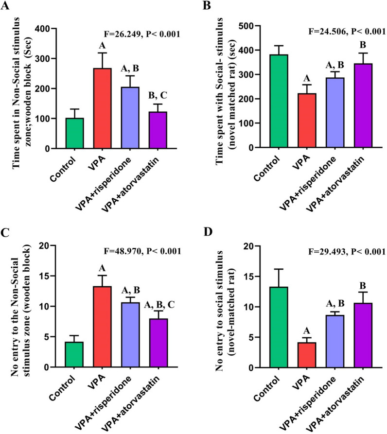
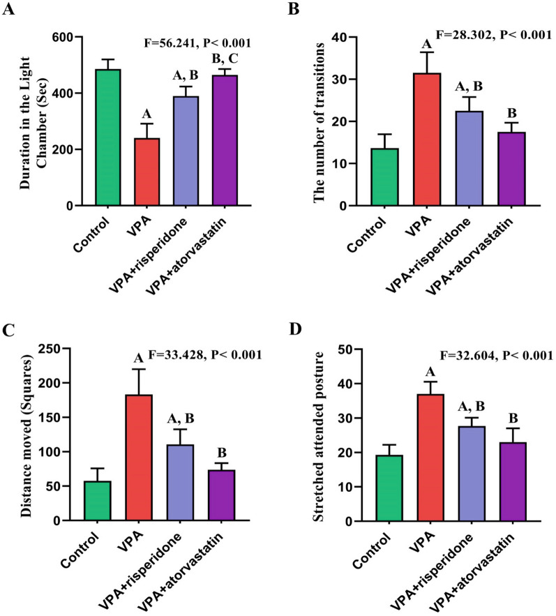
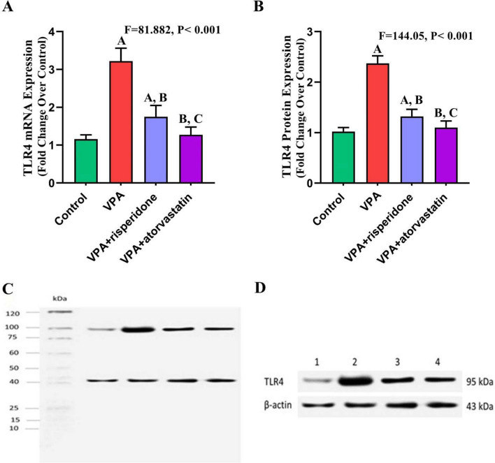
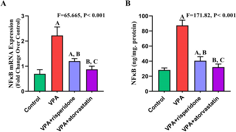
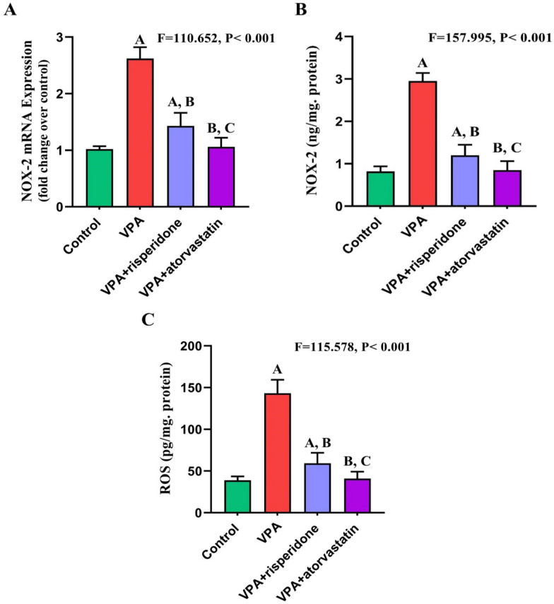
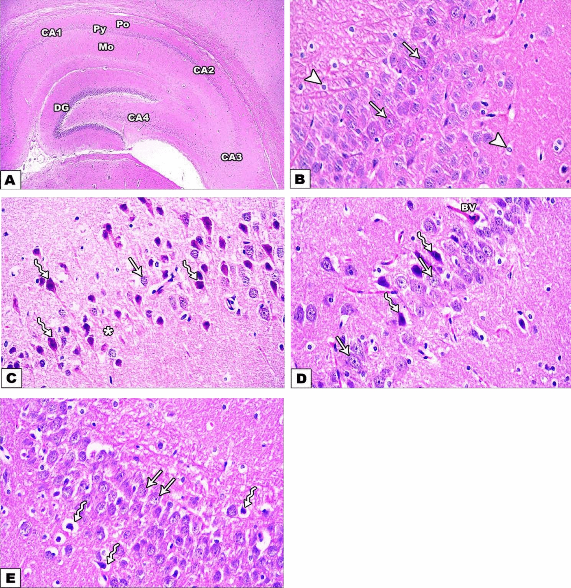
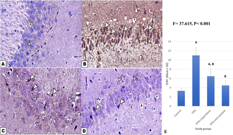
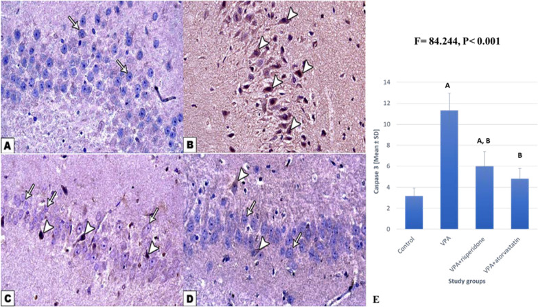
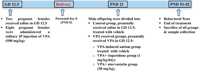
Methods: On gestational day 12.5, pregnant rats received a single IP injection of VPA (500 mg/kg), for VPA induced autism, risperidone and atorvastatin groups, or saline for control normal group. At postnatal day 21, male offsprings were randomly divided into four groups (n = 6): control, VPA induced autism, risperidone, and atorvastatin. Risperidone and atorvastatin were administered from postnatal day 21 to day 51. The study evaluated autism-like behaviors using the three-chamber test, the dark light test, and the open field test at the end of the study. Biochemical analysis of TLR4, NF-κB, NOX-2, and ROS using ELISA, RT-PCR, WB, histological examination with hematoxylin and eosin and immunohistochemical study of CAS-3 were performed.

Results: Male offspring of prenatal VPA-exposed female rats exhibited significant autism-like behaviors and elevated TLR4, NF-κB, NOX-2, ROS, and caspase-3 expression. Histological analysis revealed structural alterations. Both risperidone and atorvastatin effectively mitigated the behavioral, biochemical, and structural changes associated with VPA-induced rat model of autism. Notably, atorvastatin group showed a more significant improvement than risperidone group.

Conclusions: The research results unequivocally demonstrated that atorvastatin can modulate VPA-induced autism by suppressing inflammation, oxidative stress, and apoptosis through TLR4/NF-κB/NOX-2 signaling pathway. Atorvastatin could be a potential treatment for ASD.

Keywords: Apoptosis; Atorvastatin; Autism; NF-κB; Risperidone; TLR4; Valproic acid.


Conflict of interest statement

The authors declare no competing interests.

Figures

Fig. 1
Fig. 1
Effect of risperidone/atorvastatin treatment on the three-chamber test (time spent in non-social and social stimulus A, B number of enteries to non-social and social stimulus C, D) in prenatally VPA-exposed male offspring. Data are expressed as mean ± SD. One-way ANOVA and the post-hoc Tukey's tests were used to compare between groups. P value ≤ 0.05 is significant. A, B, and C represent significance for the control, VPA, and VPA + risperidone groups, respectively. (P value < 0.001). VPA: valproic acid
Fig. 2
Fig. 2
Effect of risperidone/ atorvastatin treatment on the dark light test; duration in the light chamber and number of transitions (A, B) and open field tests; distance traveled and stretched attended posture (C, D) tests in prenatally VPA-exposed male offspring. Data are expressed as mean ± SD. One-way ANOVA test and the post hoc Tukey’s tests were used to compare groups and P value ≤ 0.05 is significant. A, B, and C represent significance for the control, VPA, and risperidone groups, respectively. (P value < 0.001). VPA: valproic acid
Fig. 3
Fig. 3
Effect of risperidone/atorvastatin treatment on the TLR4 marker in the hippocampi of prenatally VPA-exposed male offspring. A TLR4 mRNA expression as detected by RT-PCR. B TLR4 protein relative quantitation by WB. C Representative picture for uncropped blot (whole ladder) including TLR4, β actin protein expression by western blot. D Cropped blot of TLR4, β-actin protein bands with their molecular weight (molecular weight: 95, 43 kDa, respectively). β-actin is selected as an endogenous control. Data are expressed as mean ± SD. One-way ANOVA test and the post hoc Tukey's tests were used to compare groups, A, B, and C represent significance for the control, VPA, and risperidone groups, respectively. P value ≤ 0.05 is significant (P values significance with control, VPA, VPA + risperidone group respectively are: RT-PCR; 0.001, 0.001, 0.019. WB; 0.001, 0.001, 0.027). VPA: valproic acid. TLR4, toll-like receptor 4
Fig. 4
Fig. 4
Effect of risperidone/atorvastatin treatment on NFKβ marker in the hippocampi of prenatally VPA-exposed male offspring. A NFκB mRNA expression as detected by RT-PCR. B NFκB protein level as detected by ELISA. Data are expressed as mean ± SD. One-way ANOVA and post hoc Tukey's tests were used to compare the groups. A, B, and C represent significance for the control, VPA, and risperidone groups, respectively. P value ≤ 0.05 is significant (P values significance with control, VPA, VPA + risperidone group respectively are: RT-qPCR; 0.001, 0.001, 0.049. ELISA; 0.001, 0.001, 0.042). VPA: valproic acid. NFκβ; nuclear factor kappa
Fig. 5
Fig. 5
Effect of risperidone/atorvastatin treatment on NOX2/ ROS markers in the hippocampi of prenatally VPA-exposed male offspring. A NOX2 mRNA expression as detected by RT-qPCR. B NOX2 protein level determined by ELISA. C Total ROS as determined by ELISA. Data are expressed as mean ± SD. One-way ANOVA and post hoc Tukey’s tests were used to compare the groups. A, B, and C represent significance for the control, VPA, and risperidone groups, respectively. P value ≤ 0.05 is significant (P values significance with control, VPA, VPA + risperidone group respectively are: NOX2 RT-qPCR; 0.001, 0.001, 0.007. NOX2 ELISA; 0.001, 0.001, 0.028. total ROS ELISA; 0.001, 0.001, 0.046)VPA; valproic acid. NOX2; NADPH oxidase. ROS, reactive oxygen species
Fig. 6
Fig. 6
photomicrographs of H&E stained sections of rat hippocampi of prenatally VPA-exposed male offspring.: A, B of the control group. A The Hippocampus comprises cornu ammonis regions: CA1, CA2, CA3, CA4. Each region is arranged into 3 layers: polymorphic (Po), pyramidal (Py), and molecular (Mo). The dentate gyrus (DG) appears enclosing CA4 (H& E × 40). B The pyramidal layer of the CA3 region contains large pyramidal cells with vesicular nuclei (arrows). Glial cells are also evident in molecular and polymorphic layers (arrowheads). C VPA-induced autism group: most of the pyramidal cells appear shrunken with darkly stained cytoplasm and pyknotic nuclei (zigzag arrows). Wide intercellular spaces can be observed (*). Few cells appeared normal (arrow). D Risperidone-treated group: most pyramidal cells appear normal with vesicular nuclei (arrows) except for some shrunken cells with darkly stained cytoplasm and pyknotic nuclei (zigzag arrows). Blood vessels can be seen (Bv). E Atorvastatin-treated group: numerous pyramidal cells are normal with vesicular nuclei (arrows), and only little shrunken cells appeared with darkly stained cytoplasm and pyknotic nuclei (zigzag arrows). (H& E × 400)
Fig. 7
Fig. 7
Photomicrographs of TLR4-immunostained hippocampus sections in all study groups of prenatally VPA-exposed male offspring. A control group: pyramidal cells of CA3 region show negative immune reactions (arrows). B VPA induced autism group: pyramidal cells show strong positive immune reactions (arrowheads). C VPA-risperidone treated group, and D VPA-atorvastatin treated group: most pyramidal cells show negative immune reaction (arrows) except for a few cells with positive immune reaction (arrowheads) (immunostaining for TLR4 × 400). E Number of TLR4 positive cells of the CA3 region of the hippocampus in the study groups. Data are expressed as mean ± SD. One-way ANOVA and the post hoc Tukey's tests were used to compare the groups. A and B represent significance for the control and VPA groups, respectively. P value ≤ 0.05 is significant (P values significance of number of TLR4 positive cells with control, VPA, VPA + risperidone group respectively are: 0.001, 0.001, 0.079). VPA: valproic acid. TLR4; Toll-like receptor 4
Fig. 8
Fig. 8
Photomicrographs of Caspase-3 immunohistochemically stained hippocampus sections in all study group in prenatally VPA-exposed male offsprings. A Control group: pyramidal cells of the CA3 region show negative immune reaction (arrows). B VPA-treated group: pyramidal cells show strong positive immune reactions (arrowheads). C VPA-risperidone treated group, and D VPA-atorvastatin treated group: most of the pyramidal cells show negative immune reactions (arrows) except for a few cells with positive immune reactions (arrowheads). (Immunostaining for Caspase- 3 × 400). E Caspase- 3 positive cells number of CA3 region of the hippocampus in the study groups. Data are expressed as mean ± SD. One-way ANOVA and the post-hoc Tukey's tests were used to compare the groups; P value ≤ 0.05 is significant. (P values significance of number of caspase3 positive cells with control, VPA, VPA + risperidone group respectively are: 0.001, 0.001, 0.389). VPA: valproic acid
Fig. 9
Fig. 9
Study design and experimental grouping

References

    1. Rein B, Ma K, Yan Z. A standardized social preference protocol for measuring social deficits in mouse models of autism. Nat Protoc. 2020;15(10):3464–77. - PMC - PubMed
    1. Nadeem A, et al. Toll-like receptor 4 signaling is associated with upregulated NADPH oxidase expression in peripheral T cells of children with autism. Brain Behav Immunity. 2017;61:146–54. - PubMed
    1. Ebrahimi Meimand S, Rostam-Abadi Y, Rezaei N. Autism spectrum disorders and natural killer cells: a review on pathogenesis and treatment. Expert Rev Clin Immunol. 2021;17(1):27–35. - PubMed
    1. Sabra A, Aderbal Filho S, Selma S. Autism: etiology, epidemiology, pathology, clinical aspects and treatment. Autism Open Access. 2020;10(3):253.
    1. Bergeron JD, et al. White matter injury and autistic-like behavior predominantly affecting male rat offspring exposed to group B streptococcal maternal inflammation. Dev Neurosci. 2013;35(6):504–15. - PubMed

Publication types

MeSH terms

LinkOut - more resources