. 2024 Sep 30;43(1):272.
doi: 10.1186/s13046-024-03188-4.

MBD2 regulates the progression and chemoresistance of cholangiocarcinoma through interaction with WDR5

Affiliations

MBD2 regulates the progression and chemoresistance of cholangiocarcinoma through interaction with WDR5

Da Wang et al. J Exp Clin Cancer Res. .

Abstract

Background: Cholangiocarcinoma (CCA) is a highly malignant, rapidly progressing tumor of the bile duct. Owing to its chemoresistance, it always has an extremely poor prognosis. Therefore, detailed elucidation of the mechanisms of chemoresistance and identification of therapeutic targets are still needed.

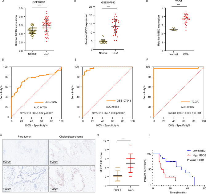
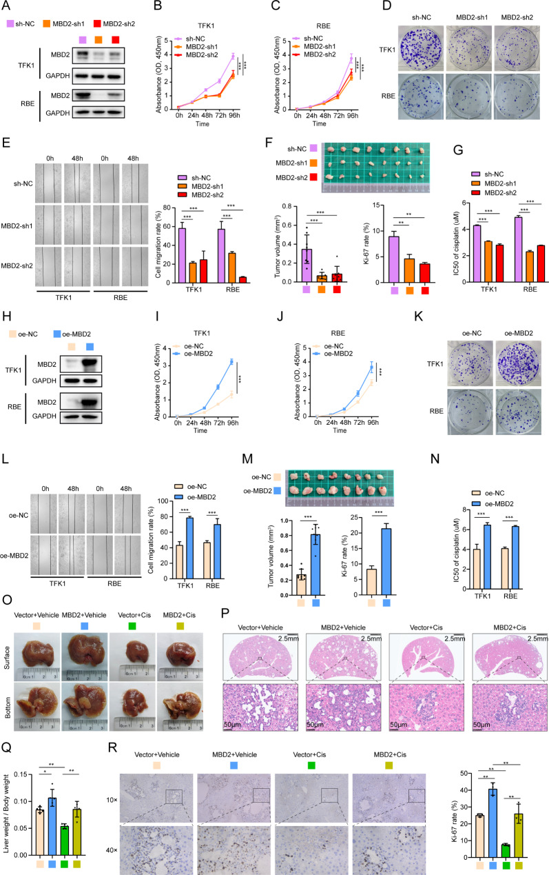
Methods: We analyzed the expression of MBD2 (Methyl-CpG-binding domain 2) in CCA and normal bile duct tissues using the public database and immunohistochemistry (IHC). The roles of MBD2 in CCA cell proliferation, migration, and chemoresistance ability were validated through CCK-8, plate cloning assay, wound healing assays and xenograft mouse model. In addition, we constructed a primary CCA mouse model to further confirm the effect of MBD2. RNA-seq and co-IP-MS were used to identify the mechanisms by how MBD2 leads to chemoresistance.

Results: MBD2 was upregulated in CCA. It promoted the proliferation, migration and chemoresistance of CCA cells. Mechanistically, MBD2 directly interacted with WDR5, bound to the promoter of ABCB1, promoted the trimethylation of H3K4 in this region through KMT2A, and activated the expression of ABCB1. Knocking down WDR5 or KMT2A blocked the transcriptional activation of ABCB1 by MBD2. The molecular inhibitor MM-102 targeted the interaction of WDR5 with KMT2A. MM-102 inhibited the expression of ABCB1 in CCA cells and decreased the chemoresistance of CCA to cisplatin.

Conclusion: MBD2 promotes the progression and chemoresistance of CCA through interactions with WDR5. MM-102 can effectively block this process and increase the sensitivity of CCA to cisplatin.

Keywords: ABCB1; Cholangiocarcinoma; H3K4me3; MBD2; WDR5.


Conflict of interest statement

The authors declare that they have no competing interests.

Figures

Fig. 1
Fig. 1
MBD2 expression is upregulated in CCA. A: Expression levels of MBD2 in GSE76297. B: Expression levels of MBD2 in GSE107943. C: Expression levels of MBD2 in TCGA. D: ROC curve of MBD2 from GSE76297. E: ROC curve of MBD2 from GSE107943. F: ROC curve of MBD2 from TCGA. G: Representative IHC images of MBD2 expression in 40 paratumor tissues and CCA tissues. H: IHC scores of MBD2 in paratumor tissues (n = 40) and CCA tissues (n = 40). I: Kaplan–Meier analysis of the overall survival rates in the low MBD2 expression group and the high MBD2 expression group. ***P < 0.001
Fig. 2
Fig. 2
MBD2 promotes the proliferation, migration and chemoresistance of CCA. A: Knockdown of MBD2 in TFK1 cells and RBE cells was validated by western blotting. B-D: The proliferation ability of MBD2-knockdown TFK1 and RBE cells was detected by a CCK-8 assay (B and C) and a plate cloning assay (D). E: Representative images of the wound healing assay used to detect the migration of MBD2-knockdown TFK1 and RBE cells and the statistical volume. F: Tumors of xenograft mice models derived from MBD2-knockdown TFK1 cells. The image below shows the statistical analysis of the tumor volume and the Ki-67 rate. G: IC50 values of MBD2 knockdown in CCA cells treated with cisplatin. H: Overexpression of MBD2 in TFK1 and RBE cells was validated by western blotting. I-K: The proliferation abilities of MBD2-overexpressing TFK1 and RBE cells were detected by a CCK-8 assay (I and J) and a plate cloning assay (K). L: Representative images of a wound healing assay in which MBD2 was overexpressed in TFK1 and RBE cells and the statistical analysis of the results. M: The tumor of xenograft mice models derived from MBD2-overexpressing TFK1 cells. The image below shows the statistical analysis of the tumor volume and the Ki-67 rate. N: IC50 values of MBD2 overexpression in TFK1 and RBE cells exposed to cisplatin. O: Representative images of livers in primary CCA mice models. Cis: cisplatin. P: HE staining of the liver in primary CCA mice models. Q: The ratio of liver weight to body weight in primary CCA mice models. R: IHC staining of Ki-67 in liver of primary CCA mice models. The right panel shows the statistical of Ki-67 rate. *P<0.05, **P<0.01, ***P<0.001
Fig. 3
Fig. 3
MBD2 regulates the expression of ABCB1 not through NURD. A: Venn diagram of the RNA-seq (GSE275477, negative control with MBD2 knockdown in TFK1 cells) with the ChIP-seq data of MBD2 from the GEO database (GSE41006). B: Volcano map of the RNA-seq data (negative control with MBD2 knockdown in TFK1 cells) with the ChIP-seq data of MBD2 from GEO database (GSE41006). C: Heatmap of the top 10 genes among up-regulated and down-regulated genes from the intersection of RNA-seq and ChIP-seq data. D-E: The protein and mRNA levels of ABCB1 was examined by western blot and RT-PCR in MBD2-knockdown and MBD2-overexpressing cells. F-H: IP was performed to detect the interaction of MBD2 or MBD2-Mut with HDAC1/2 in 293T, TFK1 and RBE cells. I: ABCB1 expression was examined by western blot and RT-PCR in oe-MBD2 and oe-MBD2-Mut cells. ***P < 0.001
Fig. 4
Fig. 4
MBD2 directly interacts with WDR5 in CCA cells. A: The gel of the negative control group and MBD2-IP group from 293T cells. The protein samples were separated by SDS-PAGE and stained with Coomassie brilliant blue. The protein masses are shown in the left. B: Co-localization of MBD2 and WDR5 was detected by immunofluorescence. C-E: The interaction of MBD2 with WDR5 was detected by IP in 293T, TFK1 and RBE cells. F: The direct interaction of MBD2 with WDR5 was confirmed by GST pull-down. G: The schematic diagram of the domain organization of MBD2 and its truncated mutants. H: 293T cells was transfected with the full length or the truncated mutants of MBD2. Co-IP was used to detect which of the truncated fragment of MBD2 bound to WDR5. I: IP was used to detect the binding ability between Flag-M1 fragments and WDR5
Fig. 5
Fig. 5
WDR5 promotes the proliferation, migration and chemoresistance of CCA. A: Western blotting was performed to detect the effect of WDR5 knockdown in TFK1 and RBE cells. B-D: The proliferation abilities of TFK1 and RBE cells with WDR5 knockdown was detected by CCK-8 assay (B and C) and plate cloning assay (D). E: Representative images of wound healing assay used to detect the migration of TFK1 and RBE cells in which WDR5 was knocked down and the statistical volume. F: Tumors of xenograft mice models derived from TFK1 cells with WDR5 knockdown. The image below shows the statistical analysis of the tumor volume and the Ki-67 rate. G: IC50 values of CCA cells in response to cisplatin upon WDR5 knockdown. H: Overexpression of WDR5 in TFK1 and RBE cells was validated by western blotting. I-K: The proliferation abilities of TFK1 and RBE cells overexpressing WDR5 were detected by CCK-8 assay (I and J) and plate cloning assay (K). L: Representative images of wound healing assay in which WDR5 was overexpressed in TFK1 and RBE cells and the statistical volume. M: The tumor of xenograft mice models derived from TFK1 cells with WDR5 overexpression. The image below is the statistical of tumor volume and Ki-67 rate. N: IC50 values of TFK1 and RBE cells response to cisplatin upon WDR5 overexpression. **P < 0.01, ***P < 0.001
Fig. 6
Fig. 6
MBD2 activates the expression of ABCB1 through the WDR5-KMT2A complex. A-B: The protein levels of ABCB1 was examined by western blot in WDR5 knockdown and overexpression cells. C: Western blotting was used to detect the effect of KMT2A knockdown in TFK1 and RBE cells. D: The protein levels of ABCB1 was examined by western blot in KMT2A knockdown cells. E-F: The protein levels of ABCB1 was detected by western blot upon overexpression of MBD2 and knockdown of WDR5. G-H: The protein levels of ABCB1 was detected by western blot upon overexpression of MBD2 and knockdown of KMT2A. I: The GEO database indicates binding peaks of MBD2, WDR5, and KMT2A on the ABCB1 gene. The below panel is the schematic diagram of binding peak of ABCB1 and the ChIP-qPCR product (P1, P2, P3, P4). J-M: The ChIP-qPCR was used to examine the binding of MBD2, WDR5 and H3K4me3 to ABCB1 in MBD2 or WDR5 overexpression TFK1 cells. Anti-Flag antibody was used for ChIP-qPCR to detect the enrichment of MBD2-Flag or WDR5-Flag in the binding region; Anti-H3K4me3 antibody was used for ChIP-qPCR to detect the enrichment of H3K4me3 in the binding region. ***P < 0.001
Fig. 7
Fig. 7
Targeted inhibition of the WDR5-KMT2A interaction by MM-102 weakens the proliferation, migration, and chemoresistance of CCA. A: The protein levels of ABCB1, MBD2 and WDR5 was examined by western blot in TFK and RBE cells supplemented with MM-102. B: The representative image of wound healing assay to detect the migration of TFK1 and RBE cells added with MM-102. The right panel is the statistical volume. C-D: The proliferation abilities of TFK1 and RBE cells supplemented with MM-102 was detected by CCK-8 assay (C) and plate cloning assay (D). E: IC50 value of TFK1 and RBE cells to cisplatin upon added with MM-102. F: Representative image of liver in primary CCA mice models treated with cisplatin or MM-102. Cis: cisplatin. G: HE staining of the liver in primary CCA mice models treated with cisplatin or MM-102. Cis: cisplatin. H: The ratio of liver weight to body weight in primary CCA mice models. I: IHC staining of Ki-67 in liver of primary CCA mice models. The right panel is the statistical of Ki-67 rate. *P < 0.05, **P < 0.01, ***P < 0.001
Fig. 8
Fig. 8
Schematic of a model for the major molecular mechanisms. MBD2 forms a complex with WDR5-KMT2A through their interaction, promoting the H3K4 trimethylation of specific regions of the ABCB1 promoter, which enhances ABCB1 expression and contributes to the chemoresistance, proliferation, and migration capabilities of cholangiocarcinoma

References

    1. Brindley PJ, Bachini M, Ilyas SI, Khan SA, Loukas A, Sirica AE, et al. Cholangiocarcinoma Nat Reviews Disease Primers. 2021;7:65. - PMC - PubMed
    1. Florio AA, Ferlay J, Znaor A, Ruggieri D, Alvarez CS, Laversanne M, et al. Global trends in intrahepatic and extrahepatic cholangiocarcinoma incidence from 1993 to 2012. Cancer. 2020;126:2666–78. - PMC - PubMed
    1. Forner A, Vidili G, Rengo M, Bujanda L, Ponz-Sarvisé M, Lamarca A. Clinical presentation, diagnosis and staging of cholangiocarcinoma. Liver International: Official J Int Association Study Liver. 2019;39(Suppl 1):98–107. - PubMed
    1. Roth GS, Neuzillet C, Sarabi M, Edeline J, Malka D, Lièvre A. Cholangiocarcinoma: what are the options in all comers and how has the advent of molecular profiling opened the way to personalised medicine ? European journal of cancer (Oxford, England: 1990). 2023; 179: 1–14. - PubMed
    1. Khan SA, Thomas HC, Davidson BR, Taylor-Robinson SD, Cholangiocarcinoma. Lancet (London England). 2005;366:1303–14. - PubMed

MeSH terms

Substances