. 2024 Sep 30;22(1):461.
doi: 10.1186/s12964-024-01839-9.

Keratinocyte derived extracellular vesicles mediated crosstalk between epidermis and dermis in UVB-induced skin inflammation

Affiliations

Keratinocyte derived extracellular vesicles mediated crosstalk between epidermis and dermis in UVB-induced skin inflammation

Yubin Li et al. Cell Commun Signal. .

Abstract

Background and rationale: Ultraviolet-B (UVB) light induces dermal inflammation, although it is mostly absorbed in the epidermis. Recent reports suggest extracellular vesicles (EVs) act as a mediator of photodamage signaling. Melatonin is reported to be a protective factor against UV-induced damage. We hypothesized that EVs derived from UVB-irradiated keratinocytes might trigger proinflammatory responses in dermal cells and tested whether melatonin can ameliorate UVB-induced inflammation.

Methods: We used UVB-irradiated HaCaT cells, primary keratinocytes and STING knock-out mice to model production of EVs under photodamaging conditions and performed immunoblotting and ELISA to measure their effect on dermal macrophages.

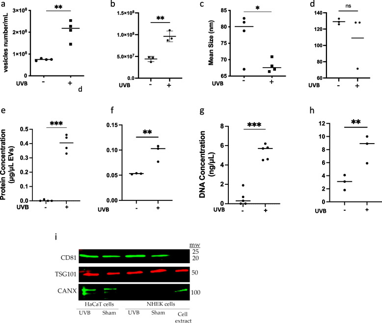
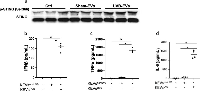
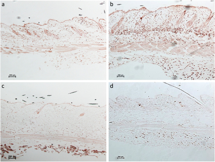
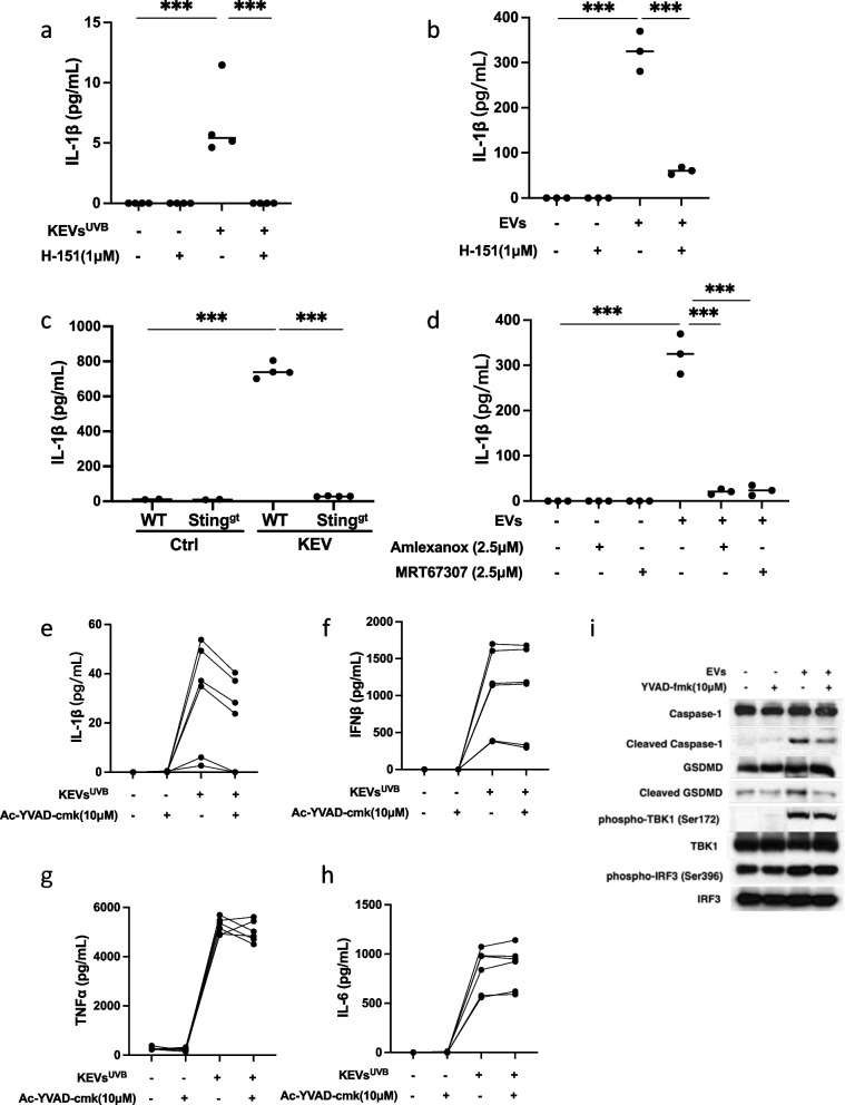
Results: UVB-irradiated keratinocytes produce an increased number of EVs that contain higher concentrations of DNA and protein compared with controls. KC-derived EVs (KEVs) induced a STING- and inflammasome-mediated proinflammatory response in macrophages in vitro, and a pronounced inflammatory infiltrate in mouse dermis in vivo. Melatonin ameliorated KEVs inflammatory effect both in vitro and in vivo.

Conclusions: This data suggests EVs are mediators in a crosstalk that takes place between keratinocytes and their neighboring cells as a result of photodamage. Further studies exploring EVs induced by damaging doses of UVB, and their impact on other cells will provide insight into photodamage and may help develop targeted therapeutic approaches.


Conflict of interest statement

The authors declare no competing interests.

Figures

Fig. 1
Fig. 1
Characterization of keratinocyte-derived EVs (KEVs). HaCaT keratinocytes were plated in 35-mm wells (5x10⁵/mL, 2mL/well) and then irradiated with 0 (-) or 75mJ/cm2 of UVB (+), NHEKs were plated in 35-mm wells (2.7x10⁶/mL, 2mL/well) and then irradiated with 0 (-) or 30 mJ/cm2 of UVB (+). 15 hours after UVB irradiation, culture supernatants were collected for isolation of KEVs. Total number of KEVs derived from both UVB-irradiated HaCaT cells (a) and primary keratinocytes (b) is higher than in sham-irradiated cells. Average size of KEVs derived from sham-irradiated HaCaT keratinocytes is larger than UVB-irradiated HaCaT keratinocytes (c), in NHEKs no significant size difference was detected (d). Protein content of KEVs derived from sham- and UVB-irradiated HaCaT (e) and NHEK (f) cells. DNA content of KEVs derived from sham- and UVB-irradiated HaCaT (g) and NHEK (h) cells. Western blot demonstrates enriched EV markers CD81 and TSG101 in representative samples. CANX was not detected in NHEKs but was detected in HaCaT cells (i). Data represent means±SDs. *p<0.05, **p<0.01, ***p<0.001 between groups as indicated
Fig. 2
Fig. 2
UVB-irradiated keratinocyte-derived EVs (KEVs-UVB) triggered more STING phosphorylation and proinflammatory cytokines in primary human PBMCs and macrophages. UVB-irradiated KEVs triggered more STING phosphorylation than non-UVB-irradiated keratinocyte-derived EVs in PBMCs (a). UVB-irradiated keratinocyte-derived EVs triggered more IFNβ (b), TNFα (c) and IL-6 (d) in macrophages. Data represent mean±SD. *p<0.05, between groups as indicated
Fig. 3
Fig. 3
KEVs-UVB induced STING-dependent proinflammatory responses in macrophages and to a lesser extent in fibroblasts.UVB-induced KEVs stimulate STING phosphorylation (a, b), IFNß (c) and IL-6 (d) in RAW264.7 cells, while H-151inhibited those effects. UVB-induced KEVs stimulate WT murine macrophages to secrete more IFNß (e), TNFα (f), and IL-6 (g) than STING KO macrophages. TBK1 inhibitors suppressed UVB-induced KEVs-stimulated RAW264.7 macrophages secretion of IFNß (h), TNFα (i), and IL-6 (j). UVB-KEVs did not trigger more IFNβ or TNFα production in human primary dermal fibroblasts (kl). UVB-KEVs did trigger increased production of IL-6 in fibroblasts, but the production was not attenuated by STING inhibition (m). Data represent mean±SD. *p<0.05, **p<0.01, ***p<0.001 between groups as indicated
Fig. 4
Fig. 4
UVB irradiation induced less dermal inflammation in STING KO (Stinggt/gt) mice than in wild-type (WT) mice. CD45 staining shows that UVB irradiation of STING KO mice stimulated the recruitment of fewer immune cells into the dermis than in WT mice. a WT-CTRL (b) WT-UVB, Sting gt/gt - CTRL (d) Sting gt/gt -UVB
Fig. 5
Fig. 5
Suppression of STING and inflammasome pathways. Pretreatment of macrophages with the STING antagonist H-151 abolished KEVs-induced IL-1β release in primary mice macrophages (a) and RAW264.7 macrophages (b). KEVs-UVB induced production of interleukin-1 β is suppressed in STING KO macrophages (c). Pretreatment of macrophages with the TBK1 antagonists suppressed KEVs-UVB-induced IL-1β release (d). UVB-induced KEVs stimulated RAW264.7 cells to secrete large amounts of IL-1β, IFNß, TNFα, and IL-6. Pretreatment of the macrophages with 0μM (–) or 10μM (+) of the pyroptosis inhibitor Ac-YVAD-cmk suppressed KEVs-UVB-triggered IL-1β production (e) but had no effect on KEV-stimulated production of the other cytokines (f-h). Similar results obtained using VX765, a second inflammasome inhibitor, are shown in Supplementary Figure 1. Immunoblot shows inhibition of the inflammasome pathway affects apoptotic inflammasome markers but does not inhibit the STING pathway (i)
Fig. 6
Fig. 6
Melatonin suppressed UVB irradiation induced KEVs release, apoptosis, inflammation, and KEVs-mediated proinflammatory responses. HaCaT cells pretreated with melatonin produce a lower number of EVs compared with control keratinocytes (a, b). Particle mean size was not significantly different (c). Treatment of primary keratinocytes resulted in similar findings (Supplementary Figure 2). PARP-1 and Caspase-3 cleavage in primary keratinocytes is attenuated by melatonin (d-f). TNFα production by macrophages stimulated by UVB-induced KEVs is attenuated by pretreatment of macrophages with melatonin in a dose dependent manner (g). Skin biopsy of C57BL/6J mice treated with melatonin prior to UVB irradiation, stained for CD45, shows less inflammatory infiltrate (h) and thinner epidermis (i). Data represent mean ± SD. *<0.05, **p<0.01, ***p<0.001

References

    1. Amaro-Ortiz A, Yan B, D’Orazio JA. Ultraviolet radiation, aging and the skin: prevention of damage by topical cAMP manipulation. Molecules. 2014;19(5):6202–19. - PMC - PubMed
    1. Arioz BI, Tastan B, Tarakcioglu E, Tufekci KU, Olcum M, Ersoy N, et al. Melatonin attenuates LPS-induced acute depressive-like behaviors and microglial NLRP3 inflammasome activation through the SIRT1/Nrf2 pathway. Front Immunol. 2019;10:1511. - PMC - PubMed
    1. Baka Z, Senolt L, Vencovsky J, Mann H, Simon PS, Kittel A, et al. Increased serum concentration of immune cell derived microparticles in polymyositis/dermatomyositis. Immunol Lett. 2010;128(2):124–30. - PubMed
    1. Bauwens E, Parée T, Meurant S, Bouriez I, Hannart C, Wéra AC, et al. Senescence induced by UVB in keratinocytes impairs amino acids balance. J Invest Dermatol. 2023;143(4):554–65. - PubMed
    1. Bennett MF, Robinson MK, Baron ED, Cooper KD. Skin immune systems and inflammation: protector of the skin or promoter of aging? J Investig Dermatol Symp Proc. 2008;13(1):15–9. - PubMed

MeSH terms

LinkOut - more resources