

Frontal Glioblastoma Presenting With Catatonia: A Case Report
- PMID: 39350835
- PMCID: PMC11441845
- DOI: 10.7759/cureus.68320
Frontal Glioblastoma Presenting With Catatonia: A Case Report
Abstract
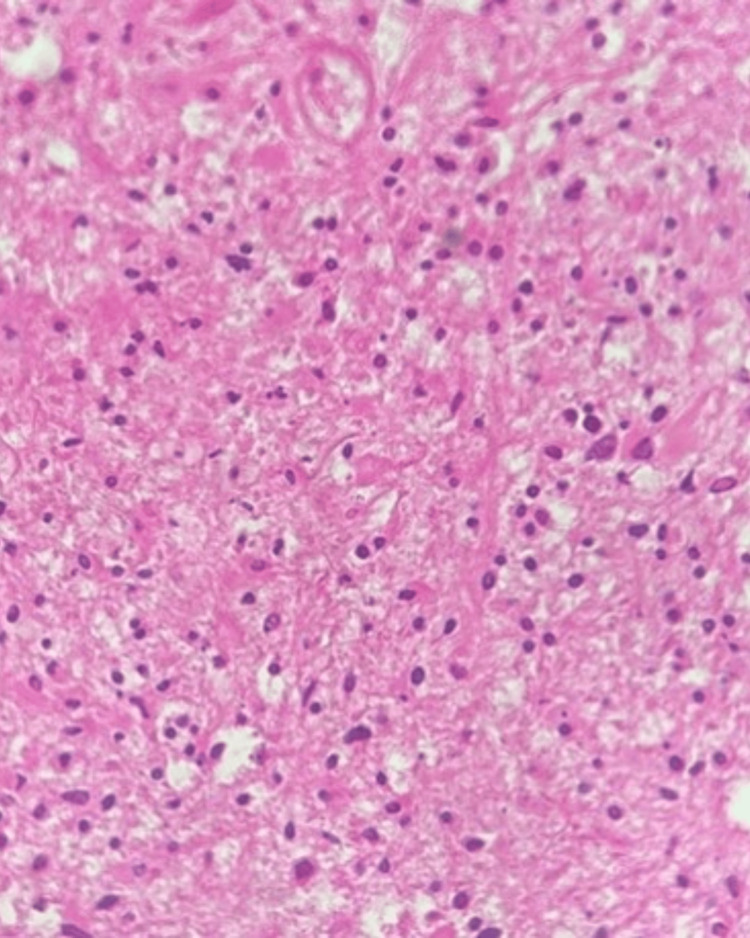
This case highlights the need for thorough medical and neurological screening before making any psychiatric diagnosis, even if the patient has a classical syndromic presentation. Presented here is a case of a female in her late 40s coming to our psychiatric outpatient with symptoms suggestive of catatonia. She was treated at a private clinic for depression. As her symptoms deteriorated, she was brought to our hospital for further management. She was diagnosed with catatonia during admission assessment. A detailed neurological assessment later revealed slight weakness in the right upper and lower limb. Following this, a CT scan was done and was reported to have a hypodense area involving the bilateral frontal and basifrontal region, more pronounced on the left side (likely to be acute/subacute). MRI was subsequently done and was found to be suggestive of glioblastoma NOS (not otherwise specified) involving the bilateral cerebral hemisphere which was later confirmed by the histopathology report.
Keywords: catatonia; catatonia in glioblastoma; catatonic symptoms; frontal glioblastoma; frontal lobe glioblastoma; glioblastoma; glioblastoma and catatonia; glioblastoma tumour; psychomotor disturbances with glioblastoma.
Copyright © 2024, Sinha et al.
Conflict of interest statement
Human subjects: Consent was obtained or waived by all participants in this study. Conflicts of interest: In compliance with the ICMJE uniform disclosure form, all authors declare the following: Payment/services info: All authors have declared that no financial support was received from any organization for the submitted work. Financial relationships: All authors have declared that they have no financial relationships at present or within the previous three years with any organizations that might have an interest in the submitted work. Other relationships: All authors have declared that there are no other relationships or activities that could appear to have influenced the submitted work.
Figures


References
-
- Jain A, Mitra P. StatPearls [Internet] Treasure Island (FL): StatPearls Publishing; 2023. Catatonic schizophrenia. - PubMed
-
- Catatonia: diagnosis, classification, and treatment. Francis A. Curr Psychiatry Rep. 2010;12:180–185. - PubMed
-
- Catatonia in DSM-5. Tandon R, Heckers S, Bustillo J, et al. Schizophr Res. 2013;150:26–30. - PubMed
Publication types
LinkOut - more resources
Full Text Sources
Research Materials