. 2024 Aug 23;27(10):110624.
doi: 10.1016/j.isci.2024.110624. eCollection 2024 Oct 18.

SARS-CoV-2 ferritin nanoparticle vaccines produce hyperimmune equine sera with broad sarbecovirus activity

Affiliations

SARS-CoV-2 ferritin nanoparticle vaccines produce hyperimmune equine sera with broad sarbecovirus activity

Elizabeth J Martinez et al. iScience. .

Abstract

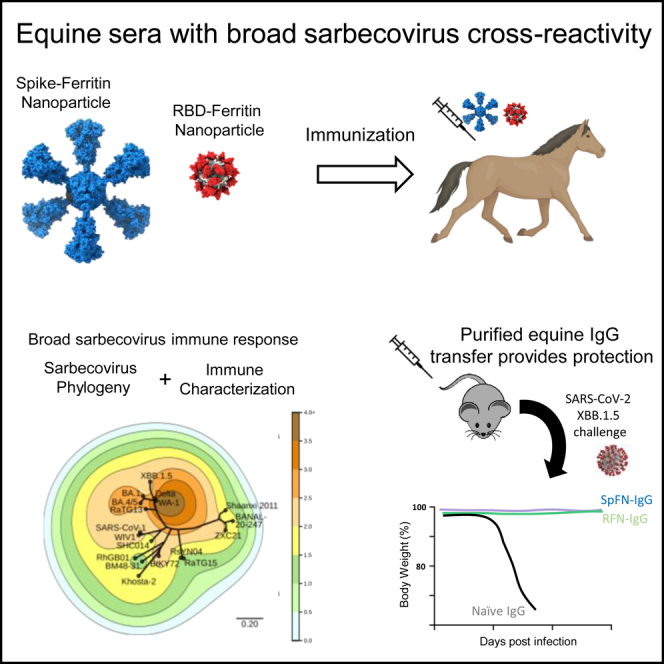
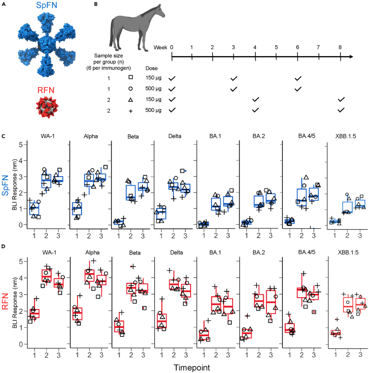
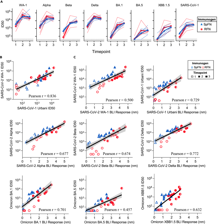
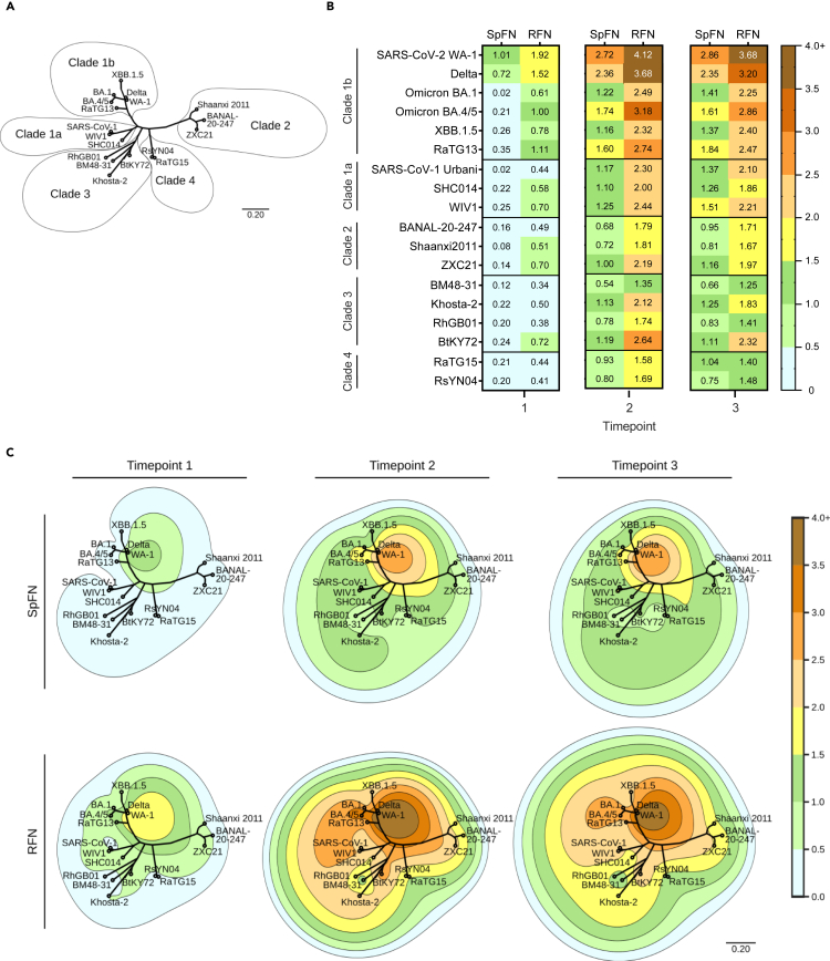
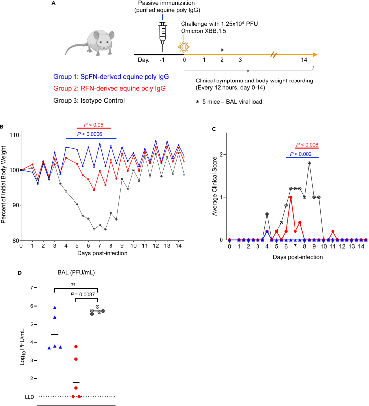
The rapid emergence of SARS-CoV-2 variants of concern (VoC) and the threat of future zoonotic sarbecovirus spillover emphasizes the need for broadly protective next-generation vaccines and therapeutics. We utilized SARS-CoV-2 spike ferritin nanoparticle (SpFN), and SARS-CoV-2 receptor binding domain ferritin nanoparticle (RFN) immunogens, in an equine model to elicit hyperimmune sera and evaluated its sarbecovirus neutralization and protection capacity. Immunized animals rapidly elicited sera with the potent neutralization of SARS-CoV-2 VoC, and SARS-CoV-1 pseudoviruses, and potent binding against receptor binding domains from sarbecovirus clades 1b, 1a, 2, 3, and 4. Purified equine polyclonal IgG provided protection against Omicron XBB.1.5 virus in the K18-hACE2 transgenic mouse model. These results suggest that SARS-CoV-2-based nanoparticle vaccines can rapidly produce a broad and protective sarbecovirus response in the equine model and that equine serum has therapeutic potential against emerging SARS-CoV-2 VoC and diverse sarbecoviruses, presenting a possible alternative or supplement to monoclonal antibody immunotherapies.

Keywords: Immunology; Virology.


Conflict of interest statement

W.H.C, A.H., P.V.T., J.L.J., K.M. and M.G.J. are named inventors on provisional patents describing SpFN molecules. S.J.K., V.D., N.L.M., and K.M. are named inventors on provisional patents describing monoclonal antibodies against coronaviruses. M.G.J. is named as an inventor on international patent application WO/2018/081318 and U.S. patents 10,960,070, and 11,964,010 entitled “Prefusion coronavirus spike proteins and their use.” K.M. is a current employee of Pfizer and may, therefore, be a shareholder. A.F., K.Mur., and J.Kau. are former or current employees of B.S.V. The other authors declare no competing interests.

Figures

None
Graphical abstract
Figure 1
Figure 1
Robust RBD binding antibody responses elicited in the equine model (A) Structural model of immunogens, Spike ferritin nanoparticle (SpFN) in blue and receptor binding domain ferritin nanoparticle (RFN) in red. (B) Immunization schema for equine hyperimmune SARS-CoV-2-reactive sera generation (n = 12). Horses C9-C14 were immunized with SpFN, and horses C15-C20 were immunized with RFN, with CFA adjuvant for prime, and IFA for the subsequent two immunizations. Immunization schedules are depicted as square (150 μg dose at 0, 3, and 6 weeks), circle (500 μg dose at 0, 3, and 6 weeks), triangle (150 μg dose at 0, 4, and 8 weeks), and plus sign (500 μg dose at 0, 4, and 8 weeks). (C and D) SARS-CoV-2 VoC RBD binding levels measured by BLI. (C) SpFN immunized group (blue), (D) RFN immunized group (red). Symbols are matched to the immunization schema shown in (B).
Figure 2
Figure 2
Sarbecovirus cross-reactive neutralization and binding capacity (A) Pseudovirus neutralization (ID50 values) of WA-1, Alpha, Beta, Delta, Omicron BA.1, Omicron BA.5, Omicron XBB.1.5, and SARS-CoV-1. Thin lines represent individual animals, and the bold line depicts the geometric mean (blue, triangle: SpFN, red, circle: RFN). (B) Correlation between SARS-CoV-2 WA-1 and SARS-CoV-1 Urbani pseudovirus neutralization titers (ID50), the black line depicts the locally weighted smoothing line and gray shading is the 95% confidence interval around the smoothing line. (C) Correlation between BLI RBD-binding responses and pseudovirus neutralization titer (ID50), for each SARS-CoV-2 VoC and SARS-CoV-1. Pearson correlation coefficient calculation (r) assumed Gaussian distribution, and a two-tailed P value was calculated using GraphPad.
Figure 3
Figure 3
Cross-reactive RBD-binding response against genetically distant sarbecoviruses (A) Phylogenetic tree, based on the RBD amino acid sequences of SARS-CoV-2 VoC and sarbecoviruses. (B) Average serum binding responses (nm) to sarbecovirus RBD molecules measured by BLI for SpFN- and RFN-immunized animals (timepoints indicated at base of the plot). (C) Contour-phylogenetic plots of binding of SpFN and RFN immunized animal sera to sarbecovirus RBD molecules. X and Y coordinates are determined by a phylogenetic tree of the RBD amino acid sequences, and contour height determined by detected BLI serum binding. Interpolation and extrapolation of binding data (elevation) between points on the phylogenetic tree was determined by a thin plate radial basis function (r2∗log(r)) using the scipy python package. For clarity, extrapolated values higher than the maximum measured binding were truncated at the maximum binding for the timepoint.
Figure 4
Figure 4
Passive immunization with purified equine IgG protect mice from SARS-CoV-2 XBB.1.5 challenge (A) Schematic of K18-hACE2 mice SARS-CoV-2 challenge study. Mice (n = 10/group, 5 female, 5 male) received an intraperitoneal injection of purified IgG from SpFN-immunized horses (blue), or RFN-immunized horses (red), or human IgG1 isotype control mAb (black), one day prior to challenge with 1.25 × 104 PFU of SARS-CoV-2 virus (Omicron XBB.1.5). (B) Body weight measurements for K18-hACE2 mice over the course of the challenge study (n = 5/group). Percentage of initial weight is plotted. Isotype control mAb (black open X circle), or SpFN-purified IgG (blue triangle) or RFN-purified IgG (red circle). Significant difference for each measurement timepoint between each group compared to the antibody isotype control group, as assessed by t-test is indicated by a horizontal line. (C) Average cinical score measurements of the K18-hACE2 study groups (n = 5/group). Isotype control mAb (black open X circle), or SpFN-purified IgG (blue triangle) or RFN-purified IgG (red circle). Significant difference for each measurement timepoint between each group compared to the antibody isotype control group, as assessed by t-test is indicated by a horizontal line above the plot. (D) SARS-CoV-2 viral loads in BAL, were measured 2 days post-challenge in a subset of animals (n = 5/group) by plaque assay. BAL (PFU/mL) viral levels in the two study groups were compared for significance against the control group using a Kruskal-Wallis ANOVA test followed by post hoc Dunn’s multiple comparison test.

References

    1. Starr T.N., Zepeda S.K., Walls A.C., Greaney A.J., Alkhovsky S., Veesler D., Bloom J.D. ACE2 binding is an ancestral and evolvable trait of sarbecoviruses. Nature. 2022;603:913–918. doi: 10.1038/s41586-022-04464-z. - DOI - PMC - PubMed
    1. Wells H.L., Letko M., Lasso G., Ssebide B., Nziza J., Byarugaba D.K., Navarrete-Macias I., Liang E., Cranfield M., Han B.A., et al. The evolutionary history of ACE2 usage within the coronavirus subgenus Sarbecovirus. Virus Evol. 2021;7 doi: 10.1093/ve/veab007. - DOI - PMC - PubMed
    1. Boni M.F., Lemey P., Jiang X., Lam T.T.Y., Perry B.W., Castoe T.A., Rambaut A., Robertson D.L. Evolutionary origins of the SARS-CoV-2 sarbecovirus lineage responsible for the COVID-19 pandemic. Nat. Microbiol. 2020;5:1408–1417. doi: 10.1038/s41564-020-0771-4. - DOI - PubMed
    1. Heffron A.S., McIlwain S.J., Amjadi M.F., Baker D.A., Khullar S., Armbrust T., Halfmann P.J., Kawaoka Y., Sethi A.K., Palmenberg A.C., et al. The landscape of antibody binding in SARS-CoV-2 infection. PLoS Biol. 2021;19 doi: 10.1371/journal.pbio.3001265. - DOI - PMC - PubMed
    1. Racine T., Denizot M., Pannetier D., Nguyen L., Pasquier A., Raoul H., Saluzzo J.F., Kobinger G., Veas F., Herbreteau C.H. In Vitro Characterization and In Vivo Effectiveness of Ebola Virus Specific Equine Polyclonal F(ab')2. J. Infect. Dis. 2019;220:41–45. doi: 10.1093/infdis/jiz068. - DOI - PubMed

LinkOut - more resources