. 2024 Sep 14:29:101246.
doi: 10.1016/j.mtbio.2024.101246. eCollection 2024 Dec.

A bioengineered tumor matrix-based scaffold for the evaluation of melatonin efficacy on head and neck squamous cancer stem cells

Affiliations

A bioengineered tumor matrix-based scaffold for the evaluation of melatonin efficacy on head and neck squamous cancer stem cells

Julia López de Andrés et al. Mater Today Bio. .

Abstract

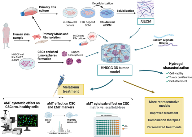
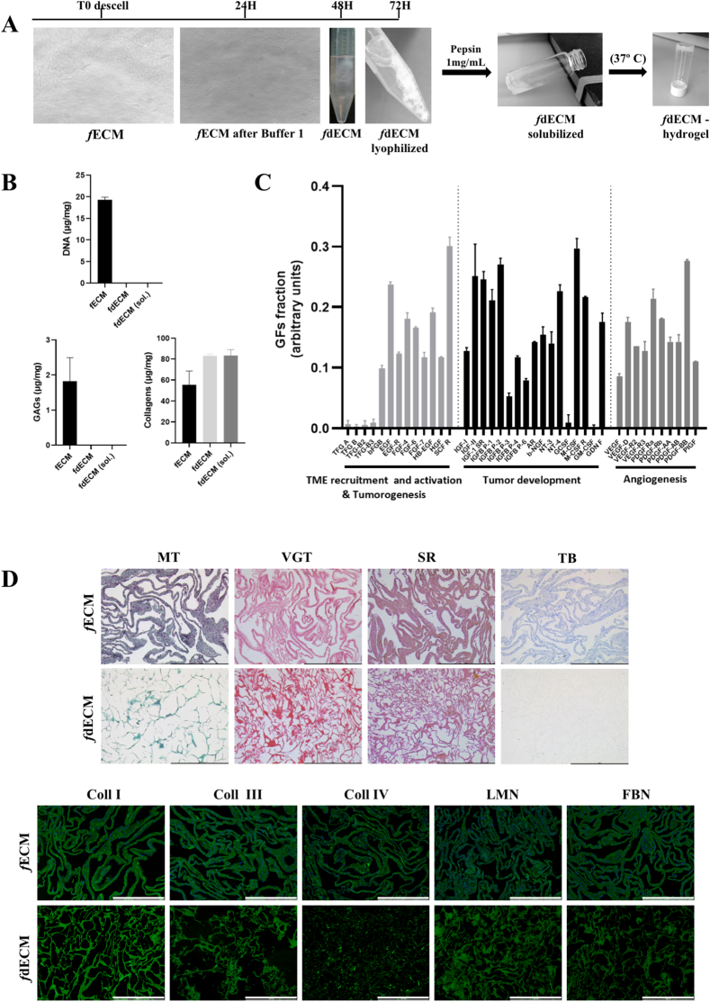
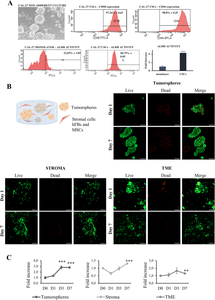
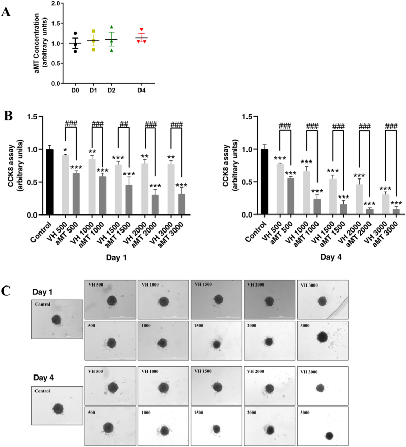
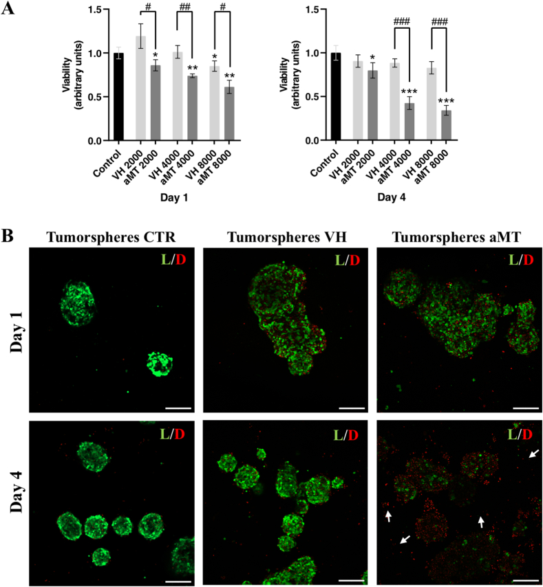
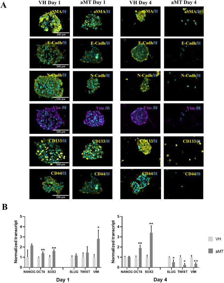
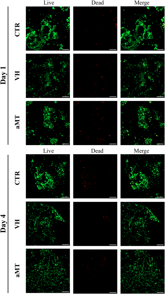
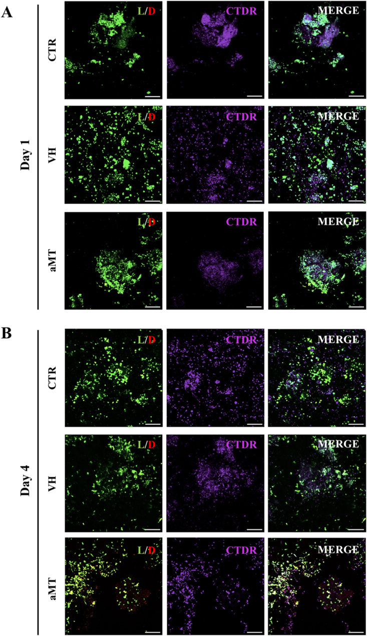
Head and neck squamous cell carcinoma (HNSCC) presents a significant challenge worldwide due to its aggressiveness and high recurrence rates post-treatment, often linked to cancer stem cells (CSCs). Melatonin shows promise as a potent tumor suppressor; however, the effects of melatonin on CSCs remain unclear, and the development of models that closely resemble tumor heterogeneity could help to better understand the effects of this molecule. This study developed a tumor scaffold based on patient fibroblast-derived decellularized extracellular matrix that mimics the HNSCC microenvironment. Our study investigates the antitumoral effects of melatonin within this context. We validated its strong antiproliferative effect on HNSCC CSCs and the reduction of tumor invasion and migration markers, even in a strongly chemoprotective environment, as it is required to increase the minimum doses necessary to impact tumor viability compared to the non-scaffolded tumorspheres culture. Moreover, melatonin exhibited no cytotoxic effects on healthy cells co-cultured in the tumor hydrogel. This scaffold-based platform allows an in vitro study closer to HNSCC tumor reality, including CSCs, stromal component, and a biomimetic matrix, providing a new valuable research tool in precision oncology.

Keywords: 3D hydrogel; 3D scaffold; Cancer stem cells; Decellularized extracellular matrix; Head and neck squamous cell carcinoma; Melatonin; Tumor microenvironment.


Conflict of interest statement

The authors declare that they have no known competing financial interests or personal relationships that could have appeared to influence the work reported in this paper.

Figures

Image 1
Graphical abstract
Fig. 1
Fig. 1
fdECM characterization. (A) Process of deposition and collection of fdECM, and its macroscopic appearance after the different decellularization, solubilization and matrix gelation treatments. (B) Quantification of residual DNA, GAGs and collagens present in the matrices obtained (fECM), after the decellularization (fdECM) and solubilization (fdECM solub.) processes. (C) Quantification of the retained GF fraction of fdECM. (D) Representative images of histological staining of fECMs and fdECMs for Masson's Trichrome (MT), Van Gieson's Trichrome (VGT), Sirius red (SR) and Toluidine Blue (TB), and immunofluorescence of collagens type I (Coll I), III (Coll III) and IV (Coll IV), fibronectin (FBN) and laminin (LMN). Scale bar: 500 μm. (For interpretation of the references to color in this figure legend, the reader is referred to the Web version of this article.)
Fig. 2
Fig. 2
HNSCC microenvironment model. (A) Characterization of Cal-27 CSCs. Image of tumorspheres grown in suspension with serum-free spheres medium (10×). Cytometry histograms of CD98 and CD44 CSCs cell-membrane markers expression. Percentage of ALDH1 activity of cells grown in monolayer or in suspension (cytometry histograms besides). (B) Schematic representation of the TME components embedded in the hydrogel. Confocal representative images of cell viability of tumorspheres, FBs and MSCs (stroma) and the co-culture of stroma and tumorspheres (TME) cultured in the hydrogel. Living cells shown in green and nuclei of death cells in red. Scale bar: 200 μm. (C) Proliferation assay of the tumorspheres, FBs and MSCs (stroma) and the co-culture of stroma and tumorspheres (TME) cultured in the hydrogel. (For interpretation of the references to color in this figure legend, the reader is referred to the Web version of this article.)
Fig. 3
Fig. 3
ct in HNSCC tumorspheres. (A) Quantification of aMT concentration in the culture medium monitored over four days. (B) Cell cytotoxicity assay of the tumorspheres at increasing concentrations of aMT, compared to their corresponding vehicle control (VH), at day 1 and 4 after application of the last dose of aMT (C) Representative images of the aMT-treated tumorspheres and their corresponding vehicle controls. The aMT doses applied were: 500, 1,000, 1,500, 2,000 and 3,000 μM. Scale bar: 1000 μm.
Fig. 4
Fig. 4
aMT antiproliferative effect in HNSCC tumorspheres viability cultured in the tumor hydrogel. (A) Cell cytotoxicity assay of tumor spheres cultured in hydrogel at increasing concentrations of aMT, compared to their corresponding vehicle control, at day 1 and 4 after application of the last dose of aMT. (B) Representative confocal images of cell viability of tumorspheres cultured in hydrogel, untreated (CTR tumorspheres), administering vehicle alone (tumorspheres VH) and with aMT 4,000 μM (tumorspheres aMT), after 1 or 4 days of treatment. White arrows point to the isolated cells from tumorspheres disaggregation. Living cells shown in green and nuclei of death cells in red. Scale bar: 200 μm. (For interpretation of the references to color in this figure legend, the reader is referred to the Web version of this article.)
Fig. 5
Fig. 5
aMT effect in HNSCC tumorspheres phenotype cultured in the tumor hydrogel. (A) Representative immunofluorescence images of CD44, CD133, αSMA, Vimentin, E-Cadherin, and N-Cadherin in the tumorspheres cultured in the hydrogel and treated with aMT 4,000 μM or vehicle, after 1 or 4 days of treatment. Scale bar: 100 μm. (B) qPCR expression analysis of NANOG, OCT4, SOX2, SLUG, TWIST, and VIMENTIN (VIM) genes in the tumor spheres cultured in the hydrogel and treated with aMT 4,000 μM or vehicle after 1 or 4 days of treatment. Normalized with endogenous GAPDH and relative to the day 1 control.
Fig. 6
Fig. 6
aMT effect in HNSCC stromal cells viability cultured in the tumor hydrogel. Representative confocal images of cell viability of FBs and MSCs (stroma) cultured in hydrogel, untreated (CTR), administering vehicle alone (VH) and aMT 4,000 μM (aMT), after 1 (A) or 4 (B) days of treatment. Living cells shown in green and nuclei of death cells in red. Scale bar: 200 μm. (For interpretation of the references to color in this figure legend, the reader is referred to the Web version of this article.)
Fig. 7
Fig. 7
aMT effect in HNSCC TME viability cultured in the tumor hydrogel. Representative confocal images of cell viability of HNSCC tumorspheres, FBs and MSCs (TME) cultured in hydrogel, untreated (CTR), administering vehicle alone (VH) and with aMT 4,000 μM (aMT), after 1 (A) or 4 (B) days of treatment. MSCs and FBs are stained with CTDR (violet). Living cells shown in green and nuclei of death cells in red. Scale bar: 200 μm. (For interpretation of the references to color in this figure legend, the reader is referred to the Web version of this article.)

Similar articles

Cited by

References

    1. Sung H., Ferlay J., Siegel R.L., Laversanne M., Soerjomataram I., Jemal A., Bray F. Global cancer statistics 2020: GLOBOCAN estimates of incidence and mortality worldwide for 36 cancers in 185 countries. CA A Cancer J. Clin. 2021;71:209–249. doi: 10.3322/CAAC.21660. - DOI - PubMed
    1. Johnson D.E., Burtness B., Leemans C.R., Lui V.W.Y., Bauman J.E., Grandis J.R. Head and neck squamous cell carcinoma. Nat. Rev. Dis. Prim. 2020 61 2020;(6):1–22. doi: 10.1038/s41572-020-00224-3. - DOI - PMC - PubMed
    1. Zhao Y., Wang C., Goel A. A combined treatment with melatonin and andrographis promotes autophagy and anticancer activity in colorectal cancer. Carcinogenesis. 2022;43:217–230. doi: 10.1093/CARCIN/BGAC008. - DOI - PMC - PubMed
    1. Florido J., Martinez-Ruiz L., Rodriguez-Santana C., López-Rodríguez A., Hidalgo-Gutiérrez A., Cottet-Rousselle C., Lamarche F., Schlattner U., Guerra-Librero A., Aranda-Martínez P., Acuña-Castroviejo D., López L.C., Escames G. Melatonin drives apoptosis in head and neck cancer by increasing mitochondrial ROS generated via reverse electron transport. J. Pineal Res. 2022;73 doi: 10.1111/JPI.12824. - DOI - PMC - PubMed
    1. Martinez-Ruiz L., Florido J., Rodriguez-Santana C., López-Rodríguez A., Guerra-Librero A., Fernández-Gil B.I., García-Tárraga P., Garcia-Verdugo J.M., Oppel F., Sudhoff H., Sánchez-Porras D., Ten-Steve A., Fernández-Martínez J., González-García P., Rusanova I., Acuña-Castroviejo D., Carriel V.S., Escames G. Intratumoral injection of melatonin enhances tumor regression in cell line-derived and patient-derived xenografts of head and neck cancer by increasing mitochondrial oxidative stress. Biomed. Pharmacother. 2023;167 doi: 10.1016/J.BIOPHA.2023.115518. - DOI - PubMed

LinkOut - more resources