. 2024 Dec;11(45):e2403849.
doi: 10.1002/advs.202403849. Epub 2024 Oct 1.

Ccl2-Induced Regulatory T Cells Balance Inflammation Through Macrophage Polarization During Liver Reconstitution

Affiliations

Ccl2-Induced Regulatory T Cells Balance Inflammation Through Macrophage Polarization During Liver Reconstitution

Rui Wang et al. Adv Sci (Weinh). 2024 Dec.

Abstract

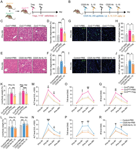
Inflammation is highlighted as an initial factor that helps orchestrate liver reconstitution. However, the precise mechanisms controlling inflammation during liver reconstitution have not been fully elucidated. In this study, a clear immune response is demonstrated during hepatic reconstitution. Inhibition of the hepatic inflammatory response retards liver regeneration. During this process, Ccl2 is primarily produced by type 1 innate lymphoid cells (ILC1s), and ILC1-derived Ccl2 recruits peripheral ILC1s and regulatory T cells (Tregs) to the liver. Deletion of Ccl2 or Tregs exacerbates hepatic injury and inflammatory cytokine release, accelerating liver proliferation and regeneration. The adoption of Tregs and IL-10 injection reversed these effects on hepatocyte regenerative proliferation. Additionally, Treg-derived IL-10 can directly induce macrophage polarization from M1 to M2, which alleviated macrophage-secreted IL-6 and TNF-α and balanced the intrahepatic inflammatory milieu during liver reconstitution. This study reveals the capacity of Tregs to modulate the intrahepatic inflammatory milieu and liver reconstitution through IL-10-mediated macrophage polarization, providing a potential opportunity to improve hepatic inflammation and maintain homeostasis.

Keywords: inflammatory milieu; liver reconstitution; macrophage polarization; regulatory T cell; type 1 innate lymphoid cell.


Conflict of interest statement

The authors declare no conflict of interest.

Figures

Figure 1
Figure 1
DEX inhibits inflammatory response and regenerative proliferation after liver injury. A) tSNE projection visualizing the total (left) and each time point (right, control, 4 and 8 weeks) of intrahepatic immune cells after CCl4 treatment (n = 3). B) Heatmap showing the marker genes among different cell subsets (n = 3). C) The cell count and fraction of indicated time point (n = 3). D) The expression of DEGs among ten major cell types (8 weeks versus control, n = 3). E) The enrichment score of DEGs in the inflammatory response and immune process (n = 3). F,G) Representative images of HE staining (F) and quantification of vacuolar degeneration (G) after saline or DEX treatment (n = 10, scale bar, 20 µm). H) The serum level of ALT at the indicated time point after PHx (n = 5). (I–K) qPCR analysis of the relative mRNA expression of Il6 (I), Tnfa (J), Ifng (K) (n = 5). L,M) Representative fluorescence images (L) and quantification (M) of Ki‐67‐positive cells (n = 5, scale bar, 20 µm). N) The ratio of liver/body weight (n = 8). O–Q) Representative images of western blotting (O) and quantification of the protein expression 24 (P) or 48 h (Q) after PHx (n = 5). Data represent three to five independent experiments. Data are shown as the mean + SEM along with individual data points and were compared using unpaired Student's t‐test (G, P,Q) or two‐way ANOVA followed by Bonferroni's multiple comparisons test (H–K, M,N). * p < 0.05, ** p < 0.01, *** p < 0.001, ns indicates p > 0.05.
Figure 2
Figure 2
Liver injury leads to ILC1‐derived Ccl2 production. A) The ILC was re‐clustering from Figure 1A. Three ILC subtypes were identified based on clustering after CCl4 treatment (n = 3). B) The cell count and fraction of indicated time point after CCl4 treatment (n = 3). (C and D) The specificity C) and intensity D) of Ccl2 expression in ILC subtypes (n = 3). E,F) Representative flow cytometry plots (E) and the statistical quantification (F) of hepatic ILC1s (CD49a+ NK1.1+) 6 h after PHx (n = 9). (G and H) Representative flow cytometry plots G) and percentage H) of the Ccl2+ cells in ILC1s (CD49a+ NK1.1+, gated from Figure 2E) (n = 5). I) The level of Ccl2 measured by ELISA (n = 8). J) qPCR analysis of the relative mRNA expression of Ccl2 in the liver (n = 7). K) The level of Ccl2 measured by ELISA in WT, Rag2 −/− (lack T and B cells), Rag2 −/− γC −/− (lack T, B, ILC1s, and NK cells) mice (n = 5). L) 1×10⁵ ILC1s (CD49a+, NK1.1+) were isolated from the regenerative liver 6 h after PHx. The western blotting of cGAS‐STING (p‐IRF3, IRF3, and STING) and Ccl2 protein expression (n = 3). M) Volcano plots of DEGs in ILC1s. The red dots indicate upregulated genes. The blue dots indicate downregulated genes (n = 4). Data represent three to five independent experiments. Data are shown as the mean + SEM along with individual data points and were compared using unpaired Student's t‐test (F,I,J) or Mann–Whitney test (H) or two‐way ANOVA followed by Bonferroni's multiple comparisons test (K). ** p < 0.01, *** p < 0.001, **** p < 0.0001.
Figure 3
Figure 3
Deletion of Ccl2 results in deteriorated hepatic inflammation and enhanced hepatocyte proliferation in acutely injured livers. A) The scRNA‐seq data were analyzed from Figure 2A.The expression of Ccl2 and Ccr2 in tSNE plot. Ccl2‐positive cell colored by red. Ccr2‐positive cell colored by green. Ccl2 and Ccr2 coexpression colored by yellow (n = 3). B) Correlation analysis between the Ccl2 and Ccr2 expression (n = 3). C,D) Wildtype (WT) and Ccl2 −/− mice were subjected to 2/3 PHx. Representative flow cytometry plots (C) and the statistical quantification (D) of hepatic ILC1s (CD49a+ NK1.1+) and Ccr2+ ILC1s 6 h after PHx (n = 5, 6). (E and F) Representative images of HE staining E) and quantification F) of vacuolar degeneration 24 h after PHx (n = 5, scale bar, 20 µm). G) Serum levels of ALT (n = 5). H–J) The relative mRNA expression of Il6 (H), Tnfa (I) and Il10 (J) (n = 5). K,L) Representative fluorescence images (K) and quantification (L) of Ki‐67‐positive cells (n = 5, scale bar, 20 µm). M) The ratio of liver/body weight (n = 6). N–P) Representative images of western blotting (N) and quantification (O,P) of the relative protein expression (n = 5). Data represent three to five independent experiments. The Spearman's correlation analysis was used in panel B. Data are shown as the mean + SEM along with individual data points and were compared using unpaired Student's t‐test (D,F,O,P) or two‐way ANOVA followed by Bonferroni's multiple comparisons test (G–J,L,M). * p < 0.05, ** p < 0.01, *** p < 0.001, **** p < 0.0001, ns indicates p > 0.05.
Figure 4
Figure 4
Treg balance hepatic inflammatory response to control liver regeneration and development. A) The liver tissue from Foxp3DTR mice after 4 weeks of DT injection. Flow chart of DT administration. B,C) Representative images of histology (B) and quantification (C) of liver/body weight ratio (n = 5, gross scale bar, 2.5 mm; HE staining scale bar, 20 µm). D,E) Representative fluorescence images (D) and quantification (E) of Ki‐67‐positive cells (n = 5, scale bar, 20 µm). F) Flow chart of anti‐CD25 antibody administration. G,H) Representative flow cytometry plots (G) and the statistical quantification (H) of hepatic Tregs (CD4+ Foxp3+) (n = 5). I,J) Representative images of HE staining (I) and quantification (J) of vacuolar degeneration (n = 5, scale bar, 20 µm). K,L) Representative fluorescence images (K) and quantification (L) of Ki‐67‐positive cells (n = 5, scale bar, 20 µm). M) The ratio of liver/body weight (n = 6). N,O) The relative mRNA expression of Il6 (N) and Tnfa (O) (n = 5). P–R) Representative images of immunoblotting (P) and quantification (Q,R) of the relative protein expression (n = 5). S) The relative mRNA expression by qPCR 6 h after PHx (n = 5). T) The relative mRNA expression of Il10 (n = 5). U,V) Representative flow cytometry plots (U) and percentage (V) of IL‐10+ cells in Tregs (CD4+ Foxp3+, gated from Figure 4G) 6 h after PHx (n = 5). W) The T cells were re‐clustering from Figure 1A. Five T cell subtypes were identified based on clustering after CCl4 treatment (n = 3). X) The cell count and fraction of indicated time point after CCl4 treatment (n = 3). Y,Z) The specificity (Y) and intensity (Z) of Il10 expression in T cell subtypes (n = 3). Data represent three to five independent experiments. Data are shown as the mean + SEM along with individual data points and were compared using unpaired Student's t‐test (C,H,J, Q–S,V) or Mann–Whitney test (E) or two‐way ANOVA followed by Bonferroni's multiple comparisons test (L–O,T). * p < 0.05, ** p < 0.01, *** p < 0.001, **** p < 0.0001, ns indicates p > 0.05.
Figure 5
Figure 5
Treg‐derived IL‐10 controls regenerative proliferation. A,B) Flow chart of Treg adoption (A) or anti‐CD25 antibody and IL‐10 administration (B). C–F) Representative images of HE staining (C,E) and quantification (D,F) of vacuolar degeneration 24 h after PHx (n = 5, scale bar, 20 µm). G–J) Representative images (G,I) and quantification (H,J) of Ki‐67 positive cells 24 h after PHx (n = 6, 7, scale bar, 20 µm). K,L) The ratio of liver/body weight (n = 6–8). M,N) Serum levels of ALT (n = 5). O–R) The relative mRNA expression of Tnfa (O,P) and Il6 (Q,R) (n = 5). Data represent three independent experiments. Data are shown as the mean + SEM along with individual data points and were compared using one‐way ANOVA followed by Bonferroni's multiple comparisons test (D,F,H,J) or two‐way ANOVA followed by Bonferroni's multiple comparisons test (K–R). * p < 0.05, ** p < 0.01, *** p < 0.001, **** p < 0.0001, comparison between the 2 indicated groups, or comparison between the Ccr2 f/f + PBS and Ccr2 cKO + PBS groups, or comparison between the Control+PBS and CD25Ab + PBS groups; # p < 0.05, ## p < 0.01, ### p < 0.001, #### p < 0.0001, comparison between the Ccr2 cKO + PBS and Ccr2 cKO + Treg, or comparison between the CD25Ab + PBS and CD25Ab + IL‐10 groups.
Figure 6
Figure 6
IL‐10 forces M2 macrophage polarization to regulate hepatocyte proliferation. A) The macrophages were re‐clustering from Figure 1A. Two macrophage subtypes were identified based on clustering after CCl4 treatment (n = 3). B) The cell count and fraction of indicated time point after CCl4 treatment (n = 3). C, D) The specificity (C) and intensity (D) of Tnf expression in T cell subtypes (n = 3). E) Go enrichment of DEGs in M1 (n = 3). F–I) Representative histogram plots (F and H) and quantification (G and I) of the fluorescence intensity of macrophage (CD11b+)‐gated IL‐6, TNF‐α and CD206 24 h after PHx (n = 5). J–M) Macrophages were isolated and incubated with IL‐10 for 48 h. The relative mRNA expression by qPCR (n = 5). N–P) Representative images of western blotting (N) and quantification (O, P) of the relative protein expression (n = 5). Q) Flow chart of co‐culture procedure in vitro. R, S) NCTC1469 cells were labeled with CFSE and co‐cultured with macrophages (isolated from the injured liver) containing PBS/IL‐10 for 2 days or 4. Representative histogram plots (R) and quantification (S) of the proliferation ratio of NCTC1469 cells (n = 5). T) The expression levels of TNF‐α in the culture supernatants were measured by ELISA (n = 5). U, V) Representative images of immunoblotting (U) and quantification (V) of the relative protein expression in NCTC1469 cells (n = 5). Data represent three independent experiments. Data are shown as the mean + SEM along with individual data points and were compared using unpaired Student's t‐test (I, V) or one‐way ANOVA followed by Bonferroni's multiple comparisons test (G, J–M, and O–P) or two‐way ANOVA followed by Bonferroni's multiple comparisons test (S, T). * p < 0.05, ** p < 0.01, *** p < 0.001, **** p < 0.0001.

References

    1. Campana L., Esser H., Huch M., Forbes S., Nat. Rev. Mol. Cell Biol. 2021, 22, 608. - PubMed
    1. Brenner C., Galluzzi L., Kepp O., Kroemer G., J Hepatol 2013, 59, 583. - PubMed
    1. Tacke F., Luedde T., Trautwein C., Clin. Rev. Allergy Immunol. 2009, 36, 4. - PubMed
    1. Li N., Hua J., Oncotarget 2017, 8, 3628. - PMC - PubMed
    1. Wu J., Zhang L., Shi J., He R., Yang W., Habtezion A., Niu N., Lu P., Xue J., EBioMedicine 2020, 58, 102920. - PMC - PubMed

MeSH terms

LinkOut - more resources