. 2024 Oct 1;134(19):e180251.
doi: 10.1172/JCI180251.

IL-7-dependent and -independent lineages of IL-7R-dependent human T cells

Carlos A Arango-Franco  1   2   3 Masato Ogishi  4 Susanne Unger  5   6 Ottavia M Delmonte  7 Julio César Orrego  3 Ahmad Yatim  4 Margarita M Velasquez-Lopera  8 Andrés F Zea-Vera  7   9   10 Jonathan Bohlen  1   2 Marwa Chbihi  2   11 Antoine Fayand  1   2 Juan Pablo Sánchez  3   12 Julian Rojas  3   12 Yoann Seeleuthner  1   2 Tom Le Voyer  1   2 Quentin Philippot  1   2 Kathryn J Payne  5   6 Adrian Gervais  1   2 Lucia V Erazo-Borrás  1   2   3 Luis A Correa-Londoño  8 Axel Cederholm  13 Alejandro Gallón-Duque  3 Pedro Goncalves  14   15 Jean-Marc Doisne  14   15 Liran Horev  16   17 Bénédicte Charmeteau-de Muylder  18 Jesús Á Álvarez  3 Diana M Arboleda  3 Lizet Pérez-Zapata  3 Estefanía Vásquez-Echeverri  3 Marcela Moncada-Vélez  3   4 Juan A López  3   12 Yolanda Caicedo  19 Boaz Palterer  7 Pablo J Patiño  3 Carlos J Montoya  20 Matthieu Chaldebas  4 Peng Zhang  1   2   4 Tina Nguyen  21   22 Cindy S Ma  21   22 Mohamed Jeljeli  23 Juan F Alzate  24 Felipe Cabarcas  24 Taushif Khan  25   26 Darawan Rinchai  4 Jean-Luc Prétet  27 Bertrand Boisson  1   2   4 Generalized Verrucosis Japanese ConsortiumNico Marr  25   26 Ruba Ibrahim  16 Vered Molho-Pessach  16 Stéphanie Boisson-Dupuis  1   2   4 Dimitra Kiritsi  28 João T Barata  29 Nils Landegren  13   30 Bénédicte Neven  2   11 Laurent Abel  1   2   4 Andrea Lisco  31 Vivien Béziat  1   2   4 Emmanuelle Jouanguy  1   2   4 Jacinta Bustamante  1   2   4   32 James P Di Santo  14   15 Stuart G Tangye  21   22 Luigi D Notarangelo  7 Rémi Cheynier  18 Ken Natsuga  33 Andrés A Arias  3   4   12 José Luis Franco  3 Klaus Warnatz  5   6 Jean-Laurent Casanova  1   2   4   34   35 Anne Puel  1   2   4
Affiliations

IL-7-dependent and -independent lineages of IL-7R-dependent human T cells

Carlos A Arango-Franco et al. J Clin Invest. .

Abstract

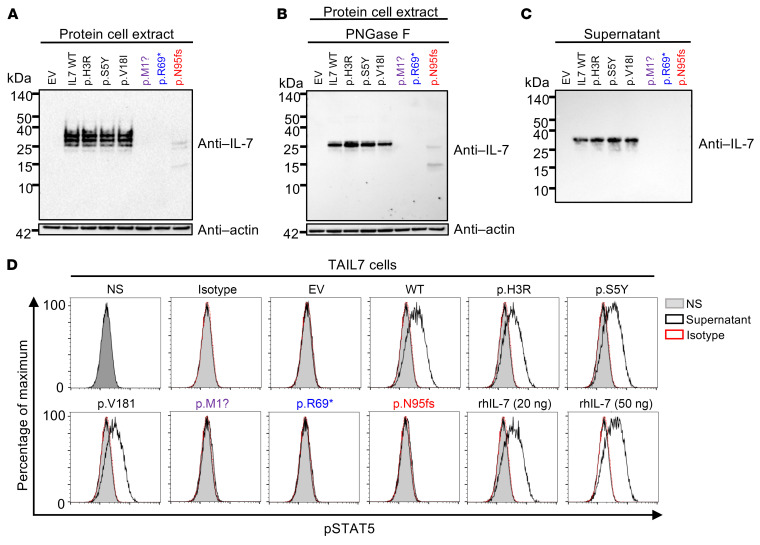
Infants with biallelic IL7R loss-of-function variants have severe combined immune deficiency (SCID) characterized by the absence of autologous T lymphocytes, but normal counts of circulating B and NK cells (T-B+NK+ SCID). We report 6 adults (aged 22 to 59 years) from 4 kindreds and 3 ancestries (Colombian, Israeli Arab, Japanese) carrying homozygous IL7 loss-of-function variants resulting in combined immunodeficiency (CID). Deep immunophenotyping revealed relatively normal counts and/or proportions of myeloid, B, NK, and innate lymphoid cells. By contrast, the patients had profound T cell lymphopenia, with low proportions of innate-like adaptive mucosal-associated invariant T and invariant NK T cells. They also had low blood counts of T cell receptor (TCR) excision circles, recent thymic emigrant T cells and naive CD4+ T cells, and low overall TCR repertoire diversity, collectively indicating impaired thymic output. The proportions of effector memory CD4+ and CD8+ T cells were high, indicating IL-7-independent homeostatic T cell proliferation in the periphery. Intriguingly, the proportions of other T cell subsets, including TCRγδ+ T cells and some TCRαβ+ T cell subsets (including Th1, Tfh, and Treg) were little affected. Peripheral CD4+ T cells displayed poor proliferation, but normal cytokine production upon stimulation with mitogens in vitro. Thus, inherited IL-7 deficiency impairs T cell development less severely and in a more subset-specific manner than IL-7R deficiency. These findings suggest that another IL-7R-binding cytokine, possibly thymic stromal lymphopoietin, governs an IL-7-independent pathway of human T cell development.

Keywords: Cytokines; Genetic diseases; Genetics; Immunology; T cell development.


Figures

Figure 1
Figure 1. Private biallelic IL7 variants in 6 patients from 4 unrelated kindred.
(A) Family pedigrees with allele segregation. The patients, in black, suffer from CID and are homozygous for the indicated IL7 alleles. The arrow indicates the proband. Each generation is designated by a Roman numeral (I, II), individuals of unknown genotype are indicated by “E?”. M, mutated; WT, wild-type. (B) Combined annotation depletion-dependent (CADD) score v1.6 (y axis) versus minor allele frequency (MAF) (x axis) plot for all nonsynonymous IL7 variants present in the homozygous state in the gnomAD database v2.1 (black dots), and the 3 IL7 variants of P1–P6 (P1 and P2, c.284del/p.N95Ifs*11, red triangle; P3, c.3G>A/p.M1?, purple triangle; and P4–P6, c.205A>T/p.R69*, blue triangle). The 99% mutation significance cutoff (MSC) is indicated (dotted line). (C) Consensus negative selection (CoNeS) of IL7 and IL7R. AR, autosomal recessive; AD, autosomal dominant. (D) Schematic representation of the IL-7 protein. Exons are indicated by Roman numerals. The signal peptide is highlighted in gray (amino acids 1–25). The positions of the variants found in the patients are indicated (P1 and P2 in red; P3 in purple, and P4–P6 in blue), together with the positions of the variants found in the homozygous state in gnomAD (in black).
Figure 2
Figure 2. The IL7 mutant alleles are biochemically deleterious in an overexpression system.
(AC) Western blot of total cell lysates (A and B) before (A), and after (B) PNGase F treatment, and of supernatants (C) from HEK293T cells transfected with C-terminally DDK-tagged IL7 WT, the patients’ or gnomAD IL7 mutant cDNAs, or with empty vector (EV). The data shown are representative of 2 independent experiments. (D) STAT5 phosphorylation in TAIL7 cells after 15 minutes of incubation with the supernatants of HEK293T cells transfected with C-terminally DDK-tagged IL7 WT, patients’ or gnomAD IL7 mutant cDNAs or with empty vector (EV), or with human recombinant IL-7 (rhIL-7, 20 or 50 ng/mL). NS, no supernatant (gray); supernatant (black line); and isotype (red dashed line). Representative data from 2 independent experiments are shown.
Figure 3
Figure 3. Lack of endogenous IL-7 detection.
(AD) IL-7 expression, as measured by ELISA on EBV-B cell supernatants (A), SV40-fibroblast supernatants (B), plasma (C), and nasopharyngeal swabs (D) from healthy donors (green circles), and the patients. Representative data from 2 independent experiments are shown. CTLs, controls. (E) STAT5 phosphorylation in TAIL7 cells after 15 minutes of incubation with supernatants from HEK293T cells transfected with IL7 WT cDNA, resting EBV-B cells from healthy controls (C1–C4) and P2, or rhIL-7, in the absence or the presence of blocking anti–IL-7 antibody. Left panel shows representative data from 3 independent experiments; right panel represents the quantification of p-STAT5 MFI normalized to the average MFI from 4 healthy controls. Nonparametric Mann-Whitney tests were used for analysis. *P < 0.05. NS, no supernatant (gray); supernatant (black line); and supernatant plus blocking anti–IL-7 antibody (α-IL-7, blue line). SN, supernatant.
Figure 4
Figure 4. Impaired development of specific T lymphocyte subsets.
(A) Counts of lymphocytes, CD3+, CD3+CD4+, CD3+CD8+, NK, and γδ T cells in the blood of the 6 (P1–P6) IL-7–deficient patients. The upper and lower limits of the normal range for age are indicated by gray boxes. (BR) Frequencies of several leukocyte subsets in IL-7–deficient patients (P1–P3 and P6) and age-matched healthy controls (green dots), as assessed by spectral flow cytometry on cryopreserved PBMCs, (B) classical monocytes (CD14+CD16), nonclassical monocytes (CD14dimCD16+), and intermediate monocytes (CD14+CD16+), (C) plasmacytoid dendritic cells (pDCs) (LinHLA-DR+CD11cCD123+), conventional DC type 1 (cDC1) (LinHLA-DR+CD11c+CD1c+CD141) and cDC2 (LinHLA-DR+CD11c+CD1cCD141+), (D) NK and NK CD56bright cells, (E) B cells and plasmablasts, (F) innate lymphoid cells (total ILCs, ILCPs [CD117+CRTh2], and ILC2s [CRTh2+]), (G) total TCR-γδ+, TCR-γδ1+, TCR-γδ2+, and TCR-γδ1+γδ2+ T cells, (H) MAIT (MR1+TCR-Vα7.2+), (I) iNKT, (J) total α/β T cells, (K) α/β CD4+ T cells, (L) α/β CD8+ T cells, among PBMCs, in controls and patients. (M and N) Frequencies of naive (CD45RA+CCR7+), central memory (CD45RACCR7+), effector memory (CD45RACCR7), and TEMRA (CD45RA+CCR7) cells among the CD4+ (M) and CD8+ (N) T cells. (O) Frequencies of the Th1 (CXCR5CXCR3+CCR4CCR6), Th1* (CXCR5CXCR3+CCR4CCR6+), Th2 (CXCR5CXCR3CCR4+CCR6), Th17 (CXCR5CXCR3CCR4+CCR6+), and Treg (CD3+CD4+CD25+CD127) subsets among CD4+ T cells in controls and patients. (P) Frequencies of Tfh (CXCR5+) cells among CD4+ T cells in controls and patients. (QR) Frequencies of α/β DN T cells (CD3+CD4CD8) (Q), and of α/β DP T cells (CD3+CD4+CD8+) (R) among the total α/β T cells of controls and patients. Nonparametric Mann-Whitney tests were used for analysis. *P < 0.05, **P < 0.01, ***P < 0.001, ****P < 0.0001.
Figure 5
Figure 5. Single-cell transcriptomic analysis.
Cryopreserved PBMCs from P1–P3 and P6, together with cryopreserved PBMCs from 2 healthy adult controls, were analyzed. The data were integrated with historical controls via Harmony (40). (A) Relative abundance of each leukocyte subset identified through clustering analysis. (B) Batch-corrected principal component analysis (PCA). The first principal components (PC1, y axis) are shown for each individual leukocyte subset. (C) Gene set enrichment analysis (GSEA). Genes were ranked based on the fold-change differences between patients and healthy controls estimated by pseudobulk differential expression analysis. Gene ranks were projected against the Hallmark gene sets (https://www.gsea-msigdb.org/gsea/msigdb/genesets.jsp?collection=H). Selected gene sets related to immune responses are shown. (D) GSEA for MYC target V1 genes in naive CD4+ and CD8+ T cells from IL-7–deficient patients (P1–P3 and P6), and 6 adult controls. The heatmap shows adjusted z score values for each sample as a color gradient from blue for transcripts that were detected but below our significance cutoff values (downregulated), through purple, to red for adjusted z score values above our significance cutoff values (upregulated). Nonparametric Mann-Whitney tests were used for analysis. *P < 0.05, **P < 0.01.
Figure 6
Figure 6. Impaired early T cell development in AR IL-7 deficiency.
(A) TCR excision circles (TRECs) in P1 (at 20 and 22 years of age), P2 (at 54 and 57 years of age), and an IL-7R–deficient patient (at 4 months of age), relative to healthy age-matched controls. The sjTREC count in blood (sjTREC/mL) is presented as a function of age. (B) Counts of recent thymic emigrant (RTE) cells, defined as CD3+CD4+CD45RA+CD31+ cells in fresh blood from P1, P2, an IL-7R–deficient patient, and age-matched healthy controls. (C) Heatmap of paired gene rearrangements of the TRAD locus in whole-blood samples from P1, P2, and 2 age-matched controls. The fold-change differences are indicated below. The red color highlights V-J gene pairings overused in patients and the blue color highlights V-J gene pairings overused in controls. The TRAV and TRDV genes known to be involved in TCR-δ rearrangements are indicated with a red rectangle. The TCR-δ1 (TRDV1:TRDJ1) rearrangement is indicated with a black rectangle. (D) TCRB and TCRA rearrangements, represented by Treemap plots, for whole-blood samples from P1, P2, and 2 age-matched healthy controls.
Figure 7
Figure 7. Peripheral T cell functions.
(A and B) Fresh PBMCs from age-matched controls (only 2 controls are shown), P1, and P2 were incubated for 4 days with (A) PHA (5 μg/mL or 20 μg/mL), or (B) with beads coated with anti-CD3 and anti-CD28 mAbs (anti-CD3 mAbs were used at concentrations of 1.25 μg/mL and 5 μg/mL). Histograms show CFSE dilution for CD3+ T lymphocytes. Representative results from 2 independent experiments are shown. (C) Intracellular IL-2 production by CD4+ or CD8+ naive or memory T cells after stimulation with PMA plus ionomycin for 4 hours and flow cytometry analysis, for IL-7–deficient patients (P4, P5, and P6) and 3 healthy controls. (D and E) Memory CD4+ T cells from healthy controls, P1, and P2 were stimulated with PMA plus ionomycin after 5 days of culture; the percentages of cells expressing IFN-γ, TNF, IL-2, and IL-10 intracellularly were determined by flow cytometry (D) and secreted cytokines were assessed by LEGENDplex (E). (F) T cell blasts from P1–P3 and 3 healthy controls were stimulated for 2 hours with an anti-CD2/anti-CD3/anti-CD28 mAb cocktail. Secreted cytokines were determined by LEGENDplex. (G and H) Bulk RNA transcriptomic analysis on sorted CD4+ and CD8+ T cell blasts from P1–P3 and 3 healthy adult controls. Principal component analysis (PCA) results are shown for sorted CD4+ and CD8+ T cell blasts in the absence of stimulation or after stimulation with the anti-CD2/anti-CD3/anti-CD28 mAb cocktail (G). Normalized enrichment score (NES) analyses are shown for unstimulated sorted CD4+ and CD8+ T cell blasts (H).

References

    1. Notarangelo LD. Combined immunodeficiencies with nonfunctional T lymphocytes. Adv Immunol. 2014;121:121–190. doi: 10.1016/B978-0-12-800100-4.00004-0. - DOI - PubMed
    1. Notarangelo LD, et al. Human inborn errors of immunity: an expanding universe. Sci Immunol. 2020;5(49):eabb1662. doi: 10.1126/sciimmunol.abb1662. - DOI - PMC - PubMed
    1. Tangye SG, et al. Human inborn errors of immunity: 2022 update on the classification from the International Union of Immunological Societies Expert Committee. J Clin Immunol. 2022;42(7):1473–1507. doi: 10.1007/s10875-022-01289-3. - DOI - PMC - PubMed
    1. Leonard WJ, et al. The γc family of cytokines: basic biology to therapeutic ramifications. Immunity. 2019;50(4):832–850. doi: 10.1016/j.immuni.2019.03.028. - DOI - PubMed
    1. Leonard WJ, et al. The molecular basis of X-linked severe combined immunodeficiency: the role of the interleukin-2 receptor gamma chain as a common gamma chain, gamma c. Immunol Rev. 1994;138:61–86. doi: 10.1111/j.1600-065X.1994.tb00847.x. - DOI - PubMed

MeSH terms