. 2024 Nov 4;221(11):e20231152.
doi: 10.1084/jem.20231152. Epub 2024 Oct 1.

Incontinentia pigmenti underlies thymic dysplasia, autoantibodies to type I IFNs, and viral diseases

Jérémie Rosain  1   2   3   4 Tom Le Voyer #  1   2   3   5 Xian Liu #  6 Adrian Gervais #  1   2 Laura Polivka #  7   8 Axel Cederholm #  9 Laureline Berteloot #  10 Audrey V Parent #  6 Alessandra Pescatore #  11 Ezia Spinosa #  11 Snezana Minic #  12   13 Ana Elisa Kiszewski #  14   15 Miyuki Tsumura #  16 Chloé Thibault #  1   2 Maria Esnaola Azcoiti #  1   2 Jelena Martinovic #  17 Quentin Philippot #  1   2 Taushif Khan #  18 Astrid Marchal #  1   2 Bénédicte Charmeteau-De Muylder #  19 Lucy Bizien #  1   2 Caroline Deswarte #  1   2 Lillia Hadjem  20 Marie-Odile Fauvarque  21 Karim Dorgham  22 Daniel Eriksson  23 Emilia Liana Falcone  24   25 Mathilde Puel  4 Sinem Ünal  1   2 Amyrath Geraldo  1   2 Corentin Le Floc'h  1   2 Hailun Li  1   2 Sylvie Rheault  25   26 Christine Muti  27 Claire Bobrie-Moyrand  28 Anne Welfringer-Morin  7 Ramsay L Fuleihan  29 Romain Lévy  1   2   3   30 Marie Roelens  2   4 Liwei Gao  1   2 Marie Materna  1   2 Silvia Pellegrini  31 Lorenzo Piemonti  31 Emilie Catherinot  32 Jean-Christophe Goffard  33 Arnaud Fekkar  1   2   34 Aissata Sacko-Sow  35 Camille Soudée  1   2 Soraya Boucherit  1   2 Anna-Lena Neehus  1   2 Cristina Has  36   37 Stefanie Hübner  36 Géraldine Blanchard-Rohner  38 Blanca Amador-Borrero  39 Takanori Utsumi  16 Maki Taniguchi  16 Hiroo Tani  40   41 Kazushi Izawa  42 Takahiro Yasumi  42 Sotaro Kanai  43 Mélanie Migaud  1   2 Mélodie Aubart  1   2   44 Nathalie Lambert  4 Guy Gorochov  22   45 Capucine Picard  2   4   46 Claire Soudais  46 Anne-Sophie L'Honneur  47 Flore Rozenberg  47 Joshua D Milner  29 Shen-Ying Zhang  1   2   3 Pierre Vabres  48 Dusan Trpinac  49 Nico Marr  18   50 Nathalie Boddaert  10 Isabelle Desguerre  44 Manolis Pasparakis  51 Corey N Miller  6 Cláudia S Poziomczyk  52 Laurent Abel  1   2   3 Satoshi Okada  16 Emmanuelle Jouanguy  1   2   3 Rémi Cheynier  19 Qian Zhang  1   2   3 Aurélie Cobat  1   2   3 Vivien Béziat  1   2   3 Bertrand Boisson  1   2   3 Julie Steffann  53 Francesca Fusco  11 Matilde Valeria Ursini  11 Smail Hadj-Rabia  7 Christine Bodemer  7 Jacinta Bustamante  1   2   3   4 Hervé Luche  20 Anne Puel #  1   2   3 Gilles Courtois #  21 Paul Bastard #  1   2   3   30 Nils Landegren #  9   54 Mark S Anderson #  6 Jean-Laurent Casanova #  1   2   3   55   56
Affiliations

Incontinentia pigmenti underlies thymic dysplasia, autoantibodies to type I IFNs, and viral diseases

Jérémie Rosain et al. J Exp Med. .

Abstract

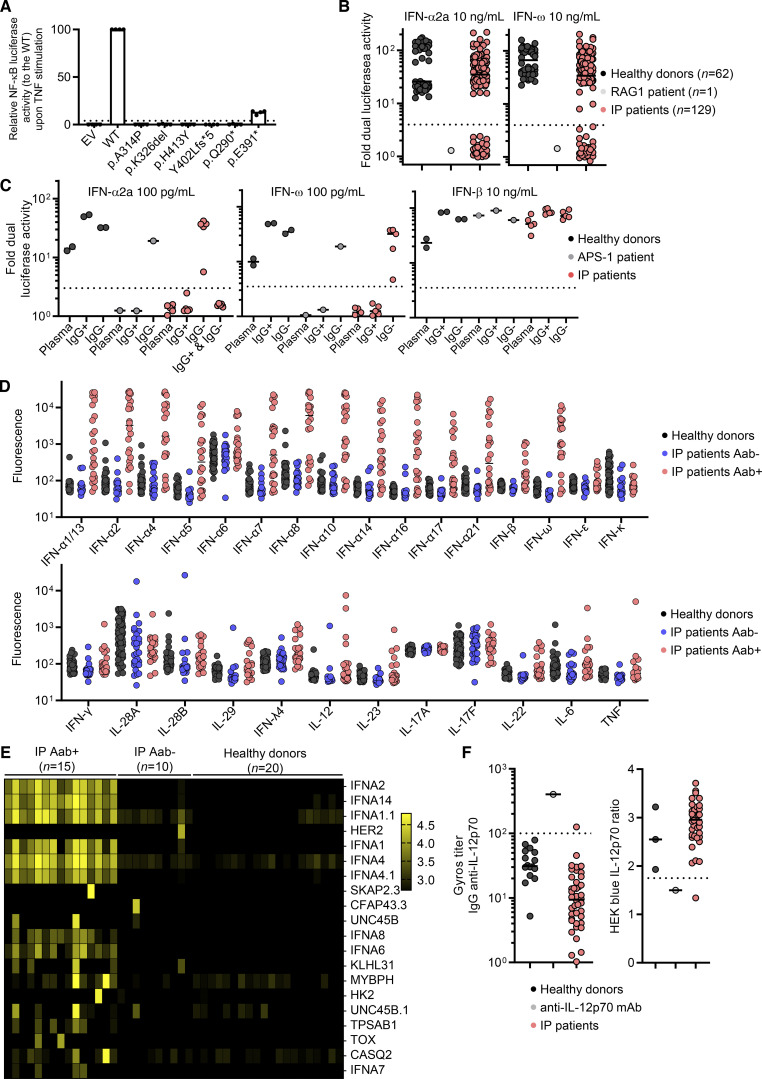
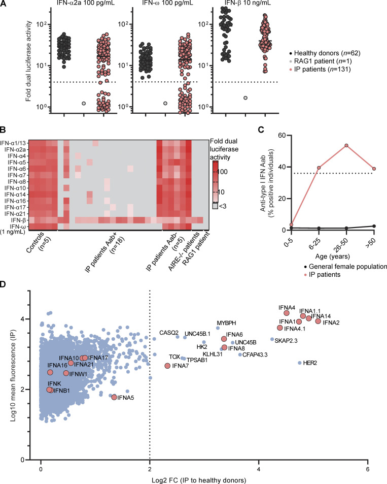
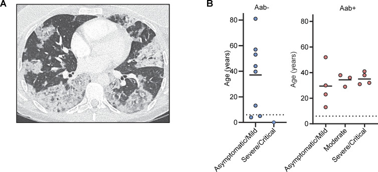
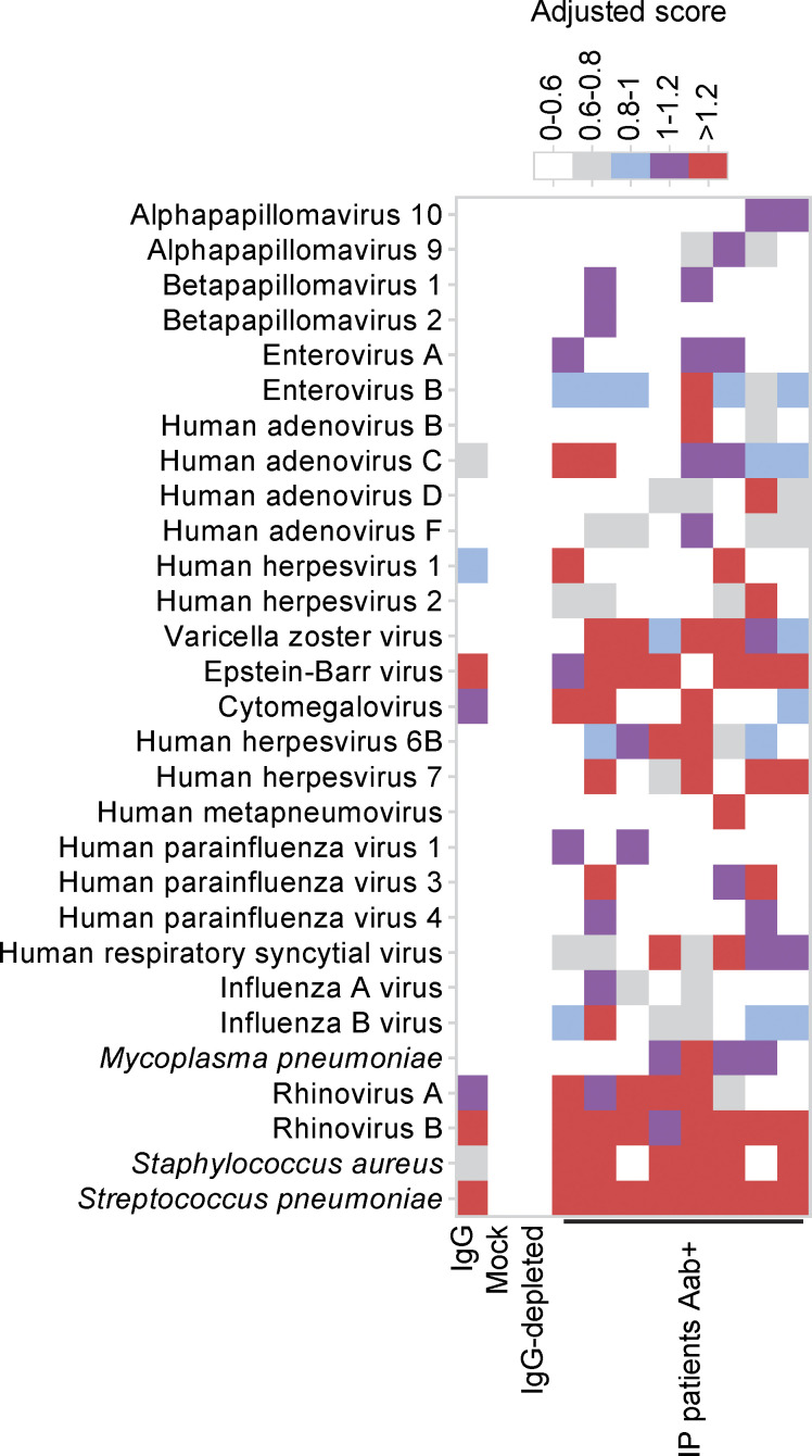
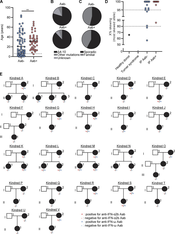
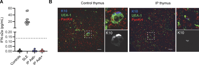
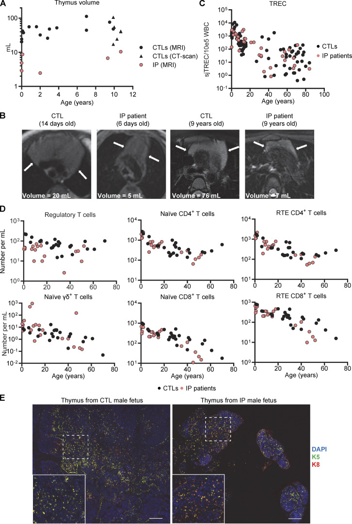
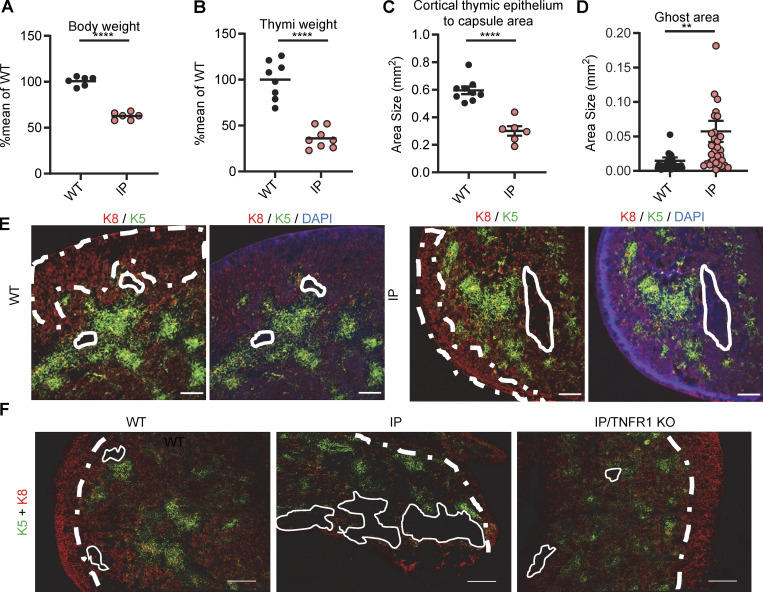
Human inborn errors of thymic T cell tolerance underlie the production of autoantibodies (auto-Abs) neutralizing type I IFNs, which predispose to severe viral diseases. We analyze 131 female patients with X-linked dominant incontinentia pigmenti (IP), heterozygous for loss-of-function (LOF) NEMO variants, from 99 kindreds in 10 countries. Forty-seven of these patients (36%) have auto-Abs neutralizing IFN-α and/or IFN-ω, a proportion 23 times higher than that for age-matched female controls. This proportion remains stable from the age of 6 years onward. On imaging, female patients with IP have a small, abnormally structured thymus. Auto-Abs against type I IFNs confer a predisposition to life-threatening viral diseases. By contrast, patients with IP lacking auto-Abs against type I IFNs are at no particular risk of viral disease. These results suggest that IP accelerates thymic involution, thereby underlying the production of auto-Abs neutralizing type I IFNs in at least a third of female patients with IP, predisposing them to life-threatening viral diseases.


Conflict of interest statement

Disclosures: A.V. Parent reported personal fees from Thymmune Therapeutics outside the submitted work; in addition, A.V. Parent had a patent to generate thymic epithelial progenitor cells in vitro licensed (Thymmune Therapeutics). J.D. Milner reported personal fees from Blueprint Pharmaceuticals and grants from Pharming outside the submitted work. M.S. Anderson reported other from Merck, Inc. and Medtronic, Inc. outside the submitted work. No other disclosures were reported.

Figures

Figure S1.
Figure S1.
Auto-antibodies to type I IFNs and other cytokines in females with incontinentia pigmenti. Related to Fig. 1. (A) Dual luciferase NF-κB activity of HEK293T cells transfected with empty vector (EV) or a vector containing WT NEMO cDNA, or various NEMO variants. Cells were stimulated with TNF. Results are normalized against the WT. The p.A314P, p.K326del, p.H413Y and p.Y402Lfs*5 variants were found in IP patients from this cohort. p.Q290* was previously described in a patient with IP and was used as an amorphic control. p.E391* was previously described in a patient with ectodermal dysplasia and immunodeficiency and was used as a hypomorphic control. Pooled results for from n = 4 independent experiments are presented. Bar is displayed at 4%. (B) Neutralization of 10 ng/ml IFN-α2a or IFN-ω by a 1:10 dilution of plasma from the indicated individuals or patients. All patients with neutralizing ratio below 3 were assessed two or three times independently. (C) Neutralization of 100 pg/ml IFN-α2a, 100 pg/ml IFN-ω, or 10 ng/ml of IFN-β by a 1:10 dilution of plasma, the purified IgG-positive fraction (IgG+), or the purified IgG-depleted fraction (IgG) from the indicated individuals or patients. (D) Bead-based protein array detecting IgG auto-Abs against the indicated cytokines in n = 93 healthy donors, and in n = 30 and n = 24 IP patients negative and positive, respectively, for the neutralization of type I IFNs by auto-Abs. (E) Anti-human IgG fluorescence signal intensities (log10) according to protein microarray (HuProt) data for all individuals plotted with correlation clusters for proteins, including the 20 proteins with the largest positive fold-change in levels. Case-control status is indicated at the top. Data displayed are merged from two independent experiments. (F) Gyros anti-IL-12p70 IgG antibody titer (left) and neutralization activity determined in HEK293-blue cells (right) for plasma from healthy donors and patients with IP.
Figure 1.
Figure 1.
Auto-Abs against type I IFNs in female patients with IP. (A) Neutralization of 100 pg/ml IFN-α2a, 100 pg/ml IFN-ω, and 10 ng/ml IFN-β by a 1:10 dilution of plasma from the indicated individuals or patients. All patients with a neutralizing ratio below 3 were assessed two or three times independently. (B) Neutralization of 1 ng/ml of 15 type I IFNs by plasma diluted 1:10. (C) The proportion of individuals with plasma-neutralizing type I IFNs by age, for female patients with IP and the general female population. (D) Anti-human IgG fluorescence signal intensities with positive fold-change in 15 IP patients with serum neutralizing type I IFNs and 10 IP patients without auto-Abs neutralizing type I IFNs, relative to 20 healthy donors. Each dot corresponds to the protein spot with the highest signal intensity among the duplicate spots for the protein concerned. Data displayed are merged from two independent experiments.
Figure 2.
Figure 2.
Viral diseases in female patients with IP. (A) Thorax-computed tomography scan for P1 who had critical COVID-19 pneumonia. (B) Severity of COVID-19 before vaccination as a function of negativity or positivity for auto-Abs against type I IFNs (Aab−, and Aab+, respectively), and age. One of the patients (P9) was excluded from this figure because she received early treatment with recombinant IFN-β (Bastard et al., 2021b).
Figure S2.
Figure S2.
Exposure to viruses and bacteria in females with incontinentia pigmenti. Related to Fig. 2. Antiviral antibody responses to species for which at least one sample tested seropositive by PhIP-Seq, based on in-house stringent cutoff values, color-coded as indicated.
Figure 3.
Figure 3.
Demographic, genetic, and familial features of female patients with IP by auto-Abs status. (A) Age distribution of IP patients negative (Aab−) or positive (Aab+) for auto-Abs against type I IFNs. (B–D) Distribution of (B) underlying NEMO variants, (C) sporadic versus familial forms, and (D) X-chromosome inactivation skewing in female patients with IP negative (Aab−) or positive (Aab+) for auto-Abs against type I IFNs. (E) Auto-Ab status for IFN-α2a and IFN-ω in multiplex IP kindreds. Statistical analysis was performed with Student’s t tests. **P < 0.01.
Figure S3.
Figure S3.
Level of circulating IFN-α2a in females with incontinentia pigmenti (IP) and pathological study of the thymus from a male fetus with IP. Fig. 4. (A) Determination of IFN-α2a levels with the Simoa platform for plasma from healthy donors (n = 10), patients with systemic lupus erythematosus (SLE, n = 10), and female patients with IP positive (n = 6) or negative (n = 12) for auto-Abs against type I IFNs. (B) Keratin 10 (K10), Ulex europaeus agglutinin 1 lectin (UEA-1), and pankeratin (PanKrt) staining of the thymus of a human male fetus with IP; comparison with an aged-matched control. Both thymi were obtained from fetuses at 19 wk of gestation with a similar estimated maceration time. Scale bar = 50 μm.
Figure 4.
Figure 4.
Thymi of IP female patients and a male fetus. (A) Thymus volume, assessed by MRI or computed tomography scan; n = 20 aged-matched controls and n = 6 IP patients. More details regarding controls and patients are provided in Table S6. (B) MRI images of thymi from IP patients and aged-matched controls of the indicated ages. (C) Quantification of TRECs in DNA extracted from the whole blood of healthy donors or patients with IP. Data were merged from three independent experiments. (D) Absolute counts of CD25hiCD127low regulatory CD4+ T cells; naïve (CD45RA+CCR7+) γδ+, CD4+, or CD8+ T cells; and recent thymic emigrants (CD31+CD45RA+CCR7+) CD4+ or CD8+ T cells determined by mass cytometry on fresh whole blood from n = 24 female healthy donors and n = 23 IP patients. Experiments were performed independently and data were merged. (E) DAPI, keratin 5 (K5), and keratin 8 staining of formalin-fixed paraffin-embedded (FFPE) tissues from the thymus of a single male fetus with IP at 19 wk of gestation and of an aged-matched control. Scale bar = 200 μm.
Figure 5.
Figure 5.
Thymi of 7.5-day-old (P7.5) mice with IP. (A and B) Weight of (A) the body and of (B) thymi from n = 9 WT mice and n = 9 IP mice. Results are expressed as a percentage of the mean for the WT in the same experiment. Data from two independent experiments are displayed. (C) Measurement of the area of the thymic cortex. (D) Measurement of the ghost area in thymi from WT and IP mice. (E) Representative confocal images of K8 (red), K5 (green), and DAPI (blue) staining of the thymus for WT and IP mice. Scale bar = 200 μm, n = 3–4 mice. Representative confocal images of K5/K8 (red), and CD4 (green) staining in WT and IP mice. Scale bar of left panel = 200 μm, Scale bar of other panel = 50 μm. n = 3–4 mice. (F) Representative images of K5/K8 (red) staining of thymi from WT mice (n = 4), Nemo+/− female mice (IP, n = 3), and Tnfr-1−/−/Nemo+/− female mice (IP/TNFR1 KO, n = 2). ns = not significant, P > 0.05; **P < 0.01; ****P < 0.0001. All experiments were performed at least two or three times independently.
Figure S4.
Figure S4.
Thymi of 7.5-day-old (P7.5) mice with IP. Related to Fig. 5. (A) Images of the thymi, hearts, and spleens of a WT mouse and an IP mouse. (B) Representative images of K5/K8 (red) staining of thymi from WT mice (n = 4), Nemo+/− female mice (IP, n = 3), Tnfr-1−/−/Nemo+/+ female mice (TNFR1 KO, n = 1), and Tnfr-1−/−/Nemo+/− female mice (IP/TNFR1 KO, n = 2). Scale bar = 200 μm. (C) Representative confocal images of K5 (red), AIRE (green), and DAPI (blue) staining in WT and IP mice. Scale bar = 20 μm, n = 3–4 mice. (D) Percentages for the thymocyte subsets extracted from the thymi of n = 14 WT mice or n = 13 IP mice. DN = double-negative (CD4CD8); TN = triple-negative (CD3CD4CD8), DP = double-positive (CD4+CD8+); ISP = immature single-positive; IE = immediate early. Statistical analysis was performed with Student’s t test. ns = not significant, P > 0.05; *P < 0.05; **P < 0.01. All experiments in mice were performed at least two or three times independently.
Figure S5.
Figure S5.
Keratin 5 and 10 staining in thymi from newborn mice. Related to Fig. 5. Keratin 5 (K5, green), keratin 10 (K10, red), and nuclear (DAPI, blue) staining of thymic sections from WT mice, IP mice, or IP/TNFR1 KO mice. Scale bar = 50 µm. Data representative from two independent experiments are displayed.

Similar articles

  • Finding NEMO in the thymus.
    Ocampo-Godinez JM, Kreins AY. Ocampo-Godinez JM, et al. J Exp Med. 2024 Nov 4;221(11):e20241590. doi: 10.1084/jem.20241590. Epub 2024 Oct 21. J Exp Med. 2024. PMID: 39432904 Free PMC article.
  • Autoantibodies against type I IFNs in humans with alternative NF-κB pathway deficiency.
    Le Voyer T, Parent AV, Liu X, Cederholm A, Gervais A, Rosain J, Nguyen T, Perez Lorenzo M, Rackaityte E, Rinchai D, Zhang P, Bizien L, Hancioglu G, Ghillani-Dalbin P, Charuel JL, Philippot Q, Gueye MS, Maglorius Renkilaraj MRL, Ogishi M, Soudée C, Migaud M, Rozenberg F, Momenilandi M, Riller Q, Imberti L, Delmonte OM, Müller G, Keller B, Orrego J, Franco Gallego WA, Rubin T, Emiroglu M, Parvaneh N, Eriksson D, Aranda-Guillen M, Berrios DI, Vong L, Katelaris CH, Mustillo P, Raedler J, Bohlen J, Bengi Celik J, Astudillo C, Winter S; NF-κB Consortium; COVID Human Genetic Effort; McLean C, Guffroy A, DeRisi JL, Yu D, Miller C, Feng Y, Guichard A, Béziat V, Bustamante J, Pan-Hammarström Q, Zhang Y, Rosen LB, Holland SM, Bosticardo M, Kenney H, Castagnoli R, Slade CA, Boztuğ K, Mahlaoui N, Latour S, Abraham RS, Lougaris V, Hauck F, Sediva A, Atschekzei F, Sogkas G, Poli MC, Slatter MA, Palterer B, Keller MD, Pinzon-Charry A, Sullivan A, Droney L, Suan D, Wong M, Kane A, Hu H, Ma C, Grombiříková H, Ciznar P, Dalal I, Aladjidi N, Hie M, Lazaro E, Franco J, Keles S, Malphettes M, Pasquet M, Maccari ME, Meinhardt A, Ikinciogullari A, Shahrooei M, Celmeli F, Frosk P, Goodnow CC, Gray PE, Bel… See abstract for full author list ➔ Le Voyer T, et al. Nature. 2023 Nov;623(7988):803-813. doi: 10.1038/s41586-023-06717-x. Epub 2023 Nov 8. Nature. 2023. PMID: 37938781 Free PMC article.
  • Preexisting autoantibodies to type I IFNs underlie critical COVID-19 pneumonia in patients with APS-1.
    Bastard P, Orlova E, Sozaeva L, Lévy R, James A, Schmitt MM, Ochoa S, Kareva M, Rodina Y, Gervais A, Le Voyer T, Rosain J, Philippot Q, Neehus AL, Shaw E, Migaud M, Bizien L, Ekwall O, Berg S, Beccuti G, Ghizzoni L, Thiriez G, Pavot A, Goujard C, Frémond ML, Carter E, Rothenbuhler A, Linglart A, Mignot B, Comte A, Cheikh N, Hermine O, Breivik L, Husebye ES, Humbert S, Rohrlich P, Coaquette A, Vuoto F, Faure K, Mahlaoui N, Kotnik P, Battelino T, Trebušak Podkrajšek K, Kisand K, Ferré EMN, DiMaggio T, Rosen LB, Burbelo PD, McIntyre M, Kann NY, Shcherbina A, Pavlova M, Kolodkina A, Holland SM, Zhang SY, Crow YJ, Notarangelo LD, Su HC, Abel L, Anderson MS, Jouanguy E, Neven B, Puel A, Casanova JL, Lionakis MS. Bastard P, et al. J Exp Med. 2021 Jul 5;218(7):e20210554. doi: 10.1084/jem.20210554. J Exp Med. 2021. PMID: 33890986 Free PMC article.
  • [Proposal for a protocol for the staging of incontinentia pigmenti in pediatric age].
    Portaleone D, Taroni F, Micheli S, Moioli M, Pedrazzini A, Cognizzoli P, Carnelli V. Portaleone D, et al. Minerva Pediatr. 2007 Jun;59(3):255-65. Minerva Pediatr. 2007. PMID: 17519871 Review. Italian.
  • NEMO gene rearrangement (exon 4-10 deletion) and genotype-phenotype relationship in Japanese patients with incontinentia pigmenti and review of published work in Japanese patients.
    Okita M, Nakanishi G, Fujimoto N, Shiomi M, Yamada T, Wataya-Kaneda M, Takijiri C, Yokoyama Y, Sunohara A, Tanaka T. Okita M, et al. J Dermatol. 2013 Apr;40(4):272-6. doi: 10.1111/1346-8138.12091. Epub 2013 Feb 11. J Dermatol. 2013. PMID: 23398170 Review.

Cited by

  • A sensitive assay for measuring whole-blood responses to type I IFNs.
    Gervais A, Le Floc'h C, Le Voyer T, Bizien L, Bohlen J, Celmeli F, Al Qureshah F, Masson C, Rosain J, Chbihi M, Lévy R, Castagnoli R, Rothenbuhler A, Jouanguy E, Zhang Q, Zhang SY, Béziat V, Bustamante J, Puel A, Bastard P, Casanova JL. Gervais A, et al. Proc Natl Acad Sci U S A. 2024 Oct;121(40):e2402983121. doi: 10.1073/pnas.2402983121. Epub 2024 Sep 23. Proc Natl Acad Sci U S A. 2024. PMID: 39312669 Free PMC article.
  • Risk of Death, Infections, and Hyperthermia in Ectodermal Dysplasias: A Nationwide Study.
    Herlin LK, Schmidt SAJ, Mogensen TH, Sommerlund M. Herlin LK, et al. Acta Derm Venereol. 2025 Jun 18;105:adv43101. doi: 10.2340/actadv.v105.43101. Acta Derm Venereol. 2025. PMID: 40530795 Free PMC article.
  • Higher COVID-19 pneumonia risk associated with anti-IFN-α than with anti-IFN-ω auto-Abs in children.
    Bastard P, Gervais A, Taniguchi M, Saare L, Särekannu K, Le Voyer T, Philippot Q, Rosain J, Bizien L, Asano T, Garcia-Prat M, Parra-Martínez A, Migaud M, Tsumura M, Conti F, Belot A, Rivière JG, Morio T, Tanaka J, Javouhey E, Haerynck F, Duvlis S, Ozcelik T, Keles S, Tandjaoui-Lambiotte Y, Escoda S, Husain M, Pan-Hammarström Q, Hammarström L, Ahlijah G, Abi Haidar A, Soudee C, Arseguel V, Abolhassani H, Sahanic S, Tancevski I, Nukui Y, Hayakawa S, Chrousos GP, Michos A, Tatsi EB, Filippatos F, Rodriguez-Palmero A, Troya J, Tipu I, Meyts I, Roussel L, Ostrowski SR, Schidlowski L, Prando C, Condino-Neto A, Cheikh N, Bousfiha AA, El Bakkouri J; COVID Clinicians; GEN-COVID Study Group; COVID Human Genetic Effort; Peterson P, Pujol A, Lévy R, Quartier P, Vinh DC, Boisson B, Béziat V, Zhang SY, Borghesi A, Pession A, Andreakos E, Marr N, Mentis AA, Mogensen TH, Rodríguez-Gallego C, Soler-Palacin P, Colobran R, Tillmann V, Neven B, Trouillet-Assant S, Brodin P, Abel L, Jouanguy E, Zhang Q, Martinón-Torres F, Salas A, Gómez-Carballa A, Gonzalez-Granado LI, Kisand K, Okada S, Puel A, Cobat A, Casanova JL. Bastard P, et al. J Exp Med. 2024 Feb 5;221(2):e20231353. doi: 10.1084/jem.20231353. Epub 2024 Jan 4. J Exp Med. 2024. PMID: 38175961 Free PMC article.
  • Sterile production of interferons in the thymus.
    Martinez RJ, Hogquist KA. Martinez RJ, et al. J Immunol. 2025 Jul 1;214(7):1468-1476. doi: 10.1093/jimmun/vkaf048. J Immunol. 2025. PMID: 40184034 Review.
  • Mutations disrupting the kinase domain of IKKα lead to immunodeficiency and immune dysregulation in humans.
    Riller Q, Sorin B, Courteille C, Ho-Nhat D, Le Voyer T, Debray JC, Stolzenberg MC, Schmutz M, Pellé O, Becquard T, Rodrigo Riestra M, Berteloot L, Migaud M, Delage L, Jeanpierre M, Boussard C, Brunaud C, Magérus A, Bretot C, Michel V, Roux C, Picard C, Masson C, Bole-Feysot C, Cagnard N, Corneau A, Meyts I, Baud V, Casanova JL, Fischer A, Dejardin E, Puel A, Boulanger C, Neven B, Rieux-Laucat F. Riller Q, et al. J Exp Med. 2025 Feb 3;222(2):e20240843. doi: 10.1084/jem.20240843. Epub 2025 Jan 15. J Exp Med. 2025. PMID: 39812688

References

    1. Abers, M.S., Rosen L.B., Delmonte O.M., Shaw E., Bastard P., Imberti L., Quaresima V., Biondi A., Bonfanti P., Castagnoli R., et al. . 2021. Neutralizing type-I interferon autoantibodies are associated with delayed viral clearance and intensive care unit admission in patients with COVID-19. Immunol. Cell Biol. 99:917–921. 10.1111/imcb.12495 - DOI - PMC - PubMed
    1. Acosta-Ampudia, Y., Monsalve D.M., Rojas M., Rodríguez Y., Gallo J.E., Salazar-Uribe J.C., Santander M.J., Cala M.P., Zapata W., Zapata M.I., et al. . 2021. COVID-19 convalescent plasma composition and immunological effects in severe patients. J. Autoimmun. 118:102598. 10.1016/j.jaut.2021.102598 - DOI - PMC - PubMed
    1. Akbil, B., Meyer T., Stubbemann P., Thibeault C., Staudacher O., Niemeyer D., Jansen J., Mühlemann B., Doehn J., Tabeling C., et al. . 2022. Early and rapid identification of COVID-19 patients with neutralizing type I interferon auto-antibodies. J. Clin. Immunol. 42:1111–1129. 10.1007/s10875-022-01252-2 - DOI - PMC - PubMed
    1. Allen, R.C., Zoghbi H.Y., Moseley A.B., Rosenblatt H.M., and Belmont J.W.. 1992. Methylation of HpaII and HhaI sites near the polymorphic CAG repeat in the human androgen-receptor gene correlates with X chromosome inactivation. Am. J. Hum. Genet. 51:1229–1239. - PMC - PubMed
    1. Alotaibi, F., Alharbi N.K., Rosen L.B., Asiri A.Y., Assiri A.M., Balkhy H.H., Al Jeraisy M., Mandourah Y., AlJohani S., Al Harbi S., et al. . 2023. Type I interferon autoantibodies in hospitalized patients with Middle East respiratory syndrome and association with outcomes and treatment effect of interferon beta-1b in MIRACLE clinical trial. Influenza Other Respir. Viruses. 17:e13116. 10.1111/irv.13116 - DOI - PMC - PubMed