. 2024 Oct 1:15:699-713.
doi: 10.18632/oncotarget.28651.

Next-generation cell-penetrating antibodies for tumor targeting and RAD51 inhibition

Affiliations

Next-generation cell-penetrating antibodies for tumor targeting and RAD51 inhibition

Madison Rackear et al. Oncotarget. .

Abstract

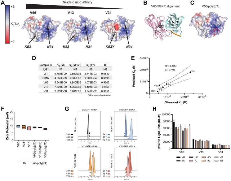
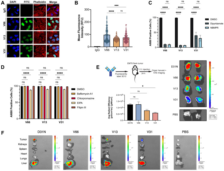
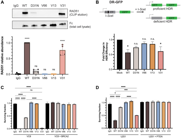
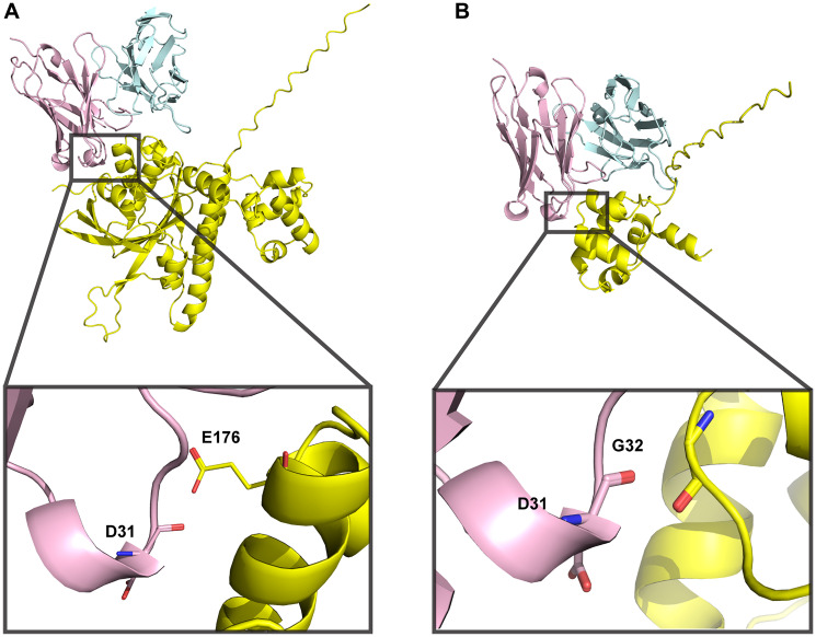
Monoclonal antibody therapies for cancer have demonstrated extraordinary clinical success in recent years. However, these strategies are thus far mostly limited to specific cell surface antigens, even though many disease targets are found intracellularly. Here we report studies on the humanization of a full-length, nucleic acid binding, monoclonal lupus-derived autoantibody, 3E10, which exhibits a novel mechanism of cell penetration and tumor specific targeting. Comparing humanized variants of 3E10, we demonstrate that cell uptake depends on the nucleoside transporter ENT2, and that faster cell uptake and superior in vivo tumor targeting are associated with higher affinity nucleic acid binding. We show that one human variant retains the ability of the parental 3E10 to bind RAD51, serving as a synthetically lethal inhibitor of homology-directed repair in vitro. These results provide the basis for the rational design of a novel antibody platform for therapeutic tumor targeting with high specificity following systemic administration.

Keywords: 3E10; RAD51; cell penetration; nucleic acid binding; nucleic acid delivery.


Conflict of interest statement

CONFLICTS OF INTEREST

E.Q. and P.M.G. are inventors on patent applications assigned to Yale University related to the work and are co-founders, hold equity in, and consult for Gennao Bio, which has licensed related intellectual property from Yale. R.A. and D.L.L. are employees of and hold equity in Gennao Bio. P.M.G. also holds equity in Patrys, Ltd., is a founder of and consultant for Cybrexa Therapeutics, and is a consultant for pHLIP Inc., none of which have a direct connection to this manuscript.

Figures

Figure 1
Figure 1. 3E10 humanization and heavy and light chain variant screening.
(A) Diagram of the 3E10 antibody engineering process. The original murine wild-type 3E10 was modified to contain a human IgG1 Fc region. This chimera was subsequently engineered with a D31N mutation in heavy chain CDR1. CDR grafting was performed to produce a fully humanized IgG1 framework; 22 variants were created by introducing point mutations into the VH and VL regions outside of the CDRs. (B) Nucleic acid affinity screening of humanized 3E10 variants. The 22 full-length antibodies were screened for their affinity to a 20-mer poly(dT) DNA oligo by ELISA for EC50 determination. All EC50s presented in the heat map are normalized to a chimeric 3E10 D31N positive control. (C) Representative ELISA assay data for poly(dT) binding by humanized V66, V13, and V31, and chimeric D31N. One biological replicate was performed. Humanized variant EC50s are 5.933, 44.34, and 685.2 nM, respectively. (D) Variable heavy chain (top) and variable light chain (bottom) sequence alignments, with deviations from WT sequence highlighted in red if substitution is increasingly anionic, blue if substitution is increasingly cationic, and grey if no significant change in formal charge at physiological pH.
Figure 2
Figure 2. In silico modeling and in vitro characterization of 3E10 humanized variants reveal correlations between nucleic acid affinity and intracellular payload delivery.
(A) In silico electrostatic modeling of V66, V13, and V31 using IgFold. Positions of N31 and K53 are labeled. (B) The crystal structure of the SLE anti-DNA antibody 5GKR overlaid with V66. Light chains are shown in pink and heavy chains are shown in teal. 5GKR was co-crystallized with 4-mer poly(dT), shown in orange. (C) Predicted binding of V66 and 4-mer poly(dT) modeled using AutoDock Vina. (D) Biolayer interferometry (BLI) assay to determine antibody equilibrium dissociation constants (KD). 20-mer biotinylated poly(dT) was adhered to a streptavidin chip. Antibody on-rates (ka) were measured for 5 min and off-rates (kd) were measured for 10 min. One biological replicate was performed. (E) Observed KD values from BLI plotted against predicted KD values calculated using AutoDock Vina. Plot shows best-fit simple linear regression (solid line) with 95% CI (dashed lines). Slope (β) and coefficient of determination (R2) are reported. (F) Assay for surface zeta potential of 3E10 human antibodies alone and antibody complexes with poly(dT), n = 3 replicates. Box plots represent upper and lower values with line at mean. (G) Representative flow cytometry traces of GFP mRNA expression at 24, 48, and 72 h. K562 cells were treated with 250 μg V66, V13, or V31 complexed with 10 μg mRNA encoding GFP. An IgG1 isotype control/GFP mRNA mixture was used as a negative control. 3 biological replicates were performed, and one representative result is shown. (H) ELISA assay for 3E10 antibody binding to mononucleotide DNA and RNA oligomers. Data are mean ± SEM, n = 3 replicates.
Figure 3
Figure 3. Characterization of 3E10 cellular penetration, mechanism of uptake, and tumor targeting.
(A) Representative confocal immunofluorescence images and of HeLa cells treated with 750 nM humanized AlexaFluor 680-labeled 3E10 variants for 24 h. (B) Quantification of (A). n ≥ 250 cells per treatment group. (C, D) Quantification of antibody uptake in K562 cells treated with inhibitors of ENT2 (C) and of canonical cellular uptake pathways (D) as assessed by flow cytometry. Cells were treated with 750 nM AlexaFluor 680-labeled antibody for 2 h. Labeled IgG1 isotype was used as a negative control. Al680: AlexaFluor 680; EIPA: 5-(N-ethyl-N-isopropyl)-amiloride; NBMPR: S-(4-nitrobenzyl)-6-thioinosine. Data are mean ± SEM, n = 3 replicates. (E) Quantification and representative IVIS images of AlexaFluor 680 fluorescence in EMT6 mouse tumors isolated from tumor-bearing mice. Mice (n = 2 per group) were treated intravenously with AlexaFluor 680-labeled antibody (100 μg) and tumors were harvested 24 h after treatment. (F) Representative IVIS images showing fluorescence signal in mice injected intravenously with AlexaFluor680-labeled antibodies. One representative image of all major organs plus EMT6 tumors is shown per treatment group.
Figure 4
Figure 4. RAD51 binding and homology-directed repair inhibition properties are retained in a fully humanized 3E10 variant.
(A) Representative western blot (top) showing RAD51 binding by antibodies as assessed following CLIP. MCF7 cells were transfected with 10 μg CMV-hRAD51 plasmid and treated with 500 nM antibodies prior to UV crosslinking and immunoprecipitation. Quantification (bottom) is of n = 4 replicates, where data are mean ± SEM. RAD51 abundance is first normalized to Fc (3E10 antibody) abundance to account for differences in antibody cellular penetration, and these values are then normalized to WT. (B) Quantification of homology-directed repair (HDR) in U2OS DR-GFP cells containing an inducible GFP reporter. Cells were treated with 1.2 μM antibody for 16–24 h and GFP expression was measured by flow cytometry. Data are mean ± SEM, n = 3 replicates. (C, D) Survival assays in isogenic cell lines. (C) VC8 ± BRCA2, and (D) U251 ± PTEN. VC8 cells were treated with 500 nM antibodies and U251 cells were treated with 1 μM antibodies for 4–5 days. Cell survival was measured using CellTiter-Glo ATP-based viability assay. Data are mean ± SEM, n = 3 replicates.
Figure 5
Figure 5. 3E10-RAD51 interaction models generated using AlphaFold 3 are of low confidence.
(A) Full-length human RAD51 sequence was used to predict docking to WT 3E10. iPTM = 0.42, pTM = 0.53. (B) N-terminal domain RAD51 sequence (aa 1–89) was used to predict docking to WT 3E10. iPTM = 0.6, pTM = 0.72. 3E10 heavy chains are shown in pink, light chains are shown in blue, and RAD51 is shown in yellow. Insets show RAD51 residues within close proximity to D31.

References

    1. Mullard A. FDA approves 100th monoclonal antibody product. Nat Rev Drug Discov. 2021; 20:491–95. 10.1038/d41573-021-00079-7. - DOI - PubMed
    1. Bagchi S, Yuan R, Engleman EG. Immune Checkpoint Inhibitors for the Treatment of Cancer: Clinical Impact and Mechanisms of Response and Resistance. Annu Rev Pathol. 2021; 16:223–49. 10.1146/annurev-pathol-042020-042741. - DOI - PubMed
    1. Ribas A, Wolchok JD. Cancer immunotherapy using checkpoint blockade. Science. 2018; 359:1350–55. 10.1126/science.aar4060. - DOI - PMC - PubMed
    1. Swain SM, Shastry M, Hamilton E. Targeting HER2-positive breast cancer: advances and future directions. Nat Rev Drug Discov. 2023; 22:101–26. 10.1038/s41573-022-00579-0. - DOI - PMC - PubMed
    1. Cunningham D, Humblet Y, Siena S, Khayat D, Bleiberg H, Santoro A, Bets D, Mueser M, Harstrick A, Verslype C, Chau I, Van Cutsem E. Cetuximab monotherapy and cetuximab plus irinotecan in irinotecan-refractory metastatic colorectal cancer. N Engl J Med. 2004; 351:337–45. 10.1056/NEJMoa033025. - DOI - PubMed

MeSH terms

LinkOut - more resources