. 2024 Oct;17(856):eado4132.
doi: 10.1126/scisignal.ado4132. Epub 2024 Oct 1.

Amyloid-β oligomers trigger sex-dependent inhibition of GIRK channel activity in hippocampal neurons in mice

Affiliations

Amyloid-β oligomers trigger sex-dependent inhibition of GIRK channel activity in hippocampal neurons in mice

Haichang Luo et al. Sci Signal. 2024 Oct.

Abstract

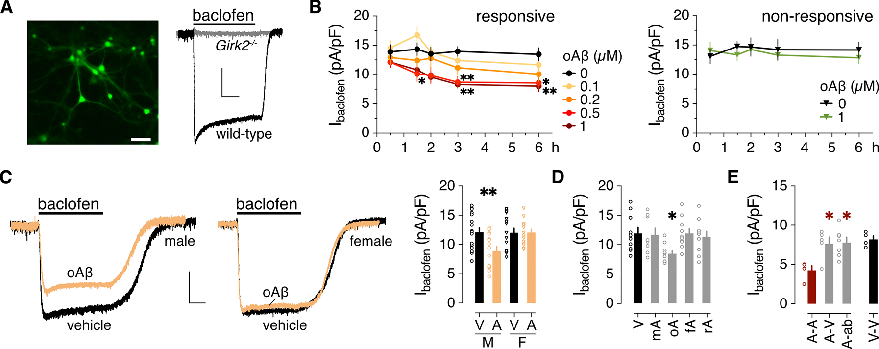
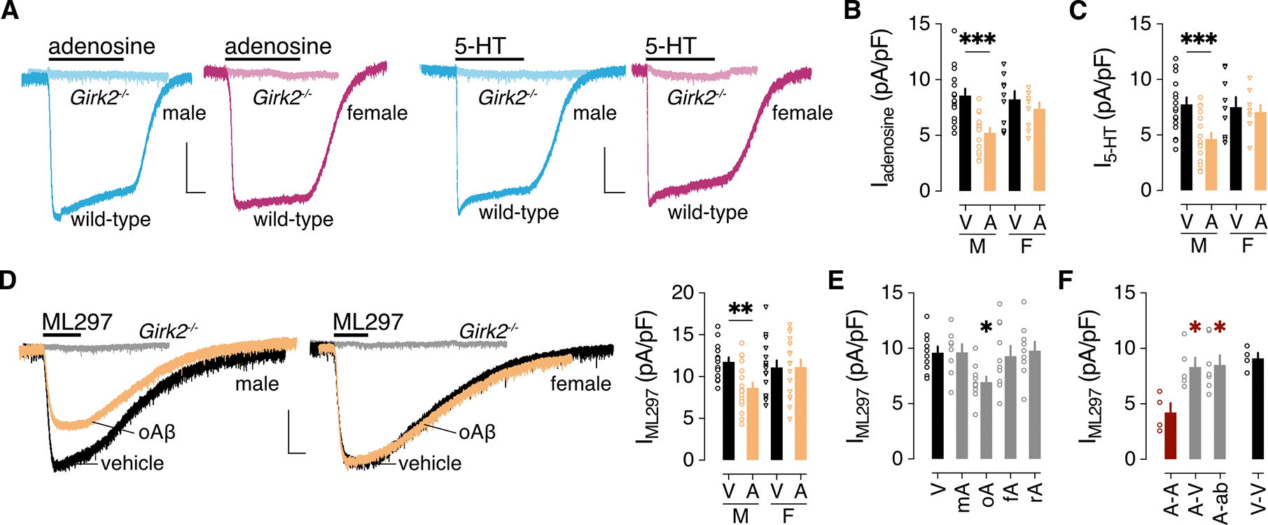
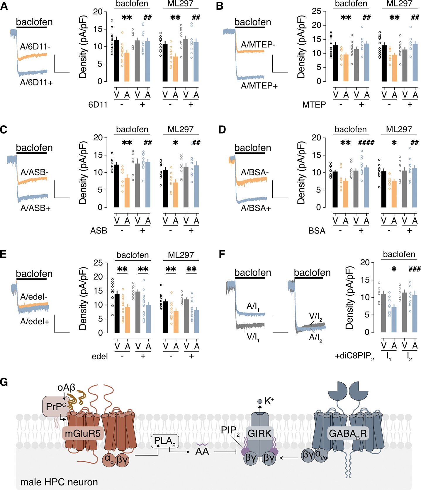
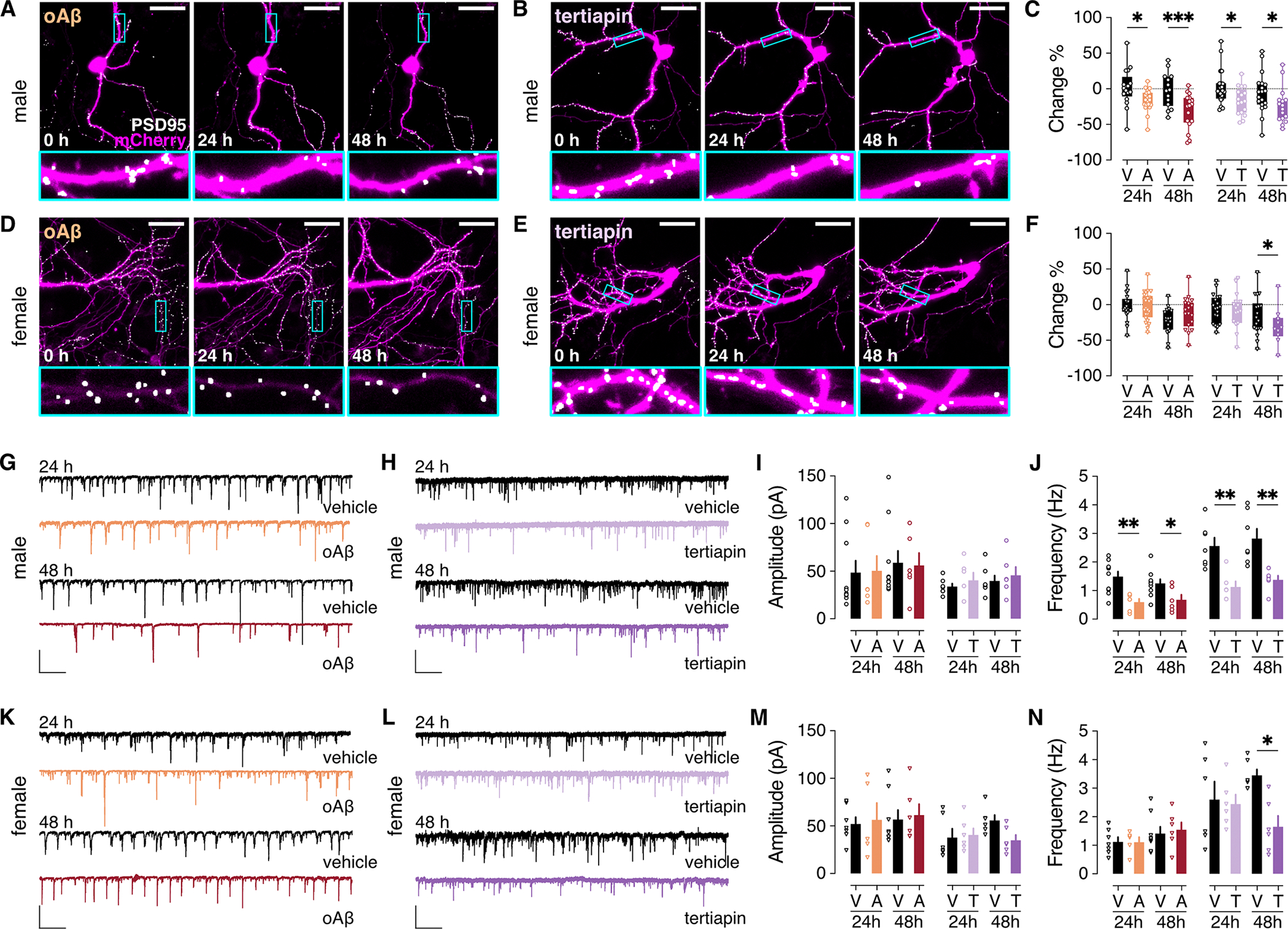
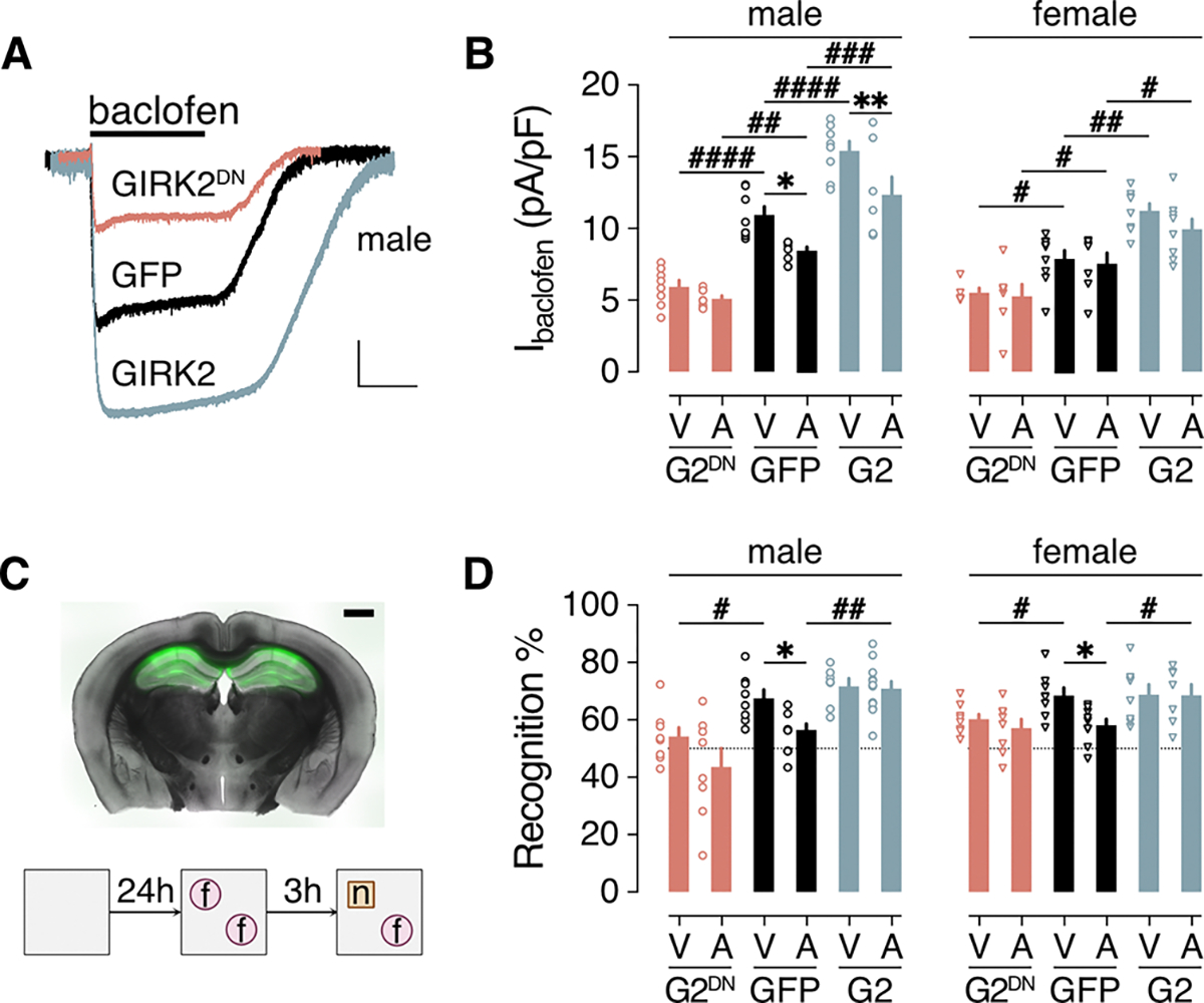
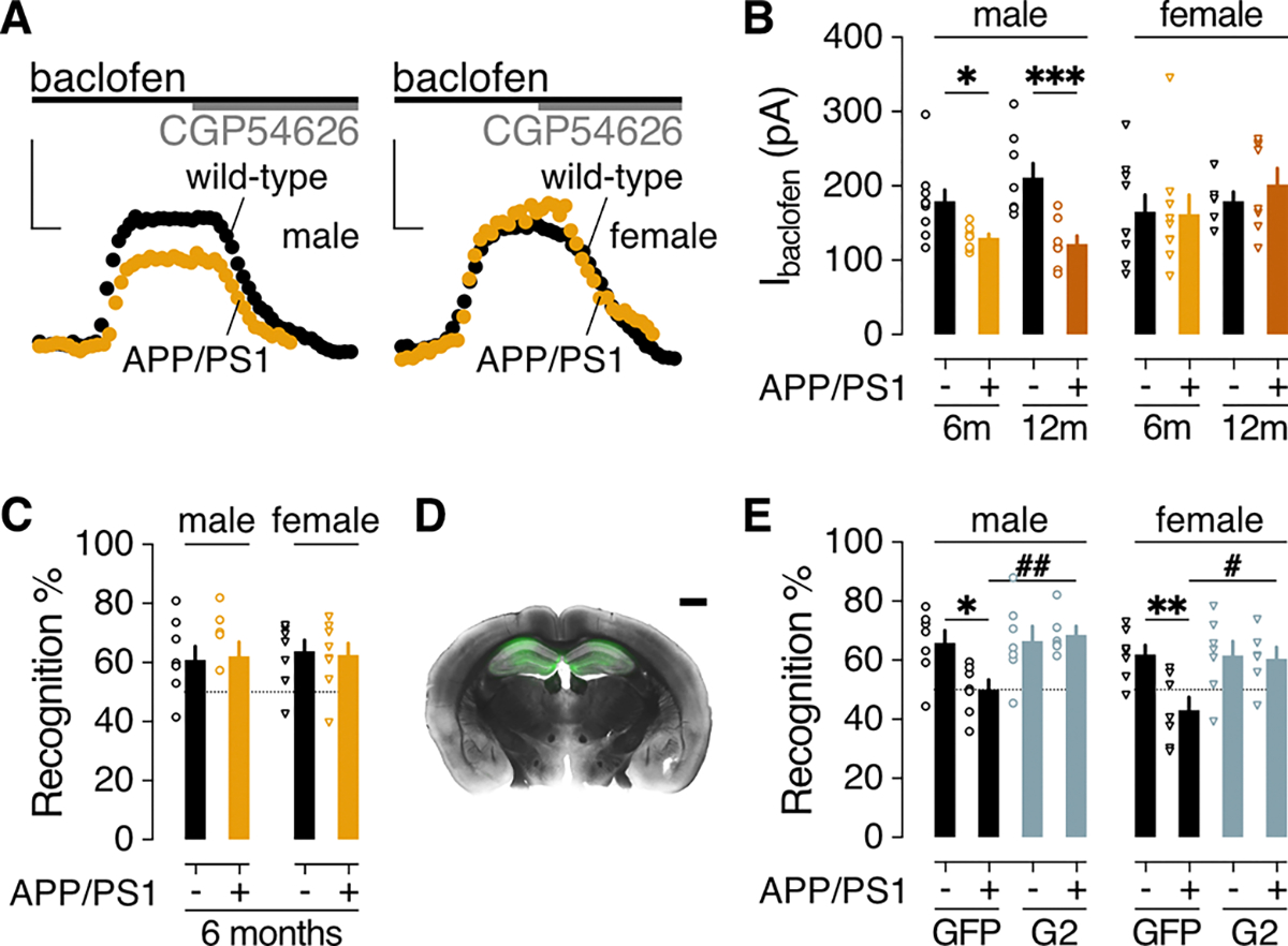
Alzheimer's disease (AD) is a progressive neurodegenerative disease characterized by amyloid plaques and cognitive decline, the latter of which is thought to be driven by soluble oligomeric amyloid-β (oAβ). The dysregulation of G protein-gated inwardly rectifying K+ (GIRK; also known as Kir3) channels has been implicated in rodent models of AD. Here, seeking mechanistic insights, we uncovered a sex-dependent facet of GIRK-dependent signaling in AD-related amyloid pathophysiology. Synthetic oAβ1-42 suppressed GIRK-dependent signaling in hippocampal neurons from male mice, but not from female mice. This effect required cellular prion protein, the receptor mGluR5, and production of arachidonic acid by the phospholipase PLA2. Although oAβ suppressed GIRK channel activity only in male hippocampal neurons, intrahippocampal infusion of oAβ or genetic suppression of GIRK channel activity in hippocampal pyramidal neurons impaired performance on a memory test in both male and female mice. Moreover, genetic enhancement of GIRK channel activity in hippocampal pyramidal neurons blocked oAβ-induced cognitive impairment in both male and female mice. In APP/PS1 AD model mice, GIRK-dependent signaling was diminished in hippocampal CA1 pyramidal neurons from only male mice before cognitive deficit was detected. However, enhancing GIRK channel activity rescued cognitive deficits in older APP/PS1 mice of both sexes. Thus, whereas diminished GIRK channel activity contributes to cognitive deficits in male mice with increased oAβ burden, enhancing its activity may have therapeutic potential for both sexes.


Conflict of interest statement

Competing interests: The authors declare that they have no competing interests.

Figures

Figure 1.
Figure 1.. oAβ suppresses GABABR-dependent signaling in male HPC neurons.
(A) Representative image of an HPC culture 4 days after infection with AAV8-CaMKIIα-GFP to highlight excitatory neurons, and representative traces of whole-cell inward currents from GFP-positive wild-type and Girk2−/− neurons following bath application of 100 μM baclofen. Scale bars: 50 μm, left; 500 pA/5 s, right. (B) Impact of oAβ pre-treatment on Ibaclofen density in “responsive” and “non-responsive” HPC cultures, collected from 8 neurons per timepoint and concentration pooled from 5 independent cultures for the responsive group, and 5 (0 μM) or 6 (1 μM) neurons per timepoint pooled from 4 independent cultures for the non-responsive group. Data were analyzed by two-way ANOVA and Dunnett’s multiple comparisons: *P<0.05 and **P<0.01 vs. 0 μM oAβ. (C) Baclofen-induced currents (Ibaclofen) in male and female HPC neurons (scale: 500 pA/5 s), and the summary of Ibaclofen densities for male (M) and female (F) neurons treated with vehicle (V) or oAβ (A: 0.5 μM for 3 h), collected from 17 (M/V), 15 (M/A), 16 (F/V), 15 (F/A) neurons across 4 independent cultures. Data were analyzed by two-way ANOVA and Šídák’s multiple comparisons, **P<0.01. (D) Ibaclofen densities following 3-hour treatment with vehicle (V, 10 cells) or 0.5 μM monomeric Aβ1–42 (mA, 8 neurons), oligomeric Aβ1–42 (oA, 10 neurons), fibrillary Aβ1–42 (fA, 10 neurons), or oligomeric Aβ42–1 (rA, 10 neurons), collected across 3 independent cultures. Data were analyzed by one-way ANOVA and Dunnett’s multiple comparisons, *P<0.05 vs. vehicle. (E) Ibaclofen densities in HPC neurons treated with 0.5 μM oAβ for 6 hours (A), measured 18 hours after replacement with media containing 0.5 μM oAβ (A-A, 4 neurons) or vehicle (A-V, 5 neurons), or anti-Aβ antibody D54D2 (A-ab; 0.16 μg/mL; 7 neurons), collected across 3 independent cultures. Data were analyzed by one-way ANOVA and Dunnett’s multiple comparisons, *P<0.05 vs. A-A. Data from a vehicle-only treatment group (V-V, 4 neurons) are presented on the right for comparison but were not included in the statistical analysis. All data are presented as mean ± SEM.
Figure 2.
Figure 2.. oAβ suppresses GIRK channel activity.
(A) Representative currents evoked by adenosine (10 μM) and 5-HT (10 μM) in HPC neurons from male (blue) and female (pink) wild-type and Girk2−/− mice. Scale bar: 500 pA/5 s. (B) Iadenosine density (pA/pF) in male (M) and female (F) HPC pyramidal neurons following a 3-hour treatment with vehicle (V) or 0.5 μM oAβ (A), collected from 15 (M/V), 15 (M/A), 9 (F/V), 9 (F/A) neurons across 4 independent cultures. Data were analyzed by two-way ANOVA and Šídák’s multiple comparisons, ***P<0.001. (C) I5-HT density (pA/pF) in male (M) and female (F) HPC pyramidal neurons following a 3-hour treatment with vehicle (V) or 0.5 μM oAβ (A), collected from 17 (M/V), 16 (M/A), 9 (F/V), 9 (F/A) neurons across 4 independent cultures. Data were analyzed by two-way ANOVA and Šídák’s multiple comparisons, ***P<0.001. (D) Representative currents evoked by ML297 (IML297, 10 μM) in HPC neurons from male and female wild-type mice following treatment with vehicle or oAβ (scale: 500 pA/10 s). Gray traces show the lack of ML297-induced current in HPC neurons from Girk2−/− mice. On the right, a summary of IML297 densities for male (M) and female (F) HPC neurons treated with vehicle (V) or oAβ (A: 0.5 μM for 3 h), collected from 14 (M/V), 19 (M/A), 15 (F/V), 16 (F/A) neurons across 4 independent cultures. Data were analyzed by two-way ANOVA and Šídák’s multiple comparisons, **P<0.01. (E) IML297 density following a 3-hour treatment with vehicle (V, 10 neurons) or 0.5 μM monomeric Aβ1–42 (mA, 8 neurons), oligomeric Aβ1–42 (oA, 10 neurons), fibrillary Aβ1–42 (fA, 10 neurons), or oligomeric Aβ42–1 (rA, 9 neurons), collected from 3 independent cultures. Data were analyzed by one-way ANOVA and Dunnett’s multiple comparisons, *P<0.05 vs. vehicle. (F) IML297 densities in HPC neurons treated with 0.5 μM oAβ for 24 hours, measured 24 hours after replacement with media containing 0.5 μM oAβ (A-A, 4 neurons) or vehicle (A-V, 5 neurons), or anti-Aβ antibody D54D2 (A-ab; 0.16 μg/mL; 7 neurons), collected from 3 independent cultures. Data were analyzed by one-way ANOVA and Dunnett’s multiple comparisons, *P<0.05 vs. A-A. Data from a vehicle-only treatment group (V-V, 4 neurons) are presented on the right for comparison but were not included in the statistical analysis. All data are presented as mean ± SEM.
Figure 3.
Figure 3.. oAβ inhibits GIRK channel activity through PrPC-mGluR5-PLA2 activation.
(A to F) Ibaclofen and IML297 in male HPC neurons incubated with vehicle (V) or 0.5 μM oAβ (A, 3 hours), with (+) or without (−) the indicated agents to interrogate underlying signaling mechanisms: (A) PrPC antibody 6D11 pretreatment (2.5 μg/mL for 30 min); (B) mGluR5 antagonist MTEP (10 μM); (C) PLA2 inhibitor ASB 14780 (ASB, 5 μM); (D) bovine serum albumin (BSA, fatty acid-free, 0.5 mg/mL); (E) PLC inhibitor edelfosine (edel, 10 μM); (F) PIP2 analog diC8PIP2 (25 μM) in the internal solution, measured immediately after whole-cell formation (I1) and then 90 s later (I2). Scale: 500 pA/5 s. Data are presented as mean ± SEM from 6 to 14 neurons in 3 to 4 independent cultures per condition. Data in (A to D) were analyzed by two-way ANOVA and Šídák’s multiple comparisons: *P<0.05 and **P<0.01 vs. V/-; ## P<0.01 and ####P< 0.0001 vs. A/−. Data in (E) were analyzed by two-way ANOVA and Šídák’s multiple comparisons: **P<0.01. Data in (F) were analyzed by two-way ANOVA with repeated measures and Šídák’s multiple comparisons: *P<0.05 vs. V/I1; ###P<0.001 vs. A/I1. (G) Schematic of the signaling pathway underlying the oAβ-induced suppression of GIRK-dependent signaling in male HPC neurons.
Figure 4.
Figure 4.. oAβ and GIRK channel inhibitor effect on excitatory synapses in cultured HPC neurons.
(A and B) Cultured HPC neurons from male mice were transfected with the plasmids pAAV-hSyn-mCherry (to fill and label neuronal structures) and pAAV-Syn-PSD95.FingR-eGFP-CCR5TC (to label excitatory post-synaptic densities). Images were taken before (t=0) and after (24 and 48 hours) addition of oAβ (1 μM; A) or the GIRK channel inhibitor tertiapin (1 μM; B) to the media. Scale bar: 30 μm. Boxed regions in upper panels are displayed at higher magnification in lower panels. (C) Percent change in excitatory synapse count 24 and 48 hours in male HPC neurons treated with oAβ (A) or vehicle (V), or tertiapin (T) or vehicle (V). oAβ data were extracted from 21 (24 hours) or 17 (48 hours) ROIs and corresponding vehicle data were extracted from 17 (24 hours) or 14 (48 hours) ROIs. Tertiapin data were extracted from 17 ROIs (24 and 48 hours) and corresponding vehicle data were extracted from 23 (24 hours) or 19 (48 hours) ROIs. Data were derived from 8 coverslips per group pooled from 4 independent cultures. (D to F) As in (A to C), in HPC neurons from female mice. oAβ n = 24 (24 hours) or 18 (48 hours) ROIs and corresponding vehicle n = 19 (24 hours) or 18 (48 hours) ROIs; tertiapin n = 16 (24 hours) or 10 (48 hours) ROIs, and corresponding vehicle n = 23 (24 hours) or 19 (48 hours) ROIs. Data were derived from 8 coverslips per group pooled from 4 independent cultures. (G and H) sEPSCs in HPC neurons from male mice, measured 24 and 48 hours after treatment with oAβ (1 μM) or vehicle (G), or the GIRK channel inhibitor tertiapin (1 μM) or vehicle (H); scale: 100 pA/5s. (I and J) sEPSC amplitude (I) and frequency (J) summary in male HPC neurons from 10 (V/24 hours), 6 (A/24 hours), 10 (V/48 hours), and 6 (A/48 hours) neurons pooled from 3 independent cultures, as well as 7 (V/24 hours), 6 (T/24 hours), 7 (V/48 hours), 6 (T/48 hours) neurons pooled from 3 independent cultures. (K to N) As in (G to J) in HPC neurons from female mice. n = 7 (V/24 hours), 5 (A/24 hours), 8 (V/48 hours), and 6 (A/48 hours) neurons pooled from 3 independent cultures, as well as 6 neurons per group (V/T/24 hours/48 hours) pooled from 3 independent cultures. Data are presented in box-and-whisker format (min-max) (C,F) or mean ± SEM (I,J,M,N). Data were analyzed by two-way ANOVA with (C,F) or without (I,J,M,N) repeated measures (mixed-effects) and Šídák’s multiple comparisons, *P<0.05,** P<0.01,***P<0.001.
Figure 5.
Figure 5.. Impact of genetic manipulation of GIRK channel activity on oAβ-induced NOR deficit.
(A) Ibaclofen traces from HPC neurons infected with AAV8-CaMKIIα-GFP (GFP), AAV8-CaMKIIα-GFP-IRES-GIRK2 (GIRK2), or AAV8-CaMKIIα-GFP-IRES-GIRK2DN (GIRK2DN). Scale bar: 500 pA/10 s. (B) Summary of Ibaclofen densities in cultured male and female HPC neurons infected with GFP, G2, or G2DN vectors, and pretreated with vehicle (V) or oAβ (A, 0.5 μM). Data were collected from 8 (V/G2DN), 8 (A/G2DN), 8 (V/GFP), 6 (A/GFP), 8 (V/G2), 7 (A/G2) male neurons in 3 independent cultures and 5 (V/G2DN), 7 (A/G2DN), 9 (V/GFP), 7 (A/GFP), 8 (V/G2), 8 (A/G2) female neurons in 3 independent cultures. (C) A representative section from an adult male C57BL/6J mouse 2 weeks after bilateral intra-HPC infusion of AAV8-CaMKIIα-GFP and oAβ (scale bar: 1 mm, top), and a schematic of the novel object recognition (NOR) task. (D) Recognition index summary for adult male and female C57BL/6J mice, 1 week following intra-HPC treatment with vehicle (V) or oAβ (A), and either GFP, GIRK2 (G2), or GIRK2DN (G2DN) vector. Group sizes were 8 (V/G2DN), 8 (A/G2DN), 8 (V/GFP), 8 (A/GFP), 7 (V/G2), 12 (A/G2) male mice and 8 (V/G2DN), 8 (A/G2DN), 8 (V/GFP), 9 (A/GFP), 7 (V/G2), 8 (A/G2) female mice. Data are presented as mean ± SEM. Data were analyzed by two-way ANOVA and either Šídák’s multiple comparisons (*P<0.05,**P<0.01, within vehicle/oAβ treatment) or Dunnett’s multiple comparisons (#P<0.05,## P<0.01,### P<0.001,####P<0.0001 within viral treatment groups vs. GFP).
Figure 6.
Figure 6.. GIRK-dependent signaling and NOR performance in APP/PS1 mice.
(A) Representative baclofen-induced outward currents (200 μM; Vhold= −60 mV) and their reversal by CGP54626 (2 μM) in dHPC CA1 pyramidal neurons from 6-month-old male and female APP/PS1 and wild-type control littermates. Scale bars: 100 pA/50 s. (B) Ibaclofen summary in dHPC CA1 pyramidal neurons from 6-month-old and 12-month-old APP/PS1 (+) and wild-type control (−) littermates. Male data were collected from 10 neurons from 3 mice (6m/−), 8 neurons from 3 mice (6m/+), 8 neurons from 2 mice (12m/−), 8 neurons from 2 mice (12m/+); female data were collected from 9 neurons from 3 mice (6m/−), 9 neurons from 2 mice (6m/+), 6 neurons from 2 mice (12m/−), 8 neurons from 2 mice (12m/+). Data were analyzed by two-way ANOVA and Šídák’s multiple comparisons: *P<0.05,***P<0.001. (C) Recognition index in the NOR test for 6-month-old male (8 mice per group) and female (8 mice per group) APP/PS1 (+) and wild-type control (−) littermates. P>0.05, unpaired t test. (D) Representative section from a 12-month-old male APP/PS1 mouse, 3 weeks after bilateral intra-HPC infusion of AAV8-CaMKIIα-GIRK2. Scale bar: 1 mm. (E) Recognition index in the NOR test for 12-month-old male APP/PS1 (+) and wild-type control (−) littermates, measured 2–3 weeks after bilateral intra-HPC infusion of GFP or GIRK2 (G2) expression vectors. Group sizes were 7 (GFP/−), 7 (GFP/+), 7 (G2/−), and 6 (G2/+) male mice and 7 (GFP/−), 7 (GFP/+), 7 (G2/−), and 6 (G2/+) female mice. Data were analyzed by two-way ANOVA and Šídák’s multiple comparisons: *P<0.05,**P<0.01 within genotype; #P<0.05,##P<0.01 within viral treatment. Data in (B, C, E) are presented as mean ± SEM.

References

    1. Scheltens P, De Strooper B, Kivipelto M, Holstege H, Chetelat G, Teunissen CE, Cummings J, van der Flier WM, Alzheimer’s disease. Lancet 397, 1577–1590 (2021). - PMC - PubMed
    1. Busche MA, Konnerth A, Neuronal hyperactivity--A key defect in Alzheimer’s disease? Bioessays 37, 624–632 (2015). - PubMed
    1. Targa Dias Anastacio H, Matosin N, Ooi L, Neuronal hyperexcitability in Alzheimer’s disease: what are the drivers behind this aberrant phenotype? Transl. Psychiatry 12, 257 (2022). - PMC - PubMed
    1. Morra JH, Tu Z, Apostolova LG, Green AE, Avedissian C, Madsen SK, Parikshak N, Hua X, Toga AW, Jack CR Jr., Schuff N, Weiner MW, Thompson PM, Alzheimer’s Disease Neuroimaging I, Automated 3D mapping of hippocampal atrophy and its clinical correlates in 400 subjects with Alzheimer’s disease, mild cognitive impairment, and elderly controls. Hum. Brain. Mapp. 30, 2766–2788 (2009). - PMC - PubMed
    1. Huijbers W, Mormino EC, Schultz AP, Wigman S, Ward AM, Larvie M, Amariglio RE, Marshall GA, Rentz DM, Johnson KA, Sperling RA, Amyloid-beta deposition in mild cognitive impairment is associated with increased hippocampal activity, atrophy and clinical progression. Brain 138, 1023–1035 (2015). - PMC - PubMed

Publication types

MeSH terms

Substances

LinkOut - more resources