. 2024 Sep;21(5):e00436.
doi: 10.1016/j.neurot.2024.e00436. Epub 2024 Oct 1.

Bioreactor-produced iPSCs-derived dopaminergic neuron-containing neural microtissues innervate and normalize rotational bias in a dose-dependent manner in a Parkinson rat model

Affiliations

Bioreactor-produced iPSCs-derived dopaminergic neuron-containing neural microtissues innervate and normalize rotational bias in a dose-dependent manner in a Parkinson rat model

Nicolas Prudon et al. Neurotherapeutics. 2024 Sep.

Abstract

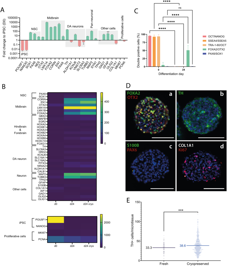
A breadth of preclinical studies now support the rationale of pluripotent stem cell-derived cell replacement therapies to alleviate motor symptoms in Parkinsonian patients. Replacement of the primary dysfunctional cell population in the disease, i.e. the A9 dopaminergic neurons, is the major focus of these therapies. To achieve this, most therapeutical approaches involve grafting single-cell suspensions of DA progenitors. However, most cells die during the transplantation process, as cells face anoïkis. One potential solution to address this challenge is to graft solid preparations, i.e. adopting a 3D format. Cryopreserving such a format remains a major hurdle and is not exempt from causing delays in the time to effect, as observed with cryopreserved single-cell DA progenitors. Here, we used a high-throughput cell-encapsulation technology coupled with bioreactors to provide a 3D culture environment enabling the directed differentiation of hiPSCs into neural microtissues. The proper patterning of these neural microtissues into a midbrain identity was confirmed using orthogonal methods, including qPCR, RNAseq, flow cytometry and immunofluorescent microscopy. The efficacy of the neural microtissues was demonstrated in a dose-dependent manner using a Parkinsonian rat model. The survival of the cells was confirmed by post-mortem histological analysis, characterised by the presence of human dopaminergic neurons projecting into the host striatum. The work reported here is the first bioproduction of a cell therapy for Parkinson's disease in a scalable bioreactor, leading to a full behavioural recovery 16 weeks after transplantation using cryopreserved 3D format.

Keywords: 3D cell format; Cell therapy; Midbrain; Off-the-shelf; Organoid; Regenerative medicine.


Conflict of interest statement

Declaration of competing interest The authors declare the following financial interests/personal relationships which may be considered as potential competing interests: Nicolas Prudon reports financial support was provided by TreeFrog Therapeutics. Erwan Bezard reports a relationship with TreeFrog Therapeutics that includes: consulting or advisory. Maxime Feyeux has patent issued to University of Bordeaux. Kevin Alessandri has patent issued to University of Bordeaux. Erwan Bezard has patent issued to University of Bordeaux. If there are other authors, they declare that they have no known competing financial interests or personal relationships that could have appeared to influence the work reported in this paper.

Figures

Image 1
Graphical abstract
Fig. 1
Fig. 1
Schematic overview of the neural microtissues bioproduction process. A: (a) hiPSCs were encapsulated using a microfluidic chip. (b) Cells were further cultured and differentiated in a 500 ​mL bioreactor. The neurodifferentiation protocol is illustrated with schematic neural microtissues and the corresponding widefield microscope pictures. (c) At harvest, the microtissues were decapsulated and cryopreserved using a CRF (Controlled Rate Freezer). B: Schematic representation and C: corresponding widefield microscope pictures of the neural microtissues across the neurodifferentiation protocol. Scale bar: 500 ​μm.
Fig. 2
Fig. 2
In vitro characterisation of the neural microtissues. A. Gene expression analysis of the fresh neural microtissue using bulk RT-qPCR. Values are represented as fold change to hiPSC (D0). Experiments were performed in technical duplicates. Data are presented as histograms (n ​= ​13 batches). Error bars are SEM. Raw expression values for the genes SLC6A3 (∗) and GFAP (∗∗) were below the detection threshold (2ΔCt ​< ​10−4) for a fraction of the bioreactors analysed (∗n ​= ​5/13, ∗∗n ​= ​6/13). Raw values are also plotted in the supplementary data (supplementary Fig. S1). B. Transcriptomics analysis of the starting hiPSC (D0), the fresh and the cryopreserved neural microtissue (D24) from the transplanted batch. The gene expression profile described in the qPCR analysis was confirmed, along with no enrichment of gene expression associated with potential forebrain (FOXG1), hindbrain (GBX2, MAFB, EGR2, HOXA2, HOXB1, HOXA3, HOXB2, HOXA4) or spinal cord (HOXB8, HOXC10) contaminants. Data are represented as TPM (transcript per million). C. Flow cytometry analysis of fresh single cells from the encapsulation day D0 (hiPSC) or at the end of the bioproduction process D24 (dissociated neural microtissue). An antibody panel was used to identify pluripotent stem cells (OCT/NANOG; SSEA4/SSEA5 and TRA-1-60/OCT), neural stem cells (PAX6/SOX1) and DA progenitors (FOXA2/OTX2). Data are presented as histograms (n ​= ​13 batches) and analysed with Sidak's multiple comparison test; error bars represent SD. ∗∗∗∗p ​< ​0,0001. D. Immunofluorescence images of cryopreserved neural microtissues from the transplanted batch (a–d). Cryopreserved neural microtissues were thawed and then fixed before the immunolabelling. Dopaminergic progenitors were detected using the markers FOXA2 and OTX2 (a) along with more mature dopaminergic neurons positive for TH (b). The absence of astrocytes and neural stem cells was confirmed, using the markers S100B and PAX6, respectively (c). No VLMCs were detected in the neural microtissues using the marker COL1A1, and cells remaining in the cell cycle were detected as positive for the marker Ki67 (d). Samples were counterstained with DAPI (a–d). Scale bar: 100 ​μm. Splits of the colour channels are available in the supplementary data (supplementary Figs. S2–8). E. In vitro quantification of DA neurons using immunofluorescence images from the transplanted batch. The numbers of TH ​+ ​cells were quantified in fresh (n ​= ​87) and cryopreserved (n ​= ​555) microtissues. Data are represented as mean in scatter-plot and analysed using Welch's t-test. ∗∗∗: p ​< ​0,001. (For interpretation of the references to colour in this figure legend, the reader is referred to the Web version of this article.)
Fig. 3
Fig. 3
Non-clinical efficacy study of neural microtissues in the hemiparkinsonian rat model. A. Distribution of animals in the different dose groups based on the estimated number of injected TH ​+ ​cells. The total number of injected TH ​+ ​cells per animal was calculated by the number of neural microtissues injected and the number of TH ​+ ​cells per microtissue (n ​= ​15) specific to each cryovial. Data are presented as mean in a scatter plot and analysed using Welch's t-test (∗∗∗: p ​< ​0.001, ∗∗∗∗: p ​< ​0.0001, ns: not significant). B. Time-based analysis of d-amphetamine-induced rotations measured pre-operatively and 4, 8, 12, 16 and 20 weeks (n ​= ​16) post-engraftment. The graft of neural microtissues achieved functional recovery of motor deficits 16 weeks after transplantation for the Fresh High (n ​= ​4, p ​< ​0,0001), the Cryopreserved High (n ​= ​5, p ​< ​0.0001) and the Cryopreserved Maximum Feasible Dose (MFD) (n ​= ​3, p ​< ​0.0001), corresponding to a total of 11 ​000 ​TH ​+ ​cells transplanted for both fresh and cryopreserved High doses and 21 ​000 ​TH ​+ ​cells for the MFD. A delay was observed for the Low dose group, corresponding to 6000 ​TH ​+ ​cells transplanted, with full functional recovery observed 20 weeks after transplantation (n ​= ​8, p ​< ​0.0001). Comparisons were made to the vehicle group using Two-Way ANOVA and Tuckey's multiple comparison test (∗: p ​< ​0.05, ∗∗∗: p ​< ​0.001, ∗∗∗∗: p ​< ​0.0001, ns: not significant). Data are presented as mean ​± ​SEM. C. Post-mortem histological analysis of the graft. Representative sections of animals 20 weeks after receiving the cryopreserved Maximum feasible dose (a–d), cryopreserved High dose (e–h), cryopreserved Low dose (i–l) or Fresh High dose (m–p). Sections were immunostained for the human marker Stem121 (b, f, j, n) and TH (c, g, k, o). Merges (a, e, i, m). Image analysis of the TH-immunolabeling was performed to better highlight reinnervation using skeletonisation (d, h, l, p). Rectangles show areas of magnification shown in the right panels, illustrating dopaminergic projections from the graft. Scale bars: 2,5 ​mm (a, e, i, m) and 100 ​μm (b-d, f-h, j-l and n-p).
figs1
Fig. S1
Raw values of the transcriptomics analysis of the final neural microtissues (D24) using qRT-PCR. Values are presented as raw (2ΔCt). The detection threshold was set at 2ΔCt < 10-4. Green and red bars correspond respectively to enriched or depleted gene expression compare to iPSC, as presented in figure 2A. Data are presented as histograms (n = 13 batches). Raw expression values for the genes SLC6A3 (∗) and GFAP (∗∗) were below the detection threshold (2ΔCt < 10-4) for a fraction of the bioreactors analyzed (∗ n = 5/13, ∗∗ n = 6/13).
figs2
Fig. S2
Immunofluorescence images of a cryopreserved neural microtissue from the transplanted batch (a-d). Neural microtissues were immunolabeled for OTX2 (b), FOXA2 (c), TPH2 (d) and counterstained with DAPI. Merge (a). Scale bar: 100 μm.
figs3
Fig. S3
Immunofluorescence images of a cryopreserved neural microtissue from the transplanted batch (a-d). Neural microtissues were immunolabeled for MAP2 (b), OLIG2 (c) and counterstained with DAPI (d). Merge (a). Scale bar: 100 μm.
figs4
Fig. S4
Immunofluorescence images of a cryopreserved neural microtissue from the transplanted batch (a-d). Neural microtissues were immunolabeled for TH (a) and counterstained with DAPI (b). Merge (c). Scale bar: 100 μm.
figs5
Fig. S5
Immunofluorescence images of cryopreserved neural microtissues from the transplanted batch (a-d). Neural microtissues were counterstained for DAPI (b) and immunolabeled for S100B (c) and PAX6 (d). Merge (a). Scale bar: 100 μm.
figs6
Fig. S6
Immunofluorescence images of a cryopreserved neural microtissue from the transplanted batch (a-d). Neural microtissues were immunolabeled for Ki67 (b), NANOG (c) and DAPI (d). Merge (a). Scale bar: 100 μm.
figs7
Fig. S7
Immunofluorescence images of a cryopreserved neural microtissue from the transplanted batch (a-d). Neural microtissues were immunolabeled for COL1A1 (b), Ki67 (c) and counterstained for DAPI (d). Merge (a). Scale bar: 100 μm.
figs8
Fig. S8
Immunofluorescence images of a cryopreserved neural microtissue from the transplanted batch (a-d). Neural microtissues were counterstained for DAPI (b) and immunolabeled for SOX9 (c) and GFAP (d). Merge (a). Scale bar: 100 μm.
figs9
Fig. S9
Epifluorescence images of the viability assay performed on thawed neural microtissues from the transplanted batch. Cryopreserved neural microtissues were thawed and stained using a viability (LiveDead) assay. Viable cells (cells with intact membranes) were stained by calcein-AM (green, A.c and B.c). Non-viable cells (cells with breached membranes) were stained by Ethidium homodimer-1 (red, A.d and B.d). A control positive for non-viable cells was performed by heating the cells (B). Merge (A.b and B.b). Non-viable cells could be detected in both conditions (A.d, B.d). However, viable cells were only detected in the thawed neural microtissues from the transplanted batch (A.b,c). Scale bar: 750 μm.

References

    1. Li W., Englund E., Widner H., Mattsson B., Van Westen D., Lätt J., et al. Extensive graft-derived dopaminergic innervation is maintained 24 years after transplantation in the degenerating parkinsonian brain. Proc Natl Acad Sci. 2016;113:6544–6549. - PMC - PubMed
    1. Olanow C.W., Goetz C.G., Kordower J.H., Stoessl A.J., Sossi V., Brin M.F., et al. A double-blind controlled trial of bilateral fetal nigral transplantation in Parkinson's disease. Ann Neurol. 2003;54:403–414. - PubMed
    1. Freed C.R., Greene P.E., Breeze R.E., Tsai W.Y., DuMouchel W., Kao R., et al. Transplantation of embryonic dopamine neurons for severe Parkinson’s disease. N Engl J Med. 2001;344:710–719. - PubMed
    1. Parmar M., Grealish S., Henchcliffe C. The future of stem cell therapies for Parkinson disease. Nat Rev Neurosci. 2020;21:103–115. - PubMed
    1. Chambers S.M., Fasano C.A., Papapetrou E.P., Tomishima M., Sadelain M., Studer L. Highly efficient neural conversion of human ES and iPS cells by dual inhibition of SMAD signaling. Nat Biotechnol. 2009;27:275–280. - PMC - PubMed

LinkOut - more resources