

doi: 10.1038/s41467-024-52265-x.
Cryo-EM structure of Alzheimer's disease tau filaments with PET ligand MK-6240
Affiliations
- PMID: 39353896
- PMCID: PMC11445244
- DOI: 10.1038/s41467-024-52265-x
Item in Clipboard
Cryo-EM structure of Alzheimer's disease tau filaments with PET ligand MK-6240
Nat Commun.
.
Abstract
Positron Emission Tomography (PET) ligands have advanced Alzheimer's disease (AD) diagnosis and treatment. Using autoradiography and cryo-EM, we identify AD brain tissue with elevated tau burden, purify filaments, and determine the structure of second-generation high avidity PET ligand MK-6240 at 2.31 Å resolution, which bound at a 1:1 ratio within the cleft of tau paired-helical filament (PHF), engaging with glutamine 351, lysine 353, and isoleucine 360. This information elucidates the basis of MK-6240 PET in quantifying PHF deposits in AD and may facilitate the structure-based design of superior ligands against tau amyloids.
© 2024. The Author(s).
Conflict of interest statement
The authors declare no competing interests.
Figures
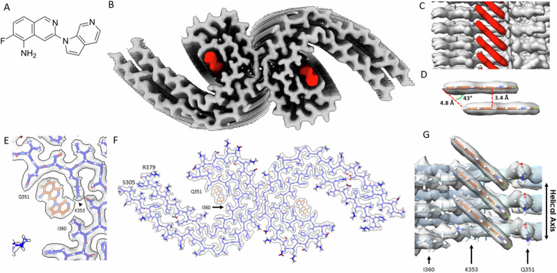
A Molecular structure of MK-6240. B Cryo-EM map of AD tau PHFs (Grey) with bound MK-6240 (Red). C Side-view depiction of MK-6240 (red) situated within the binding pocket of the cleft within the AD tau PHF (Grey). MK-6240 adopted a stacked arrangement perpendicular to the fibril axis. One MK-6240 molecule spanned approximately two tau monomer rungs. D Isolated atomic representation of MK-6240 with its binding orientation. Peripheral atoms (alpha-carbon Hydrogen, left; and Fluorine, right) exhibit a 4.8 Å distance between them. Between heterocycles or parallel atoms within MK-6240 molecules, a ~ 3.3 Å distance is observed. E Close-up depiction of the binding site, accentuating the proximity of the three amino acids (Q351, K353, and I360) to MK-6240. F Atomic model showcasing the MK-6240 binding site on paired helical filaments. Cryo-EM density (white) is juxtaposed with the atomic model of the tau fold (blue) and MK-6240 (orange). G Side-view, zoomed-in perspective (from within the cleft) of the MK-6240 binding site, revealing the ~1:1 stoichiometry and the angle that aligns individual MK-6240 molecules with both themselves and the filament axis.
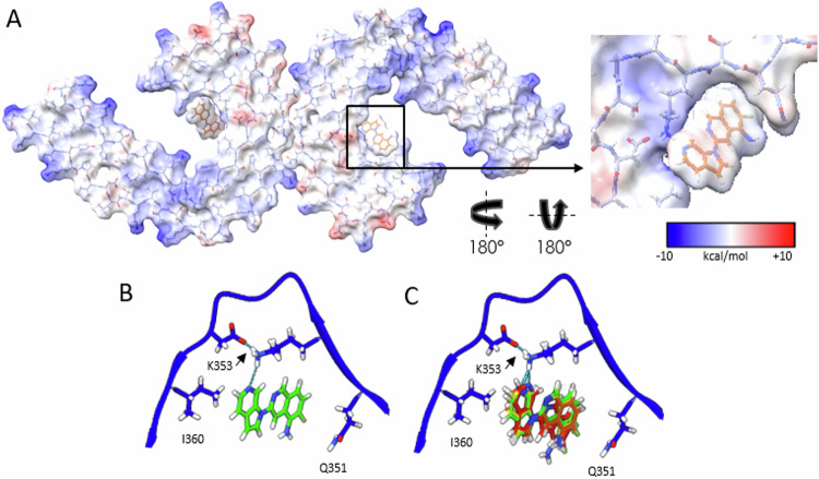
A Coulombic energy map depicting the cryo-EM density of the binding pocket, with amino acids from the tau fold and MK-6240. B Optimal pose resulting from MK-6240 binding via symmetry docking (illustrated within the protein chain modelled to the cryo-EM structure, key amino acids are highlighted), revealing hydrogen bonding involving the secondary amine of the azaindole ring and K353. Minimized energetic scores align with the manually docked pose of MK-6240 in the cryo-EM density as shown in (B). C The top four poses (Green > Yellow > Orange > Red) generated through symmetry docking, showcasing robust alignment and a predilection for the orientation of the secondary amine of the azaindole ring system to facilitate interaction with K353. Flexibility variations toward the solvent-exposed region are observed among these poses.
Update of
-
Cryo-EM structure of Alzheimer's disease tau filaments with PET ligand MK-6240.bioRxiv [Preprint]. 2023 Sep 22:2023.09.22.558671. doi: 10.1101/2023.09.22.558671. bioRxiv. 2023. Update in: Nat Commun. 2024 Oct 1;15(1):8497. doi: 10.1038/s41467-024-52265-x. PMID: 37790438 Free PMC article. Updated. Preprint.
References
Publication types
MeSH terms
Substances
Associated data
- Actions
- Actions
- Actions
Grants and funding
- R21 AG080497/AG/NIA NIH HHS/United States
- MOP-11-51-31, FRN, 152985, PI:PR-N/Gouvernement du Canada | Canadian Institutes of Health Research (Instituts de Recherche en Santé du Canada)
- 2020-VICO-279314/Fonds de Recherche du Québec - Santé (Fonds de la recherche en sante du Quebec)
- NIRG-12-259245, PR-N/ALZ/Alzheimer's Association/United States
- R21AG080497/U.S. Department of Health & Human Services | NIH | National Institute on Aging (U.S. National Institute on Aging)
- R35GM122481/Foundation for the National Institutes of Health (Foundation for the National Institutes of Health, Inc.)
- R35 GM122481/GM/NIGMS NIH HHS/United States
- NIRG-12-259245/ALZ/Alzheimer's Association/United States
- RP170644/Cancer Prevention and Research Institute of Texas (Cancer Prevention Research Institute of Texas)
