. 2024 Oct 2;13(1):36.
doi: 10.1038/s41389-024-00532-3.

The branched N-glycan of PD-L1 predicts immunotherapy responses in patients with recurrent/metastatic HNSCC

Affiliations

The branched N-glycan of PD-L1 predicts immunotherapy responses in patients with recurrent/metastatic HNSCC

Huai-Cheng Huang et al. Oncogenesis. .

Abstract

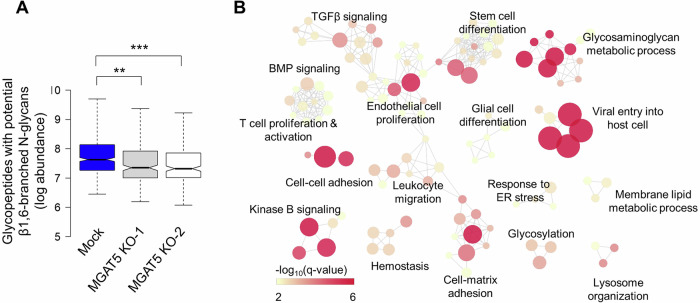
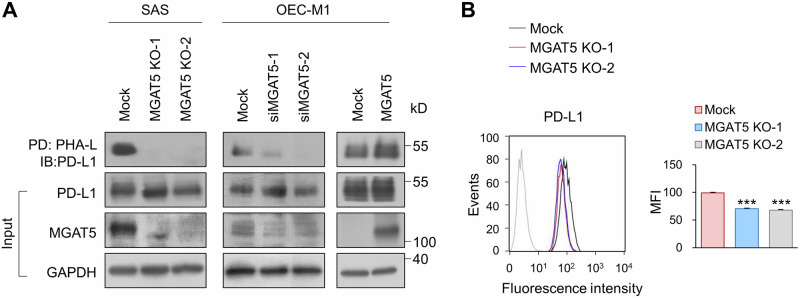
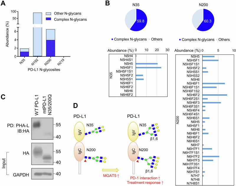
Immunotherapy has revolutionized cancer treatment, but the lack of a reliable predictive biomarker for treatment response remains a challenge. Alpha-1,6-Mannosylglycoprotein 6-β-N-Acetylglucosaminyltransferase 5 (MGAT5) is a key regulator of complex N-glycan synthesis, and its dysregulation is associated with cancer progression. The lectin Phaseolus vulgaris leukoagglutinin (PHA-L) specifically binds to mature MGAT5 products. Previous studies have indicated elevated PHA-L staining in head and neck squamous cell carcinoma (HNSCC), which implies increased activity of MGAT5. However, the specific role of MGAT5 in HNSCC remains unclear. In this study, we found significantly higher PHA-L staining and MGAT5 expression in HNSCC tumors compared to adjacent non-tumor tissues. Using a mass spectrometry (MS)-based glycoproteomic approach, we identified 163 potential protein substrates of MGAT5. Functional analysis revealed that protein substrates of MGAT5 regulated pathways related to T cell proliferation and activation. We further discovered that PD-L1 was among the protein substrates of MGAT5, and the expression of MGAT5 protected tumor cells from cytotoxic T lymphocyte (CTL) killing. Treatment of nivolumab alleviated the protective effects of MGAT5 on CTL activity. Consistently, patients with MGAT5-positive tumors showed improved responses to immunotherapy compared to those with MGAT5-negative tumors. Using purified PD-L1 from HNSCC cells and a glycoproteomic approach, we further deciphered that the N35 and N200 sites carry the majority of complex N-glycans on PD-L1. Our findings highlight the critical role of MGAT5-mediated branched N-glycans on PD-L1 in modulating the interaction with the immune checkpoint receptor PD-1. Consequently, we propose that MGAT5 could serve as a biomarker to predict patients' responses to anti-PD-1 therapy. Furthermore, targeting the branched N-glycans at N35 and N200 of PD-L1 may lead to the development of novel diagnostic and therapeutic approaches.


Conflict of interest statement

The authors declare no competing interests.

Figures

Fig. 1
Fig. 1. IHC of MGAT5 and its N-glycan products in HNSCC tumors and adjacent non-tumor tissues.
A Left, IHC score of PHA-L in 13 paired HNSCC tumors (T) and adjacent non-tumor tissues (N). Data are presented as mean ± SEM. **P < 0.01, analyzed using two-tailed paired Student’s t-test. Right, representative images of PHA-L staining. Scale bars, 50 μm. B Left, Western blot analysis of PHA-L staining and MGAT5 expression in 6 paired HNSCC tumors (T) and adjacent non-tumor tissues (N). GAPDH was the internal control. Right, quantification of PHA-L staining and MGAT5 expression was performed by the ImageJ software. *P < 0.05 and **P < 0.01 analyzed using two-tailed paired Student’s t-test.
Fig. 2
Fig. 2. Identification of MGAT5 protein substrates.
A Abundance of glycopeptides with complex N-glycans Mock or MGAT5 knockout (KO) SAS cells (1 × 107) were snap frozen and sent for mass spectrometry. Abundance of β1,6-branched N-glycopeptides was shown in log scale. **P < 0.01 and ***P < 0.001. B Functional map analysis of potential MGAT5 protein substrates. The over-represented Gene Ontology (GO) terms of 163 protein substrates of MGAT5 (with a P < 0.01) were organized into a network, where each node represented a specific GO term, and an edge signified that two GO terms share common genes.
Fig. 3
Fig. 3. Effect of MGAT5 on PD-L1 properties.
A Lectin pull-down assay. The level of branched N-glycans on PD-L1 was assessed using PHA-L pull-down assay. Mock or two independent clones of MGAT5 knockout (MGAT5 KO-1 and MGAT5 KO-2) SAS cells were used. To knock down MGAT5, OCE-M1 cells were transfected with non-targeting siRNA or siRNA against MGAT5 (siMGAT5-1 or siMGAT5-2). To overexpress MGAT5, OEC-M1 cells were transfected with an empty vector (Mock) or MGAT5/pcDNA3.1. Cell lysates (500 μg) were incubated with PHA-L conjugated agarose beads overnight and the proteins captured by PHA-L were analyzed using Western blot analysis. PD, pull-down. IB, immunoblot. Input PD-L1, MGAT5 and GAPDH in the cell lysates were shown in the lower panel. Data are representative of three independent experiments. B Flow cytometry of PD-L1. Effect of MGAT5 on the surface expression of PD-L1. Cells (1 × 106) were stained with an anti-PD-L1 antibody (#329701, BioLegend) on ice for 30 min. After washing, cells were incubated with an Alexa Fluor 488-conjugated goat anti-mouse IgG for another 30 min. Fluorescence was detected by a flow cytometer. Left, histograms of fluorescence intensity of PD-L1 in Mock and MGAT5 knockout (KO) cells. Right, mean fluorescence intensity (MFI) of Mock and MGAT5 KO cells. Data are presented as mean ± SD. ***P < 0.001, analyzed using two-tailed Student’s t-test. Data are representative of three independent experiments.
Fig. 4
Fig. 4. CTL assays of SAS and Jurkat cells with control IgG or nivolumab.
A Schematic diagram of CTL assays. Jurkat cells were activated with 100 ng/mL PMA and 5 µg/mL PHA-L for 24 h. The IncuCyte® Caspase 3/7 dyes were added to the culture medium 24 h before tumor cells were cocultured with activated Jurkat cells. The IncuCyte® dyes are designed to become fluorescent upon cleavage by active Caspase 3/7 in apoptotic cells. B Immunofluorescence microscopy. Mock or MGAT5 knockout (MGAT5 KO-2) cells were cocultured with activated Jurkat cells for 6 h. The effector-to-target ratio was 1:5. The fluorescent signal was detected via fluorescence microscopy and quantified using the ImageJ software. Left, representative images of apoptotic SAS cells incubated with activated Jurkat cells and with control IgG or nivolumab (20 ug/ml). Scale bars, 50 μm. Right, quantitative results of immunofluorescent apoptotic cells. Data are presented as mean ± SD. *P < 0.05, analyzed using two-tailed Student’s t-test. NS not significant. Data are representative of three independent experiments.
Fig. 5
Fig. 5. Correlation of MGAT5 expression or TPS with treatment responses to anti-PD-1 therapy.
A Representative IHC images of MGAT5 expression in HNSCC tumors. Scale bars, 50 μm. B Fisher’s exact tests analyzing correlation of MGAT5 expression or tumor proportion score (TPS) with treatment responses (n = 40). Treatment response was evaluated based on response evaluation criteria in solid tumors (RECIST) criteria. Stable disease, partial response, and complete remission were considered good treatment responses. PPV positive predictive value. NPV negative predictive value. C Kaplan–Meier analysis for progression-free survival and overall survival of patients treated with anti-PD-1 antibodies (n = 40). Patients were divided according to their MGAT5 expression or TPS status.
Fig. 6
Fig. 6. Site-specific N-glycosylation on PD-L1 protein.
A Abundance (% to total) of glycopeptides with complex N-glycans and other N-glycopeptides at N35, N192, N200, and N219 of PD-L1. B Abundance and structure of glycopeptides with complex N-glycans at N35 and N200 of PD-L1. The abundance (% to glycans at the indicated site) of glycopeptides with complex N-glycans was demonstrated in the upper panel. High confident glycopeptides with complex N-glycans at N35 and N200 of PD-L1 with PEP2D < 0.01 were demonstrated in the lower panel. N, HexNAc; H, Hex; F, Fucose; S, Sialic acid. Of note, LC-MS/MS analysis cannot differentiate two types of tri-antennary structures, namely 2,2’,6’-triantenna and 2,4,2’-triantenna, with the former being products of MGAT5. C PHA-L pull-down assay. PD-L1 knockout SAS cells were transfected with wild-type (WT) or mutant (mt)N35/200Q PD-L1. Cell lysates (500 μg) were incubated with PHA-L conjugated agarose beads overnight, and the proteins captured by PHA-L were analyzed using Western blot analysis. PD, pull-down. IB, immunoblot. Input PD-L1-HA and GAPDH in the cell lysates were shown in the lower panel. D MGAT5 specifically contributes to formation of the β1,6-branched N-glycan at N35 and N200 of PD-L1. These site-specific branched N-glycans enhance the ability of PD-L1 to inhibit CTL activity. Thereafter, tumors with MGAT5 expression, which indicates the presence of the branched N-glycan at N35 and N200, shows better response to the anti-PD-1 therapy than those without MGAT5 expression.

References

    1. Mody MD, Rocco JW, Yom SS, Haddad RI, Saba NF. Head and neck cancer. Lancet. 2021;398:2289–99. - PubMed
    1. Hamoir M, Schmitz S, Suarez C, Strojan P, Hutcheson KA, Rodrigo JP, et al. The current role of salvage surgery in recurrent head and neck squamous cell carcinoma. Cancers. 2018;10:267. - PMC - PubMed
    1. Morris LGT, Chandramohan R, West L, Zehir A, Chakravarty D, Pfister DG, et al. The molecular landscape of recurrent and metastatic head and neck cancers: insights from a precision oncology sequencing platform. JAMA Oncol. 2016;3:244–55. - PMC - PubMed
    1. Ferris RL, Blumenschein G Jr., Fayette J, Guigay J, Colevas AD, Licitra L, et al. Nivolumab for recurrent squamous-cell carcinoma of the head and neck. N Engl J Med. 2016;375:1856–67. - PMC - PubMed
    1. Burtness B, Harrington KJ, Greil R, Soulières D, Tahara M, de Castro G Jr., et al. Pembrolizumab alone or with chemotherapy versus cetuximab with chemotherapy for recurrent or metastatic squamous cell carcinoma of the head and neck (KEYNOTE-048): a randomised, open-label, phase 3 study. Lancet. 2019;394:1915–28. - PubMed

LinkOut - more resources