. 2024 Oct 1;14(1):22859.
doi: 10.1038/s41598-024-72714-3.

The activation of the Notch signaling pathway by UBE2C promotes the proliferation and metastasis of hepatocellular carcinoma

Affiliations

The activation of the Notch signaling pathway by UBE2C promotes the proliferation and metastasis of hepatocellular carcinoma

Ping Zhan et al. Sci Rep. .

Abstract

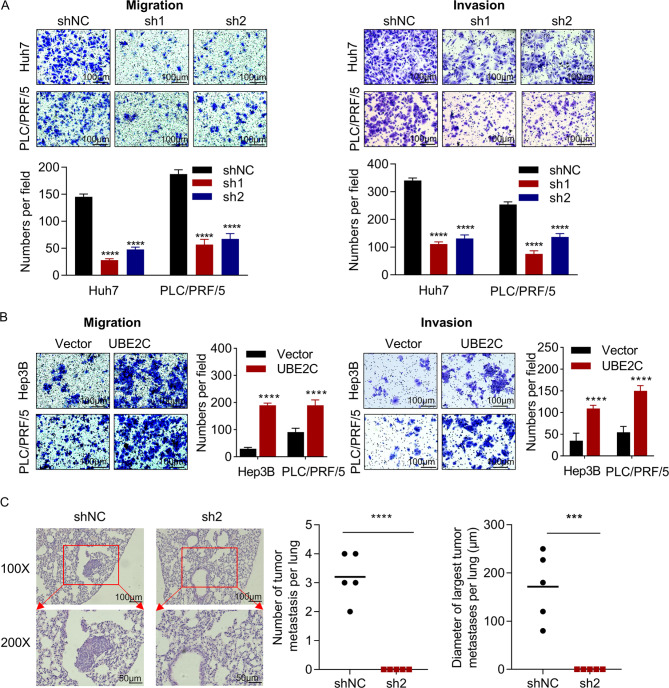
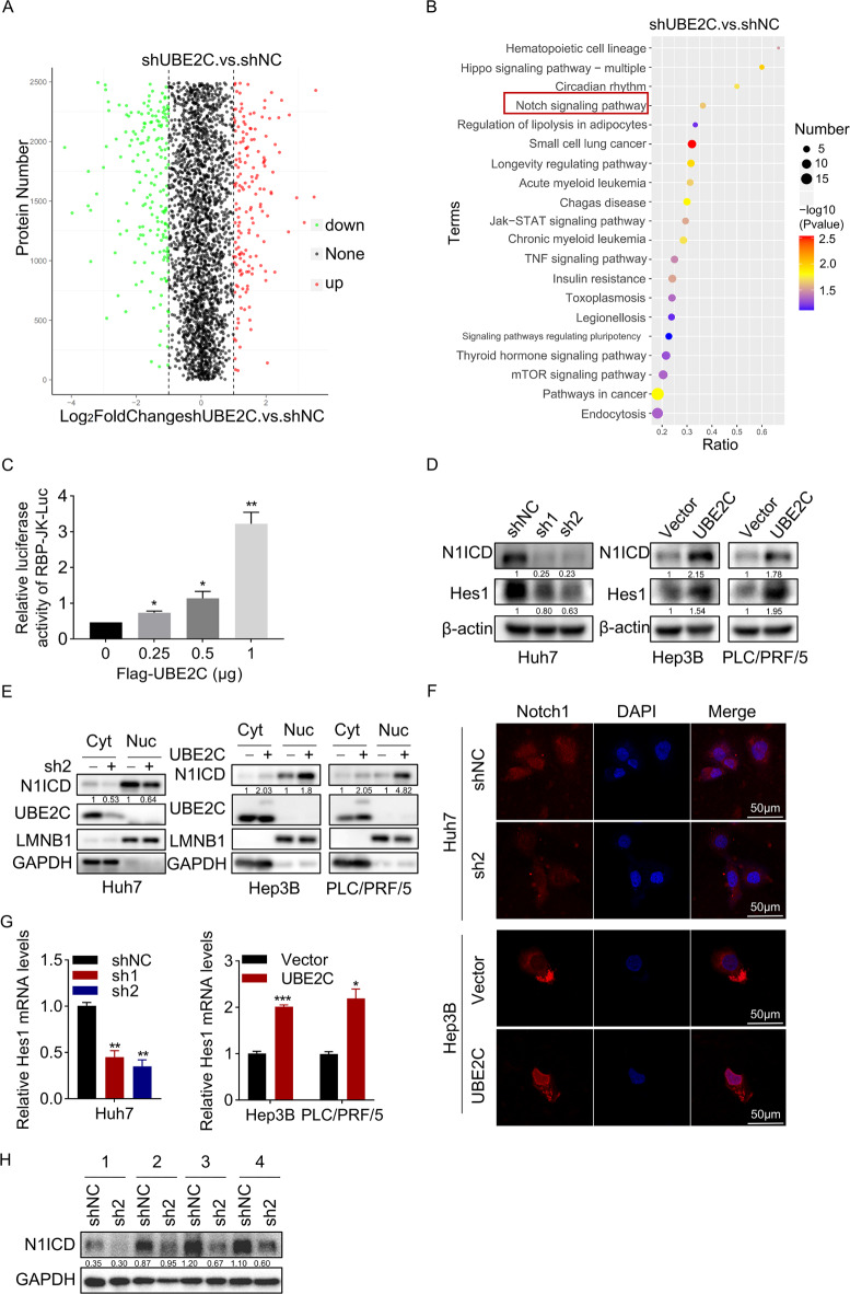
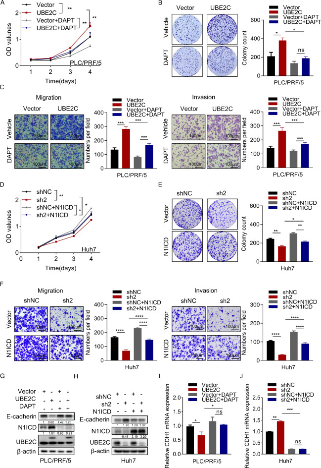
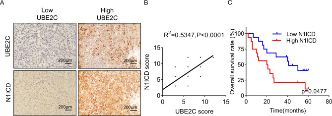
UBE2C, a ubiquitin-conjugating enzyme, functions as an oncogene in different types of human cancers. Nonetheless, the exact influence of UBE2C on the development of HCC via regulation of ubiquitination remains uncertain. Here, we found that UBE2C displayed elevated levels of expression in HCC and was associated with an unfavorable prognosis, as evidenced by the analysis of the TCGA database and the examination of clinical specimens. The role of UBE2C in HCC revealed its ability to promote the growth and metastasis of HCC. Mechanistically, UBE2C activated Notch signaling, as evidenced by the upregulation of N1ICD and Hes1, crucial components of the Notch pathway, and activation of the RBP-JK luciferase reporter by UBE2C. Finally, rescue experiments demonstrated that the oncogenic role of UBE2C was eliminated through treatment with the Notch inhibitor DAPT, while overexpression of N1ICD alleviated the anticarcinogenic impact of knockdown of UBE2C. Altogether, the results of our study indicate that UBE2C plays a role in the activation of Notch signaling and could potentially serve as a viable target for therapeutic interventions in HCC.

Keywords: EMT; Notch signaling; Ubiquitination.


Conflict of interest statement

The authors declare no competing interests.

Figures

Fig. 1
Fig. 1
UBE2C is an indicator of poor prognosis of HCC patients. (A) The mRNA level of UBE2C in 48 hepatocellular carcinoma (HCC) tissues was compared to that in corresponding non-tumor tissues using RT-PCR. (B) The expression level of UBE2C mRNA in paired HCC and non-tumor tissues was obtained from the TNMplot database. (C) The expression level of UBE2C mRNA in unpaired HCC and normal tissues was obtained from the TNMplot database. (D) The protein expression levels of UBE2C in 30 paired HCC tumor (T) and adjacent normal (N) tissues were determined through western blot analysis. (E) The levels of UBE2C protein in 82 paired HCC tissues and adjacent normal tissues were assessed using immunohistochemistry. Scale bar, 200 μm. (F) Kaplan-Meier representation of the overall survival and disease-free survival of two groups of patients with high (n = 43) or low (n = 39) UBE2C expression in HCC tissues. Statistical analysis was conducted using the log-rank test. (G) Kaplan-Meier representation of the overall survival of two groups of patients from the TCGA database with high or low UBE2C expression in HCC tissues. Split patients by upper tertile. Statistical analysis was conducted using the log-rank test (****p < 0.0001).
Fig. 2
Fig. 2
UBE2C promotes HCC growth. (A) Huh7 and PLC/PRF/5 cells were transduced with lentiviral particles carrying shRNAs specifically targeting UBE2C. The efficiency of knockdown was assessed through western blot analysis. (B) The impact of UBE2C knockdown on cell proliferation was evaluated using the CCK-8 assay in Huh7 and PLC/PRF/5 cells. (C) The influence of UBE2C knockdown on colony formation was examined in Huh7 and PLC/PRF/5 cells. (D) PLC/PRF/5 and Hep3B cells were transduced with lentiviral particles expressing either the vector or UBE2C-Flag (containing 3×Flag tags). The effectiveness of overexpression was determined by western blot analysis. (E) The effects of UBE2C overexpression on cell proliferation were assessed using the CCK-8 assay in PLC/PRF/5 and Hep3B cells. (F) The impact of UBE2C overexpression on colony formation was examined in PLC/PRF/5 and Hep3B cells. (G) Control and UBE2C knockdown Huh7 cells were subcutaneously injected into nude mice to observe tumor growth (*p < 0.05, **p < 0.01, ***p < 0.001, ****p < 0.0001).
Fig. 3
Fig. 3
UBE2C promotes HCC metastasis. (A) The impact of UBE2C knockdown on cellular migration and invasion was assessed using the transwell assay in Huh7 and PLC/PRF/5 cells. Cell quantification was conducted under a microscope in five randomly chosen fields. Scale bar, 100 μm. (B) The influence of UBE2C overexpression on cellular migration and invasion was examined through the transwell assay in PLC/PRF/5 and Hep3B cells. Cell quantification was performed under a microscope in five randomly selected fields. Scale bar, 100 μm. (C) The accompanying visuals depict pulmonary metastatic lesions formed by control and UBE2C knockdown Huh7 cells. The quantity and size of lung metastatic tumors in each group were measured. Scale bar, 50 μm and 100 μm ( ***p < 0.001, ****p < 0.0001).
Fig. 4
Fig. 4
UBE2C induces EMT in HCC cells. (A) CDH1 mRNA expression was evaluated through RT-PCR in control HCC cells and those with UBE2C knockdown or overexpression. (B) The impact of UBE2C knockdown or overexpression on the expression of EMT markers (E-cadherin, ZO-1, and Vimentin) was analyzed using western blot. (C) Immunofluorescence images depicting the levels of E-cadherin, ZO-1, and Vimentin in HCC cells with UBE2C knockdown or overexpression. Scale bar, 50 μm. (D) The expression of EMT markers (E-cadherin, ZO-1, and Vimentin) in xenograft tumors formed by control Huh7 cells and those with UBE2C knockdown (*p < 0.05, **p < 0.01).
Fig. 5
Fig. 5
UBE2C activates Notch signaling. (A) The levels of proteins exhibiting differential ubiquitination, regulated by UBE2C, were examined through UbiScan ubiquitination proteomics analysis. (B) KEGG pathway analysis was performed on the UbiScan ubiquitination proteomics data. (C) The activity of the Notch signaling reporter (RBP-JK-Luc) was assessed using a dual luciferase reporter assay after transfecting varying doses of the UBE2C overexpression plasmid into HEK293T cells. (D) Western blot analysis was conducted to examine the expression of Hes1 and N1ICD in HCC cells with either UBE2C knockdown or overexpression. (E) The subcellular localization of N1ICD was assessed in HCC cells with UBE2C knockdown or overexpression. LMNB1 served as the internal control for nuclear protein, while GAPDH served as the internal control for cytoplasmic protein. (F) Immunofluorescence staining was performed to visualize Notch1 in HCC cells with UBE2C knockdown or overexpression. Scale bar, 50 μm. (G) RT-PCR was used to assess the expression of Hes1 mRNA in HCC cells with UBE2C knockdown or overexpression. (H) The expression of N1ICD was evaluated in xenograft tumors formed by control and UBE2C knockdown Huh7 cells (*p < 0.05, **p < 0.01, ***p < 0.001).
Fig. 6
Fig. 6
UBE2C exerts oncogenic effects through Notch pathway. (A) Control and UBE2C-overexpressing PLC/PRF/5 cells were subjected to treatment with either the vehicle or DAPT (25 µM). Subsequently, a CCK-8 assay was employed to assess cellular proliferation.(B) The colony formation ability of control and UBE2C-overexpressing PLC/PRF/5 cells, following treatment with either the vehicle or DAPT (25 µM). (C) Control and UBE2C-overexpressing PLC/PRF/5 cells were exposed to treatment with either the vehicle or DAPT (25 µM) for a duration of 48 h. Subsequently, a transwell assay was conducted to evaluate cellular migration and invasion. Scale bar, 100 μm. (D) Control and UBE2C-knockdown Huh7 cells were transiently transferred with lentiviral particles expressing either the vector or Flag-N1ICD for 48 h. Subsequently, a CCK-8 assay was employed to assess cellular proliferation. (E) The colony formation ability of control and UBE2C-knockdown Huh7 cells, following transiently transferred with the vector or Flag-N1ICD plasmids for 48 h. (F) Control and UBE2C-knockdown Huh7 cells were transiently transferred with lentiviral particles expressing either the vector or Flag-N1ICD for 48 h. Subsequently, a transwell assay was conducted to evaluate cellular migration and invasion. Scale bar, 100 μm. (G) Control and UBE2C-overexpressing PLC/PRF/5 cells were treated with either the vehicle or DAPT (25 µM) for a duration of 48 h. Following this, a western blot analysis was performed to examine the expression levels of N1ICD and E-cadherin. (H) Control and UBE2C-knockdown Huh7 cells were transiently transferred with lentiviral particles expressing either the vector or Flag-N1ICD for 48 h. Following this, a western blot analysis was performed to examine the expression levels of N1ICD and E-cadherin. (I) Control and UBE2C-overexpressing PLC/PRF/5 cells were treated with either the vehicle or DAPT (25 µM) for a duration of 48 h. Subsequently, an RT-PCR analysis was conducted to assess the expression of CDH1. (J) Control and UBE2C-knockdown Huh7 cells were transiently transferred with lentiviral particles expressing either the vector or Flag-N1ICD for 48 h. Subsequently, an RT-PCR analysis was conducted to assess the expression of CDH1. (*p < 0.05, **p < 0.01, ***p < 0.001,****p < 0.0001).
Fig. 7
Fig. 7
Correlation between UBE2C and N1ICD in clinical samples. (A) Representative immunohistochemical images stained with UBE2C and N1ICD in HCC tissues. Scale bar, 200 μm. (B) The analysis of the correlation between UBE2C and N1ICD expression in 32 HCC tissues based on IHC scores. (C) Kaplan-Meier representation of the overall survival of two groups of patients with high (n = 16) or low (n = 16) N1ICD expression in HCC tissues. Statistical analysis was conducted using the log-rank test.

References

    1. Siegel, R. L., Miller, K. D. & Jemal, A. Cancer statistics, 2020. CA Cancer J. Clin.70, 7–30. 10.3322/caac.21590 (2020). - PubMed
    1. Ryu, S. H., Jang, M. K., Kim, W. J., Lee, D. & Chung, Y. H. Metastatic tumor antigen in hepatocellular carcinoma: golden roads toward personalized medicine. Cancer Metastasis Rev.33, 965–980. 10.1007/s10555-014-9522-4 (2014). - PubMed
    1. Lambert, A. W., Pattabiraman, D. R. & Weinberg, R. A. Emerging biological principles of metastasis. Cell168, 670–691. 10.1016/j.cell.2016.11.037 (2017). - PMC - PubMed
    1. Rape, M. Ubiquitylation at the crossroads of development and disease. Nat. Rev. Mol. Cell Biol.19, 59–70. 10.1038/nrm.2017.83 (2018). - PubMed
    1. Hu, H. & Sun, S. C. Ubiquitin signaling in immune responses. Cell Res.26, 457–483. 10.1038/cr.2016.40 (2016). - PMC - PubMed

MeSH terms