. 2024 Oct 1;8(1):216.
doi: 10.1038/s41698-024-00725-4.

The role of TERT C228T and KDM6A alterations and TME in NMIBC treated with BCG

Affiliations

The role of TERT C228T and KDM6A alterations and TME in NMIBC treated with BCG

Qi-Dong Xia et al. NPJ Precis Oncol. .

Abstract

We aimed to investigate the genomic and tumor microenvironmental (TME) profiles in non-muscle invasive bladder cancer (NMIBC) and explore potential predictive markers for Bacillus Calmette-Guérin (BCG) treatment response in high-risk NMIBC patients (according to European Association of Urology (EAU) risk stratification). 40 patients with high-risk NMIBC (cTis-T1N0M0) who underwent en bloc resection followed by BCG instillation were retrospectively enrolled. Surgical samples were subjected to Next Generation Sequencing (NGS) and multiplex immunofluorescence (mIF) assay. Genomic profiling revealed high prevalences of alterations in TERT (55%), KDM6A (32.5%), FGFR3(30%), PIK3CA (30%), TP53(27.5%) and ARID1A (20%). TME analysis showed different proportions of macrophages, NK cells, T cells subsets in tumoral and stromal compartment. Multivariate analysis identified TERT C228T and alteration in KDM6A as two independent factors associated with inferior RFS. The study comprehensively depicted the genomic and TME profiles in NMIBC and identified potential predictive biomarkers for BCG treatment.


Conflict of interest statement

The authors declare no competing interests.

Figures
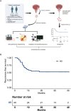
Fig. 1
Fig. 1. Overall study design and patient prognosis.
A Schematic diagram of the study flow. B The recurrence-free survival analysis for all patients with NMIBC in this cohort.
Fig. 2
Fig. 2. Genomic landscape of NMIBC in small Chinese cohort.
A An overview of the genomic landscape of NMIBC in small Chinese cohort. B Correlations of genomic alterations between these genes. C The percentage of patients with EP300 mutations and wildtype in different genders (female and male). D The percentage of patients with FGFR3 mutations and wildtype in different genders (female and male). E The percentage of patients with PIK3CA mutations and wildtype in different tumor grades (low, intermediate and high grade).
Fig. 3
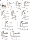
Fig. 3. The exploration of multidimensional biomarkers of the efficacy of postoperative BCG treatment.
A Comparison of TMB between patients in the recurrence and non-recurrence groups. B Survival analyses for patients in high and low TMB groups using Kaplan–Meier curves. C Survival analyses for patients in ARID1A mutations and wildtype groups using Kaplan–Meier curves. D Survival analyses for patients with ARID1A truncation and without ARID1A truncation using Kaplan–Meier curves. E Survival analyses for patients in ARID1B mutations and wildtype groups using Kaplan–Meier curves. F Survival analyses for patients in TERT mutations and wildtype groups using Kaplan–Meier curves. G Survival analyses for patients in TERT promoter mutations and wildtype groups using Kaplan–Meier curves. H Survival analyses for patients with TERTp C250T and without TERTp C250T using Kaplan–Meier curves. I Survival analyses for patients with TERTp C228T and without TERTp C228T using Kaplan–Meier curves. J Survival analyses for patients in KDM6A mutations and wildtype groups using Kaplan–Meier curves. K Survival analyses for patients in low and high HRD score groups using Kaplan–Meier curves.
Fig. 4
Fig. 4. Screening for prognosis-related genetic alterations after BCG treatment.
A Hazard ratio of HRD score, TERTp C228T and KDM6A mutation after multivariate Cox regression analysis (*p < 0.05). B Survival analyses for comparison between wild type and mutation groups (left) and comparison between co-mutation and single mutation groups (right) using Kaplan–Meier curves. The mutation groups showed significantly worse relapse free survival than wild type groups (p = 0.0027). C The percentage of patients with PIK3CA mutations and wildtype in different groups (wild type, single mutation and co-mutation). D Comparison of tumor purity between patients in different groups (wild type, single mutation and co-mutation). E Comparison of ploidy between patients in different groups (wild type, single mutation and co-mutation). F Comparison of HRD score between patients in different groups (wild type, single mutation and co-mutation).
Fig. 5
Fig. 5. The landscape of tumor microenvironment in bladder cancer.
A Relative abundance of different immune cell types in the parenchymal of tumor in different patients. B The percentage of different immune cell types in the parenchymal of tumor. C Relative abundance of different immune cell types in the stroma of tumor in different patients. D The percentage of different immune cell types in the stroma of tumor. E Comparison of the percentage of different markers in the stroma and tumor area. F Correlations of different immune cells between stroma and tumor area. G Survival analyses for high and low CD3+ cells infiltration in the tumor area using Kaplan–Meier curves. H Survival analyses for high and low CD56bright NK cells infiltration in the tumor area using Kaplan–Meier curves. I Survival analyses for high and low CD68+ cells infiltration in the stroma area using Kaplan–Meier curves.
Fig. 6
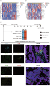
Fig. 6. Case report of a typical patient.
A Heatmap of different immune cells infiltration in patients grouped by KDM6A mutations and TERTp C228T in the tumor area. B Heatmap of different immune cells infiltration in patients grouped by KDM6A mutations and TERTp C228T in the stroma area. C Multiplex immunofluorescence images of immune cell infiltration in patient numbered RS21083210FFP.

References

    1. Babjuk, M. et al. European Association of Urology Guidelines on Non-muscle-invasive Bladder Cancer (Ta, T1, and Carcinoma in Situ). Eur. Urol.81, 75–94 (2022). - PubMed
    1. Antoni, S. et al. Bladder cancer incidence and mortality: a global overview and recent trends. Eur. Urol.71, 96–108 (2017). - PubMed
    1. Lobo, N. et al. Epidemiology, screening, and prevention of bladder cancer. Eur. Urol. Oncol.5, 628–639 (2022). - PubMed
    1. Li, Z., Zhou, Z., Cui, Y. & Zhang, Y. Systematic review and meta-analysis of randomized controlled trials of perioperative outcomes and prognosis of transurethral en-bloc resection vs. conventional transurethral resection for non-muscle-invasive bladder cancer. Int. J. Surg.104, 106777 (2022). - PubMed
    1. Lenis, A. T., Lec, P. M., Chamie, K. & Mshs, M. D. Bladder cancer: a review. Jama324, 1980–1991 (2020). - PubMed

LinkOut - more resources