. 2024 Nov;67(11):2481-2493.
doi: 10.1007/s00125-024-06274-6. Epub 2024 Oct 1.

Progression to type 1 diabetes in the DPT-1 and TN07 clinical trials is critically associated with specific residues in HLA-DQA1-B1 heterodimers

Affiliations

Progression to type 1 diabetes in the DPT-1 and TN07 clinical trials is critically associated with specific residues in HLA-DQA1-B1 heterodimers

Lue Ping Zhao et al. Diabetologia. 2024 Nov.

Abstract

Aims/hypothesis: The aim of this work was to explore molecular amino acids (AAs) and related structures of HLA-DQA1-DQB1 that underlie its contribution to the progression from stages 1 or 2 to stage 3 type 1 diabetes.

Methods: Using high-resolution DQA1 and DQB1 genotypes from 1216 participants in the Diabetes Prevention Trial-Type 1 and the Diabetes Prevention Trial, we applied hierarchically organised haplotype association analysis (HOH) to decipher which AAs contributed to the associations of DQ with disease and their structural properties. HOH relied on the Cox regression to quantify the association of DQ with time-to-onset of type 1 diabetes.

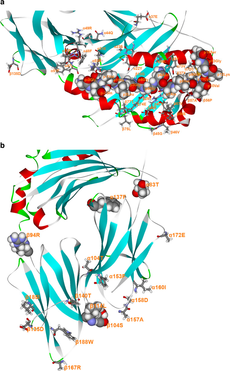
Results: By numerating all possible DQ heterodimers of α- and β-chains, we showed that the heterodimerisation increases genetic diversity at the cellular level from 43 empirically observed haplotypes to 186 possible heterodimers. Heterodimerisation turned several neutral haplotypes (DQ2.2, DQ2.3 and DQ4.4) to risk haplotypes (DQ2.2/2.3-DQ4.4 and DQ4.4-DQ2.2). HOH uncovered eight AAs on the α-chain (-16α, -13α, -6α, α22, α23, α44, α72, α157) and six AAs on the β-chain (-18β, β9, β13, β26, β57, β135) that contributed to the association of DQ with progression of type 1 diabetes. The specific AAs concerned the signal peptide (minus sign, possible linkage to expression levels), pockets 1, 4 and 9 in the antigen-binding groove of the α1β1 domain, and the putative homodimerisation of the αβ heterodimers.

Conclusions/interpretation: These results unveil the contribution made by DQ to type 1 diabetes progression at individual residues and related protein structures, shedding light on its immunological mechanisms and providing new leads for developing treatment strategies.

Data availability: Clinical trial data and biospecimen samples are available through the National Institute of Diabetes and Digestive and Kidney Diseases Central Repository portal ( https://repository.niddk.nih.gov/studies ).

Keywords: Amino acids; HLA; Immunogenetics; Islet autoimmunity; Progression; Seroconversion; Type 1 diabetes.


Figures

Fig. 1
Fig. 1
Fan-representation from phylogenic analysis of all heterodimeric HLA-DQ haplotypes based on similarity measurements between AA sequences of all individual haplotypes. A DQ haplotype is highlighted green or red if corresponding p values are <0.05 and HRs are ≥1, with red representing a risk haplotype and green representing a protective haplotype; a DQ haplotype is highlighted black if p values are ≥0.05 and the remaining DQ haplotypes are highlighted grey if they have fewer than ten copies. The brown arrow indicates the threshold for identifying a sub-phylogenic tree, resulting in clusters of heterodimeric DQ haplotypes. Note that all possible heterodimeric haplotypes are included, even though some may not be permissible
Fig. 2
Fig. 2
(a) Detailed view of the α1β1 domain of the HLA-DQ8-hybrid peptide insulin C-peptideIAPP GQVELGGGNAVEVCK (anchors in bold, p11C mutated to form a disulphide bond with mutated DQα72C) complex (from https://www.rcsb.org/structure/6XCP, accessed 29 March 2024), showing all residues involved in type 1 diabetes progression or prevention thereof, among participants of the DPT-1 and TN07 trials, as revealed by HOH. Included are all residues in absolute LD to the involved residues among all the HLA-DQs. Residue β135D is added for orientation purposes. Depiction convention: protein backbone is shown in flat ribbons (α-helix, red; β-sheet, turquoise; β-turn, green; random coil, grey); antigenic peptide is shown in space-filling mode, and DQ residues are shown in stick mode (carbon, grey; oxygen, red; nitrogen, purple; hydrogen, white; sulphur, yellow). It is quite significant, in our view, that most of the depicted DQ8α1β1 residues line up pockets 4, 6, 7 and 9, or potentially can make contact with cognate TCR residues. Residues β26L, β30Y, β38A, β52P, β55P and β71T are covered mostly by the antigenic peptide or other DQ residues, in this orientation, hence cannot be seen. (b) Detailed view of α2β2 domain of HLA-DQ8–insulin C-peptideIAPP hybrid peptide GQVELGGGNAVEVCK (anchors in bold) complex (from same source as above), in an orientation where the presumed interaction with CD4 would be such that the latter molecule has its long symmetry axis in a near vertical position. Residues α83T, α137F and β94R are in space-filling mode and shown only for orientation purposes; otherwise, the molecular depiction conventions are as in (a). The loop β105–113 is missing in the crystal structure, so the two immediately preceding residues, β104S and β114L, are shown in space-filling mode. Of the residues shown by HOH to be important for progression to type 1 diabetes or protection therefrom, plus those in absolute LD with the former, we note that α157 is involved in the putative cognate TCR-induced homodimerisation of MHC II molecules that promotes the binding of homodimer CD4 to such a complex, leading to T cell activation [10, 21]. In the respective β2 domain, residues β135 and β140 are part of the β134–148 stretch involved in CD4 binding, while residue β167R is part of the RGD loop found in all the known structures of HLA-DQ molecules possessing this tripeptide sequence. It is likely that residues involved in CD4 binding in the α2β2 domain, or others very close to them, interact with the inhibitory CD4 T cell membrane molecule LAG-3, which is inhibitory to the action of MHC II molecules [24]. Note that residues α172E and β167R have only partially determined crystallographic structure. Both figures drawn via the DSViewer Pro v.6 of Accelrys

References

    1. Patterson CC, Karuranga S, Salpea P et al (2019) Worldwide estimates of incidence, prevalence and mortality of type 1 diabetes in children and adolescents: Results from the International Diabetes Federation Diabetes Atlas, 9th edition. Diabetes Res Clin Pract 157:107842. 10.1016/j.diabres.2019.107842 - PubMed
    1. Katsarou A, Gudbjornsdottir S, Rawshani A et al (2017) Type 1 diabetes mellitus. Nat Rev Dis Primers 3:17016. 10.1038/nrdp.2017.16 - PubMed
    1. Skyler JS (2013) Primary and secondary prevention of Type 1 diabetes. Diabet Med 30(2):161–169. 10.1111/dme.12100 - PMC - PubMed
    1. Butty V, Campbell C, Mathis D, Benoist C, Group DPTS (2008) Impact of diabetes susceptibility loci on progression from pre-diabetes to diabetes in at-risk individuals of the diabetes prevention trial-type 1 (DPT-1). Diabetes 57(9):2348–2359. 10.2337/db07-1736 - PMC - PubMed
    1. Zhao LP, Skyler J, Papadopoulos GK et al (2022) Association of HLA-DQ heterodimer residues -18beta and beta57 with progression from islet autoimmunity to diabetes in the diabetes prevention trial-type 1. Diabetes Care 45(7):1610–1620. 10.2337/dc21-1628 - PMC - PubMed