. 2024 Oct 2;7(1):1239.
doi: 10.1038/s42003-024-06951-7.

Broadly potent spike-specific human monoclonal antibodies inhibit SARS-CoV-2 Omicron sub-lineages

Affiliations

Broadly potent spike-specific human monoclonal antibodies inhibit SARS-CoV-2 Omicron sub-lineages

Melanie R Walker et al. Commun Biol. .

Abstract

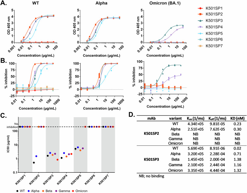
The continuous emergence of SARS-CoV-2 variants of concern has rendered many therapeutic monoclonal antibodies (mAbs) ineffective. To date, there are no clinically authorized therapeutic antibodies effective against the recently circulating Omicron sub-lineages BA.2.86 and JN.1. Here, we report the isolation of broad and potent neutralizing human mAbs (HuMabs) from a healthcare worker infected with SARS-CoV-2 early in the pandemic. These include a genetically unique HuMab, named K501SP6, which can neutralize different Omicron sub-lineages, including BQ.1, XBB.1, BA.2.86 and JN.1, by targeting a highly conserved epitope on the N terminal domain, as well as an RBD-specific HuMab (K501SP3) with high potency towards earlier circulating variants that was escaped by the more recent Omicron sub-lineages through spike F486 and E484 substitutions. Characterizing SARS-CoV-2 spike-specific HuMabs, including broadly reactive non-RBD-specific HuMabs, can give insight into the immune mechanisms involved in neutralization and immune evasion, which can be a valuable addition to already existing SARS-CoV-2 therapies.


Conflict of interest statement

The authors declare no competing interests.

Figures

Fig. 1
Fig. 1. Binding and competition of HuMabs specific for SARS-CoV-2 spike.
Seven HuMabs (each represented by a different color as shown in the key) were isolated from subject K501 and spike-specific binding by ELISA (A) and competition with ACE2 (B) were performed and inhibition (IC50) potency determined (C). HuMabs were sequenced and germline usage is shown in (D).
Fig. 2
Fig. 2. RBD-specific cross-reactive binding, inhibition and affinity of spike-specific HuMabs.
HuMabs K501SP1 – K501SP7 (each represented by a different color as shown in the key) were tested for binding (A) and competition with ACE2 (B) against the RBD of five SARS-CoV-2 variants (WT, Alpha and Omicron (BA.1), see Fig. S5 for Beta and Gamma). IC50 values were calculated for competition assays from technical triplicates over two separate experiments and are shown for all five variants in (C). Each variant tested is represented by a different color as shown in the key. Grey shading separates the different HuMabs and the dotted horizontal line shows the highest concentration of HuMab tested. Affinity was measured against each variant for HuMabs K501SP2 and K501SP3 as shown in (D) (see Fig. S6 for curve fitting). The VoCs that do not bind are represented as NB (no binding). Throughout all graphs, data is represented as individual data points.
Fig. 3
Fig. 3. Cross reactive neutralization of eight SARS-CoV-2 isolates by spike-specific HuMabs.
A HuMabs K501SP1 – K501SP7 and positive control Bebtelovimab (each represented by a different color as shown in the key) were tested for neutralization activity against ten SARS-CoV-2 isolates. B Calculated IC50’s are plotted with each SARS-CoV-2 isolate represented by a different color, as shown in the key. Grey shading separates the different HuMabs and the dotted horizontal line shows the highest concentration of HuMab tested. Positive control Bebtelovimab was tested at no greater than 10 µg/mL. Throughout all graphs, data is represented as individual data points.
Fig. 4
Fig. 4. Investigation into the epitope of K501SP6.
A The Cryo-EM structure of K501SP6 binding to BA.1 Omicron spike with protomer 1, 2, and 3 in light blue, yellow, and grey, respectively. The NTD is shown in green and K501SP6 heavy and light chains in purple and pink, respectively. B Model of K501SP6 Fab-bound NTD with the local refine electron density map outline shown. C Epitope-paratope interface of K501SP6 Fab (heavy chain in purple, and light chain in pink) and the NTD (green). Interacting residues between HumAb and NTD as estimated by LigPlot+ are shown. Oxygen and nitrogen atoms are depicted in red and blue, respectively. D Logo plot of the epitope based on the lineages used for the neutralization assays (WebLogo 3.7.11). Residues interacting with heavy and light chains are marked with a purple and pink bar, respectively.
Fig. 5
Fig. 5. Substitutions responsible for resistance to K501SP3.
A HuMab K501SP3 was tested for neutralization against a panel of recombinant (square) WT D614G (DK-AHH1) viruses carrying the RBD of Alpha, Delta and Omicron sub-lineages BA.1, BA.2 and BA.5. This was compared to non-recombinant (circle) viruses for each variant represented by a different color as shown in key. B Neutralization (IC50) of HuMab K501SP3 was also tested against a panel 4 recombinant viruses containing key BA.5 substitutions (represented by different colors). C Cryo-EM electron density map of spike trimer and HuMab K501SP3. Low-resolution is outlined and higher resolution is shown in full color. Spike trimer PDB model (PDB ID:7DK6) and the Alphafold model of K501SP3 Fab are docked into the electron density map. Protomer one, two, and three of the spike trimer are shown in blue, yellow, and grey respectively. RBDs are shown with orange arrows, Fab one is shown in purple and Fab two in red, with the heavy chain in the darker color. A zoom-in of the binding region is provided to the right where the RBM is shown in dark green and the substitutions responsible for abolishing BA.5 neutralization from K501SP3 compared to the WT are shown in lime.
Fig. 6
Fig. 6. Sequence alignments of HuMabs K501SP3 and K501SP6 with antibodies of the same gene usage.
A, B The sequences of antibodies that utilize the same immunoglobulin heavy- and light-chain variable region genes as HuMabs K501SP3 and K501SP6, respectively. The CDRH3 and CDRL3 regions are shown with a black line and labeled. Panel A shows the top 10/81 antibodies determined by sequence identity that had identical IGHV, IGHJ, IGLV, IGLJ gene usage and CDRH3 length as HuMab K501SP3. Therapeutic antibody Tixagevimab is also shown. Panel B shows antibodies with the same IGHV, IGLV and CDRH3 length or identical IGHV, IGHJ, IGLV, IGLJ gene usage but different CDRH3 length as HuMab K501SP6. See also Table S4 and S5. C The genetic features of EUA mAbs. Those which carry the same genetic features as K501SP3 or K501SP6 are highlighted in bold.

References

    1. Sohrabi, C. et al. World Health Organization declares global emergency: A review of the 2019 novel coronavirus (COVID-19). Int J. Surg.76, 71–76 (2020). - PMC - PubMed
    1. WHO. WHO Coronavirus (COVID-19) Dashboard https://covid19.who.int/2022
    1. WHO. Global excess deaths associated with COVID-19, January 2020 - December 2021. who.int; 2022 May 2022.
    1. WHO. Tracking SARS-CoV-2 variants https://www.who.int/activities/tracking-SARS-CoV-2-variants2022.
    1. Wibmer, C. K. et al. SARS-CoV-2 501Y.V2 escapes neutralization by South African COVID-19 donor plasma. Nat. Med27, 622–625 (2021). - PubMed

MeSH terms

Supplementary concepts