. 2025 Jan;20(1):132-143.
doi: 10.1038/s41565-024-01747-6. Epub 2024 Oct 1.

Combinatorial design of siloxane-incorporated lipid nanoparticles augments intracellular processing for tissue-specific mRNA therapeutic delivery

Affiliations

Combinatorial design of siloxane-incorporated lipid nanoparticles augments intracellular processing for tissue-specific mRNA therapeutic delivery

Lulu Xue et al. Nat Nanotechnol. 2025 Jan.

Abstract

Systemic delivery of messenger RNA (mRNA) for tissue-specific targeting using lipid nanoparticles (LNPs) holds great therapeutic potential. Nevertheless, how the structural characteristics of ionizable lipids (lipidoids) impact their capability to target cells and organs remains unclear. Here we engineered a class of siloxane-based ionizable lipids with varying structures and formulated siloxane-incorporated LNPs (SiLNPs) to control in vivo mRNA delivery to the liver, lung and spleen in mice. The siloxane moieties enhance cellular internalization of mRNA-LNPs and improve their endosomal escape capacity, augmenting their mRNA delivery efficacy. Using organ-specific SiLNPs to deliver gene editing machinery, we achieve robust gene knockout in the liver of wild-type mice and in the lungs of both transgenic GFP and Lewis lung carcinoma (LLC) tumour-bearing mice. Moreover, we showed effective recovery from viral infection-induced lung damage by delivering angiogenic factors with lung-targeted Si5-N14 LNPs. We envision that our SiLNPs will aid in the clinical translation of mRNA therapeutics for next-generation tissue-specific protein replacement therapies, regenerative medicine and gene editing.


Conflict of interest statement

Competing interests: L.X. and M.J.M. are inventors on a patent filed by the Trustees of the University of Pennsylvania (International Patent Application No. PCT/US23/66564) describing the lipid nanoparticle technology in this study. J.M.W. is a paid advisor to and holds equity in iECURE, Passage Bio and the Center for Breakthrough Medicines (CBM). He also holds equity in the former G2 Bio asset companies and Ceva Santé Animale. He has sponsored research agreements with Alexion Pharmaceuticals, Amicus Therapeutics, CBM, Ceva Santé Animale, Elaaj Bio, FA212, Foundation for Angelman Syndrome Therapeutics, former G2 Bio asset companies, iECURE and Passage Bio, which are licensees of Penn Technology. J.M.W., L.W. and C.C.W are inventors on patents that have been licensed to various biopharmaceutical companies and for which they may receive payments. D.W. is named on patents that describe the use of nucleoside-modified mRNA as a platform to deliver therapeutic proteins and vaccines. M.J.M., D.W. and M.-G.A. are also named on patents describing the use of lipid nanoparticles and lipid compositions for nucleic acid delivery. The other authors declare no competing interests.

Figures

Extended Data Fig. 1 |
Extended Data Fig. 1 |. Formulation parameters and characterization of SiLNPs.
a, SiLNPs formulation parameters. Siloxane-incorporated lipidoids, DOPE, cholesterol, and C14PEG2K with molar ratio of 35%, 16%, 46.5%, and 2.5% were used for SiLNPs formulation. b, Representative cryogenic transmission electron microscopy (cryo-TEM) image of SiLNP morphology. Scale bar: 100 nm. c, Hydrodynamic size distribution of representative SiLNP.
Extended Data Fig. 2 |
Extended Data Fig. 2 |. Blood chemistry evaluation of mice after administration of Si6-C14b LNP co-delivering Cas9 mRNA and TTR sgRNA.
(a) AST, (b) ALT, (c) BUN, and (d) Creatinine levels of blood samples obtained from mice treated with PBS and Si6-C14b LNP (RNA dose: 3 mg kg−1). Data are presented as mean ± s.e.m. (n = 3 mice for PBS treated groups; n =4 for LNP treated groups).
Fig. 1 |
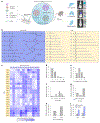
Fig. 1 |. A combinatorial library of siloxane-incorporated ionizable lipids with tunable structures for tissue-specific mRNA delivery.
a, SiLNPs were formulated using a microfluidic mixing device each with a siloxane-incorporated lipidoid, helper lipid (DOPE), cholesterol and PEG-lipid (C14PEG2K). The resulting SiLNPs with different siloxane-incorporated lipidoid structures mediate in vivo tissue-specific mRNA delivery to the liver, lungs and spleen. b, Structures of the 12 siloxane amines and 21 alkyl tails used for combinatorial design and synthesis of the 252 siloxane-incorporated lipidoids. c, A heat map of luciferase expression following treatment of HepG2 cells with SiLNPs (5,000 cells, 10 ng luciferase mRNA, n = 3 biological independent samples). Hits were defined as siloxane-incorporated lipidoids with relative luminescence units greater than 200. d, Relative hit rate of SiLNPs by the number of silicon atoms per siloxane-incorporated lipidoid. e, Relative hit rate of SiLNPs by tail substitution number. f, Relative hit rate of SiLNPs by tail length. g, Relative hit rate of SiLNPs by tail type (epoxide-, ester- and amide bond-based tails). h, Relative hit rate of SiLNPs by siloxane amine core morphology among the core morphology-associated formulations. i, Relative hit rate of SiLNPs with and without the incorporation of sulfur atoms into the starting siloxane amines. Adding sulfur substantially enhanced in vitro mRNA delivery efficacy. 2Si-X represents siloxane-incorporated lipidoids with 2 Si atoms and 1 amine group (Si1- versus Si2-), and X-2Si-X represents siloxane-incorporated lipidoids with 2 Si atoms and 2 amine groups (Si5- versus Si6-).
Fig. 2 |
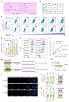
Fig. 2 |. Siloxane moiety incorporation improves cellular internalization and endosomal escape.
a, Chemical structures and ALog P value of Si5-N14 and 213-N14 lipidoids. ALog P was predicted from atomic physiochemical properties. b, Size, mRNA EE and zeta potential (ζ) of Si5-N14 and 213-N14 LNPs formulated with Cy5-tagged mRNA. c, Representative gating strategy for identifying Cy5-tagged mRNA-LNPs endocytosed by immortalized human lung microvascular endothelial cells (iMVECs). d, Cy5+ iMVECs treated with Si5-N14 and 213-N14 LNPs encapsulating Cy5-tagged mRNA. e, Cy5 MFI of iMVECs treated with Si5-N14 and 213-N14 LNPs at different post-treatment time points. iMVECs were treated with Si5-N14 and 213-N14 LNPs delivering Cy5-tagged mRNA at an mRNA dose of 200 ng ml−1. f, Relative fluorescence intensity versus post-treatment time demonstrated not only faster but also greater endocytosis of Si5-N14 LNPs than 213-N14 LNPs. Curves were calculated from e. g, Schematic illustrating differences in lipid packing and the effect on membrane fluidity. The radii of the amine heads for the Si5-N14 and 213-N14 lipidoids were calculated based on molecular dynamic simulations. Incorporation of the siloxane domain increases the radius of the amine head, which may result in looser lipid packing for improved membrane fluidity for nucleic acid delivery. h, Membrane fluidity (1/P) of Si5-N14 and 213-N14 LNPs was measured by fluorescence polarization. i, Representative confocal laser scanning microscopy images of cellular uptake and endosomal escape of Si5-N14 and 213-N14 LNPs. iMVECs cells were treated with Cy5-tagged mRNA-LNPs (mRNA dose 600 ng ml−1) for 3 h before staining with LysoTracker Green and Hoechst 33342. Scale bars, 50 μm. j,k, Haemolysis of Si5-N14 and 213-N14 LNPs at pH 5.5 (j) and 7.4 (k). Red blood cells (RBCs) were incubated with LNPs at 37 °C for 1 h before the supernatant was transferred into a clear bottom 96-well plate (insert pictures) to determine the adsorption at 540 nm. Statistical significance in d, h, j and k was calculated using an unpaired Student’s t-test. ****P < 0.0001; ***P < 0.001; *P < 0.05; P > 0.05, not significant. Data are presented as mean ± s.e.m. (d,f,h,j,k, n = 3 biological independent samples).
Fig. 3 |
Fig. 3 |. In vivo structure–activity studies of siloxane-incorporated lipidoid formulations for mRNA delivery and organ selectivity to the liver, lungs and spleen.
a, In vivo evaluation of 36 representative SiLNPs encapsulating FLuc mRNA (dose 0.25 mg kg−1). Representative bioluminescence IVIS images of various organs 6 h after i.v. injection of SiLNPs to C57BL/6J mice. H, heart; Li, liver; S, spleen; Lu, lungs; K, kidneys. bd, Quantified luciferase expression in the liver (b), lungs (c) and spleen (d) from the 36 representative SiLNPs. The pie charts in bd represent in vivo organ specificity for the top-performing liver-, lung- and spleen-targeted SiLNP formulations. ej, Tissue-specific hit rates. Hits were defined as LNPs that enabled luminescence intensity greater than 106 p s−1 (total flux). Hit rate by tail length for the liver (e), lungs (f) and spleen (g). Hit rate by tail substitution number for the liver (h), lungs (i) and spleen (j). Note that LNP formulations with a tail substitution number of 8 did not generate lungs and spleen hits. Data are presented as mean ± s.e.m. (n = 3 mice).
Fig. 4 |
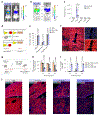
Fig. 4 |. Liver-targeted mRNA delivery and CRISPR–Cas9 gene editing by SiLNPs.
a, Whole body imaging of luciferase expression by Si6-C14b and MC3 LNPs 6 h post-injection (FLuc mRNA, 0.15 mg kg−1, n = 3 mice). b, Ex vivo imaging of luciferase expression in organs from mice depicted in a (n = 3 mice). H, heart; Li, liver; S, spleen; Lu, lungs; K, kidneys. c, Quantification of luciferase expression in organs from b (n = 3 mice). d, Schematic of the Ai14 mouse model, which demonstrates that tdTomato expression is induced upon intracellular Cre mRNA delivery for excision of the stop cassette. e, Quantification of the percentage of tdTomato+ cells from PBS-, MC3 LNP- and Si6-C14b LNP-treated Ai14 mice via flow cytometry (Cre mRNA, 0.3 mg kg−1, n = 3 mice). LSECs, liver sinusoidal endothelial cells. f, Representative immunostaining of liver histology shows tdTomato fluorescence. DAPI was used for nuclear staining. Vascular endothelial cadherin (VECad) was used for labelling LSECs. Scale bar, 100 μm. g, Schematic representation of CRISPR–Cas9 gene editing of transthyretin amyloidosis (TTR). C57BL/6J mice were systemically injected with a single dose of LNPs co-formulated with Cas9 mRNA and TTR sgRNA (wt/wt, 4/1). LNPs co-delivering Cas9 mRNA/scrambled sgRNA control were used as negative controls. Serum was collected 1 day before and 7 days post-injection. h, Serum TTR concentration in mice following treatment from g. i, TTR on-target indel frequency in the liver following treatment from g (h,i, n = 3 mice for the Si6-C14b LNP control group; n = 5 mice for the MC3 and Si6-C14b LNP editing group). j, Reduction of TTR transcript was visualized by in situ hybridization of liver sections from mice treated with PBS or Si6-C14b LNPs encapsulating Cas9 mRNA and TTR sgRNA. Scale bars, 200 μm. Statistical significance in c was calculated using an unpaired Student’s t-test. Statistical significance in e, h and i was calculated using one-way ANOVA, followed by Dunnett’s multiple comparison test. ****P < 0.0001; ***P < 0.001; **P < 0.01. Data are presented as mean ± s.e.m.
Fig. 5 |
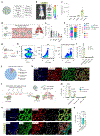
Fig. 5 |. Lung-targeted mRNA delivery and CRISPR–Cas9 gene editing by SiLNPs.
a, Characterization of the Si5-N14 LNP formulated with FLuc mRNA. b, Luciferase expression imaging from Si5-N14 LNPs 6 h post-injection (FLuc mRNA, 0.3 mg kg−1). H, heart; Li, liver; S, spleen; Lu, lungs; K, kidneys. c, Quantification of luciferase expression in organs from mice depicted in b. d, Schematic representation of the interaction of Si5-N14 LNPs with proteins in blood vessels. e, Quantification of the top five proteins in the corona of the Si5-N14 LNP. Vtn, vitronectin; Alb, serum albumin; Apob, apolipoprotein B-100; C3, complement C3; Hbb-b1, haemoglobin subunit beta-1. f, The top 20 most abundant corona proteins were categorized by molecular weight and isoelectric point. g, Ai14 mice were treated with Si5-N14 LNPs formulated with Cre mRNA for 3 days before analysis (Cre mRNA, 0.3 mg kg−1). h, Representative gating strategy to identify tdTomato+ ECs (CD45/CD31+/tdTomato+). i, Percentage of tdTomato+ cells in the lung by flow cytometry. j, Distribution of tdTomato+ cells in each cell type. k, Representative immunostaining demonstrating substantial co-localization of tdTomato+ cells and an EC marker, platelet endothelial cell adhesion molecule 1 (PECAM1). DAPI was used for nuclear staining. Scale bars, 50 μm. l, Schematic demonstration of in vivo gene editing in the lungs of transgenic GFP mice treated with Si5-N14 LNPs co-formulated with Cas9 mRNA and GFP sgRNA (4 injections, RNA dose 0.5 mg kg−1 per injection). PBS or Si5-N14 LNPs co-delivering Cas9 mRNA/scrambled sgRNA were used as negative controls. m, Quantification of the percentage of GFP cells in the lungs by flow cytometry. n, Representative immunostaining showed GFP knockout in lung ECs. DAPI was used for staining nuclei. PECAM1 was used for labelling ECs. ERG was used for staining EC nuclei. Scale bars, 50 μm. o, RT–qPCR analysis of GFP in sorted ECs. Statistical significance in i was calculated using an unpaired Student’s t-test. Statistical significance in m and o was calculated using one-way ANOVA, followed by Dunnett’s multiple comparison test. ****P < 0.0001; ***P < 0.001; **P < 0.01; *P < 0.05. Data are presented as mean ± s.e.m. (n = 3 mice).
Fig. 6 |
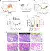
Fig. 6 |. Lung-targeted SiLNPs for efficient vascular repair.
a, Schematic illustration of endothelial repair for lung recovery through LNP-mediated delivery of mRNA encoding angiogenic factors in a viral infection lung damage model. b, Schematic timeline for LNP administration and sampling. Influenza virus A/H1N1/PR/8 was administered intranasally at 50–60 TCID50 units to female C57BL/6J mice. After injection, mice were treated with control (PBS or FLuc mRNA Si5-N14 LNPs, n = 3 mice) or FGF-2 mRNA Si5-N14 LNPs (0.5 mg kg−1, n = 4 mice) on day 15, and lungs were collected on day 25. Dexamethasone-21-phosphate (DEX) was administered intraperitoneally (i.p., 2 mg kg−1) to mice 30 min before LNP administration. c,d, Time course changes in weight loss (c) and capillary oxygen saturation (d) were observed in virus-infected C57BL/6J mice treated with either control (PBS or FLuc mRNA Si5-N14 LNPs) or FGF-2 mRNA Si5-N14 LNPs. e, Analysis of body weight and blood oxygen levels on day 25 after treatment with either control (PBS or FLuc mRNA Si5-N14 LNPs) or FGF-2 mRNA Si5-N14 LNPs to lung-damaged mice. f, Histological changes in the lungs of mice after receiving control (PBS or FLuc mRNA Si5-N14 LNPs) or FGF-2 mRNA Si5-N14 LNPs 25 days after infection. White areas in H&E stained sections are pulmonary alveoli, airway and large vessels, while dark spots represent the nuclei. Accumulated dark regions indicate large amounts of immune cell infiltration, leading to damaged inflammatory areas. Scale bars, 100 μm. Statistical significance in e was calculated using one-way ANOVA, followed by Dunnett’s multiple comparison test. *P < 0.05; P > 0.05, not significant. Data are presented as mean ± s.e.m.

References

    1. Chaudhary N, Weissman D & Whitehead KA mRNA vaccines for infectious diseases: principles, delivery and clinical translation. Nat. Rev. Drug Discov 20, 817 (2021). - PMC - PubMed
    1. Pardi N, Hogan MJ, Porter FW & Weissman D mRNA vaccines—a new era in vaccinology. Nat. Rev. Drug Discov 17, 261 (2018). - PMC - PubMed
    1. Sahin U, Karikó K & Türeci O mRNA-based therapeutics—developing a new class of drugs. Nat. Rev. Drug Discov 13, 759 (2014). - PubMed
    1. Mendes BB et al. Nanodelivery of nucleic acids. Nat. Rev. Methods Prim 2, 24 (2022). - PMC - PubMed
    1. Pastor F et al. An RNA toolbox for cancer immunotherapy. Nat. Rev. Drug Discov 17, 751 (2018). - PubMed

LinkOut - more resources