. 2024 Oct 1;25(1):63.
doi: 10.1186/s12865-024-00656-6.

Inhibition of CEACAM1 expression in cytokine-activated neutrophils using JAK inhibitors

Affiliations

Inhibition of CEACAM1 expression in cytokine-activated neutrophils using JAK inhibitors

Haruki Matsumoto et al. BMC Immunol. .

Abstract

Objectives: Carcinoembryonic-antigen-related cell-adhesion molecule 1 (CEACAM1) is an adhesion molecule that acts as a coinhibitory receptor in the immune system. We previously demonstrated that CEACAM1 is predominantly expressed on peripheral blood neutrophils in patients with RA. The aim of the present study was to investigate the effects of Janus kinase inhibitors (JAKi) on cytokine-activated human neutrophils and CEACAM1 expression.

Methods: Peripheral blood neutrophils were obtained from healthy subjects. Isolated neutrophils were stimulated with tumor necrosis factor-alpha (TNF-α) or granulocyte-macrophage colony-stimulating factor (GM-CSF) in the presence or absence of JAKi. The expression of CEACAM1 in peripheral blood neutrophils was analyzed by flow cytometry. Protein phosphorylation of signal transducer and activator of transcription (STAT)1, STAT3, and STAT5 was assessed by western blot using phospho-specific antibodies.

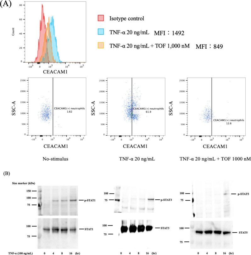
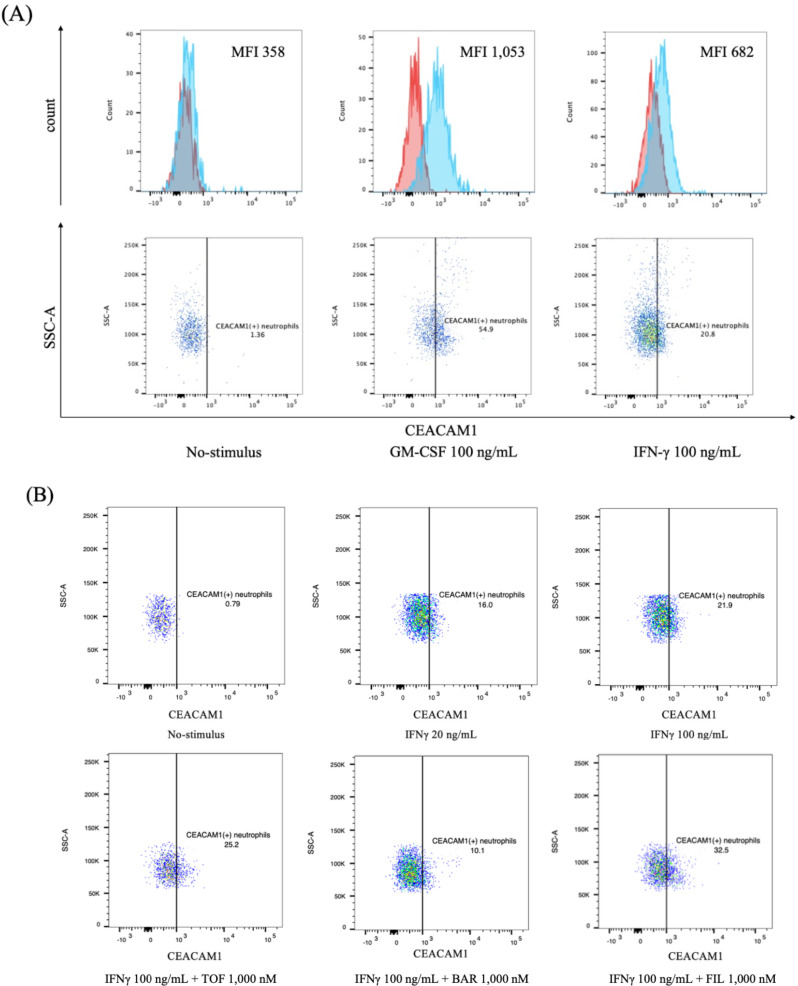
Results: We found that TNF-α-induced CEACAM1 expression was marginally suppressed after pretreatment with pan-JAK inhibitor, tofacitinib. Moreover, TNF-α induced STAT1 and STAT3 phosphorylation at the late stimulation phase (4 to 16 h). The expressions of CEACAM1 on neutrophils were markedly up-regulated by GM-CSF not by interleukin (IL)-6 stimulation. All JAKi inhibited GM-CSF-induced CEACAM1 expressions on neutrophils, however, the inhibitory effects of baricitinib were larger compared to those of tofacitinib or filgotinib. Moreover, CEACAM1 was marginally upregulated in interferon (IFN)-γ stimulated neutrophils. Similarly, JAKi inhibited IFN-γ-induced CEACAM1 expressions on neutrophils.

Conclusions: We demonstrated that JAKi prevent GM-CSF-induced CEACAM1 expression in neutrophils, and JAKi-induced inhibition depends on their selectivity against JAK isoforms. These findings suggest that JAKi can modulate the expression of CEACAM1 in cytokine-activated neutrophils, thereby limiting their activation.

Keywords: Carcinoembryonic-antigen-related cell-adhesion molecule 1; Granulocyte-macrophage colony-stimulating factor neutrophils; Janus kinase inhibitors; Rheumatoid arthritis; Tumor necrosis factor-alpha.


Conflict of interest statement

KM has received research grants from Chugai, Pfizer, AbbVie, and Eli Lilly. The rest of the authors declare that they have no competing interests.

Figures

Fig. 1
Fig. 1
TNF-α induces CEACAM1 expression, and STAT 1 and STAT3 phosphorylation at the late stimulation phase in neutrophils. (A) Flow cytometry overlay histograms for CEACAM1 expression on CD14(-)CD16(+) neutrophils. Representative histograms showing the percentage of CEACAM1(+) neutrophils stimulated with TNF-α with or without tofacitinib pretreatment. CEACAM1 expression was detected using anti–CEACAM1 antibody (blue shaded histogram) or isotype control antibody (red shaded histograms). There were marginal reductions in the number of CEACAM1(+) neutrophils by pretreatments with tofacitinib (orange shaded histograms). Data are representative of three independent experiments. (B) Neutrophil lysated were analyzed by western blot using anti-phosphatase STATs (pSTATs) (pSTAT 1, 3, and 5 at upper panel) or anti-STATs (STAT 1, 3, and 5 at lower panel). TNF-α induces STAT1 and STAT3 phosphorylation in neutrophils after 8 h from the stimulation with TNF-α. Whereas TNF-α induced STAT5 phosphorylation was barely observed after TNF-α stimulation. Data are representative of three independent experiments. Full-length blots are presented in Supplemental Fig. 2
Fig. 2
Fig. 2
A GM-CSF induces of CEACMA1 expression on neutrophils in a dose-dependent manner, and JAKi inhibit GM-CSF-induced CEACAM1 expressions on neutrophils. (A) CEACAM1 expressions on neutrophils were assessed by flow cytometry. Neutrophils were stimulated with the indicated concentrations of GM-CSF. Flow cytometry overlay histograms for CEACAM1 expression on neutrophils in each culture condition were demonstrated. Data are representative of three independent experiments. (B) Neutrophils were pretreated with JAKi (tofacitinib, baricitinib, upadacitinib) at the indicated concentrations for 1 h and then stimulated with GM-CSF (100 ng/ml) for 24 h. CEACAM1 expressions on neutrophils were assessed by flow cytometry. Percentage of CEACAM1-positive neutrophils were evaluated by flow cytometry for CEACAM1 (percentage of positive cells) in GM-CSF-stimulated neutrophils pretreated with the indicated concentrations of JAKi (tofacitinib, baricinib and filgotinib). Statistics were performed using Kruskal-Wallis test. Two independent experiments were combined. Values represent the mean ± SD of two independent experiments
Fig. 3
Fig. 3
JAKi inhibited IFN-γ-induced CEACAM1 expressions on neutrophils. (A) Neutrophils were stimulated with the indicated concentrations of GM-CSF or IFN-γ and CEACAM1 expressions on neutrophils were assessed by flow cytometry. CEACAM1 expression was induced by IFN-γ, however its induction levels were lower compared to those by GM-CSF. (B) Neutrophils were pretreated with JAKi (tofacitinib, baricitinib, upadacitinib) at the indicated concentrations for 1 h and then stimulated with IFN-γ (100 ng/ml) for 24 h. CEACAM1 expressions on neutrophils were assessed by flow cytometry. The percentage of CEACAM1-positive neutrophils was evaluated by flow cytometry. Three experiments were performed by using different neutrophils, and a representative result is shown
Fig. 4
Fig. 4
CEACAM1 expressions on different cytokines-stimulated neutrophils. (A) Neutrophils were stimulated with TNF-α, IFN-γ, and GM-CSF for 24 h and CEACAM1 expressions on neutrophils were assessed by flow cytometry. Compared with each cytokine, no statistically significant differences were found, but the stimulation with IFN-γ resulted in the low expression of CEACAM1. Statistics were performed using Kruskal-Wallis test. Two independent experiments were combined. Values represent the mean ± SD of two independent experiments. (B) Neutrophils were stimulated with TNF-α, IFN-γ, and GM-CSF for 24 h. Two experiments were performed by using different neutrophils, and representative results of high dose cytokine stimulations were shown

References

    1. Kobayashi SD, Voyich JM, Burlak C et al. Neutrophils in the innate immune response. Arch Immunol Ther Exp (Warsz). 2005;53:505–17. - PubMed
    1. Kolaczkowska E, Kubes P. Neutrophil recruitment and function in health and inflammation. Nat Rev Immunol. 2013;13:159–75. - PubMed
    1. Wright HL, Moots RJ, Edwards SW. The multifactorial role of neutrophils in rheumatoid arthritis. Nat Rev Rheumatol. 2014;10:593–601. - PubMed
    1. Cecchi I, Arias de la Rosa I, Menegatti E, Roccatello D, Collantes-Estevez E, Lopez-Pedrera C, et al. Neutrophils: novel key players in rheumatoid arthritis. Current and future therapeutic targets. Autoimmun Rev. 2018;17(11):1138–49. - PubMed
    1. Matsumoto H, Fujita Y, Onizawa M, Saito K, Sumichika Y, Yoshida S, et al. Increased CEACAM1 expression on peripheral blood neutrophils in patients with rheumatoid arthritis. Front Immunol. 2022;13:978435. - PMC - PubMed

Publication types

MeSH terms

LinkOut - more resources