Review
. 2022 Mar 3:2:825891.
doi: 10.3389/fnume.2022.825891. eCollection 2022.

Normal Variants and Pitfalls of 18F-FDG PET/CT Imaging in Pediatric Oncology

Affiliations
Review

Normal Variants and Pitfalls of 18F-FDG PET/CT Imaging in Pediatric Oncology

Khushica Purbhoo et al. Front Nucl Med. .

Abstract

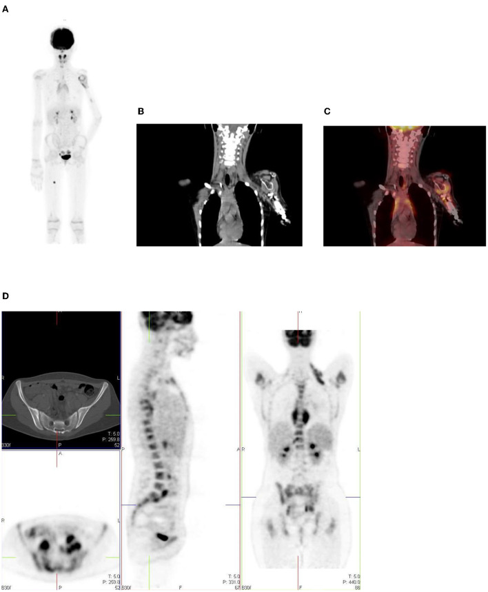
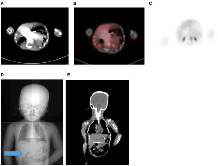
Positron emission tomography (PET) with 2-[fluorine-18] fluoro-2- deoxy-D-glucose (FDG) is a well-established modality that is used in adult oncologic imaging. Its use in pediatric oncology has increased over time. It enables increased diagnostic accuracy due to the combination of functional and morphologic imaging, resulting in optimal patient management. However, the clinician should be aware that the normal distribution of FDG uptake in children differs from adults. Also, even though FDG is used widely in oncology, it is not tumor specific. Uptake of FDG may be seen in numerous benign conditions, including inflammation, infection, and trauma. Proper interpretation of pediatric FDG PET/CT studies requires knowledge of the normal distribution of FDG uptake in children, and an insight into the physiologic variants, benign lesions, and PET/CT related artifacts. Understanding the potential causes of misinterpretation increases the confidence of image interpretation, reduce the number of unnecessary follow-up studies, optimize treatment and more importantly, reduce the radiation exposure to the patient. We review and discuss the physiological distribution of FDG uptake in children, the variation in distribution, lesions that are benign that could be misinterpreted as malignancy, and the various artifacts associated with PET/CT performed in pediatric oncology patients. We add a pictorial illustration to prompt understanding and familiarity of the above-mentioned patterns. Therefore, we believe that this review will assist in reducing possible mistakes by reading physicians and prevent incorrect interpretation.

Keywords: 18F-FDG PET/CT; normal variant; oncology; pediatric nuclear medicine; physiological.


Conflict of interest statement

The authors declare that the research was conducted in the absence of any commercial or financial relationships that could be construed as a potential conflict of interest.

Figures

Figure 1
Figure 1
Motion artifact of the brain on axial PET and PET/CT images (A,B). No obvious pathology is seen on the non- contrast axial CT component (C) of the PET study.
Figure 2
Figure 2
Physiologic uptake of 18F-FDG. Axial, sagittal and coronal FDG PET/CT (left) and PET images (right) show physiologic uptake in a 13 year old boy. Normal FDG uptake is seen in the brain, heart, liver, spleen, colon, urinary bladder and the bone marrow. Note the marked intense activity in the brain compared with the activity in the rest of the body.
Figure 3
Figure 3
Axial CT, fused PET/CT and PET only images showing physiologic uptake in the medial and lateral ocular muscles.
Figure 4
Figure 4
Axial CT (A) and fused PET/CT (B) images showing symmetric increased uptake in the tonsils (white arrows) and bilateral mild uptake in the parotid glands (blue arrows).
Figure 5
Figure 5
12 year old male referred for the evaluation of active vasculitis. FDG PET study showing diffuse symmetrical increased uptake in the thymus seen on the axial, sagittal and coronal views (inverted ‘'V”).
Figure 6
Figure 6
Diffuse uptake in the breast tissue in a 17-year-old female with embryonal rhabdomyosarcoma referred for interim PET study. CT (A) PET (B) and fused PET/CT (C) showing mild low grade uptake in bilateral breast tissue.
Figure 7
Figure 7
(A–C) Axial fused FDG PET/CT (A), CT (B) and PET (C) images showing a 16 year old male who was well fasted and shows no/minimal cardiac uptake. (D) Transverse, sagittal and coronal PET/CT in a 13 year old boy with intense physiological FDG uptake in the left ventricle of the myocardium.
Figure 8
Figure 8
A 15-year-old male known with Hodgkin's lymphoma, referred for a restaging PET/CT post chemotherapy. Sagittal PET (A) showing linear uptake anterior to the thoracic spine corresponding to uptake in the esophagus (blue arrow), likely attributed to reflux or esophagitis. The CT (B) and fused PET/CT (C) will assist in this interpretation too.
Figure 9
Figure 9
6-year-old female with Ewing's sarcoma of the left proximal tibia. Axial PET (A), CT (B) and fused PET/CT (C) showing mild diffuse uptake in the gastroesophageal junction (white arrow and crosshairs) that is physiological.
Figure 10
Figure 10
Axial PET, fused FDG PET/CT and CT showing increased FDG uptake along the wall of a contracted stomach, which is physiological.
Figure 11
Figure 11
FDG PET/CT (A) axial, (B) sagittal and (C) coronal images show markedly increased FDG uptake in the normal right colon (long arrow). Note the more intense uptake in the cecum due to the higher amount of lymphoid tissue (short arrow).
Figure 12
Figure 12
(A–C) Normal physiological liver to spleen ratio. Physiological liver uptake is always more than the spleen uptake, except in certain pathologies like lymphomatous involvement of the spleen, HIV or in patients who are post chemotherapy or after the administration of granulocyte colony stimulating factor. Note the physiological the uptake in the gastric wall in a contracted stomach. Note that in (A) and (C), non-contrast CT has a limited sensitivity in showing splenic lesions, however the uptake in the spleen is greater than the physiological liver uptake and the presence of splenomegaly is compatible with pathologic involvement. (D–F) Diffuse increased uptake in the spleen, greater than the physiological liver uptake in a 10-year-old female with diffuse large B cell lymphoma (DLBCL) prior to chemotherapy. Staging PET/CT showed stage 4 disease with nodal disease above and below the diaphragm, bone marrow and splenic involvement. (B) PET and (C) fused PET/CT showing increased uptake in the spleen and bone marrow compared to the physiological uptake in the liver. Note that in (F), a non-contrast CT has a limited sensitivity in showing splenic lesions, however the uptake in the spleen is greater than the physiological liver uptake and the presence of splenomegaly is compatible with pathologic involvement.
Figure 13
Figure 13
12 year old female referred for evaluation of active vasculitis. Incidental finding of linear uptake in the conus medullaris at T11-L1 level is physiological (Blue arrowhead). Also note the diffuse uptake in the muscles of the chest and lower limb due to playing sport the day before the injection. Other causes of diffuse muscle uptake may include high glucose level prior to injection of FDG.
Figure 14
Figure 14
MIP and coronal FDG PET/CT and PET images showing physiological activity in the left renal collecting system, left ureter and urinary bladder. The MIP image is helpful in differentiating tracer excretion in the ureter from nodal pathology. Identifying the uptake to the ureter on CT also helps in differentiating from nodal pathology.
Figure 15
Figure 15
A 16 year old male with Hodgkin's lymphoma referred to evaluate the end of treatment response to chemotherapy. Pubertal boys may have diffuse, increased and symmetrical uptake in the testes. No abnormality was seen at CT.
Figure 16
Figure 16
15 year old male with stage 3 Hodgkin's lymphoma referred for follow up FDG PET post chemotherapy. Coronal PET (B), and PET/CT (C) showing bilateral symmetrical uptake in the neck and paravertebral region of the cervical and thoracic spine. Crosshairs localized to fat density on CT (A). FDG uptake in brown adipose tissue is commonly seen in children and adolescents. Many pediatric patients have mild brown fat uptake in the neck or supraclavicular regions. However, intense uptake may include pericardiac and perirenal brown adipose tissue. 18F-FDG uptake in activated brown fat may obscure sites of pathologic FDG uptake and decrease confidence in the interpretation of the study.
Figure 17
Figure 17
(A–C) 9 year old female with high grade osteosarcoma. Coronal PET image (A) shows normal pediatric red marrow distribution. There is homogenous FDG uptake in the proximal humeri, proximal and distal femurs, and proximal tibias reflecting normal FDG uptake in red bone marrow. This uptake is usually minimal or absent in adults due to the conversion of red marrow to yellow marrow, which is less metabolically active. Pathology in the left shoulder with increased FDG uptake in the left humeral head and proximal humerus is compatible with orthopedic hardware (B). CT image (B) showing the prosthesis and fused PET/CT image (C) showing the peri-prosthetic FDG uptake. Note however, active disease cannot entirely be excluded. Due to pain in the left arm, the patient was unable to extend and rotate the left hand for correct imaging position. (D) 16 year old female with newly diagnosed Hodgkin's lymphoma. Staging PET shows increased uptake in the marrow, greater than the liver with areas of inhomogeneous and focal uptake, indicating bone marrow infiltration. The CT did not show anatomical lesions (Top left). Note the left neck (site of biopsy confirmed disease) and intrathoracic nodal disease.
Figure 18
Figure 18
(A–C) 2 year old male with neuroblastoma who had a staging PET/CT. Artifact in the abdomen due to metal component of nasogastric tube in-situ resulting in streak artifact on CT (A) and fused PET/CT (B). Note the PET only image shows that there is normal excretion in the renal calyces with physiological low grade uptake in the liver. Coronal CT scannogram (D) shows the tube extending into the abdomen (Blue arrow) with streak artifact on coronal CT (E).

References

    1. Stauss J, Franzius C, Pfluger T, Juergens KU, Biassoni L, Begent J, et al. Guidelines for 18F-FDG PET and PET-CT imaging in paediatric oncology. Eur J Nucl Med Mol Imaging. (2008) 35:1581–8. 10.1007/s00259-008-0826-x - DOI - PubMed
    1. Shammas A, Lim R, Charron M. Pediatric FDG PET/ CT: physiologic uptake, normal variants, and benign conditions. Radiographics. (2009) 29:1467–86. 10.1148/rg.295085247 - DOI - PubMed
    1. Zhuang H, Alavi A. Evolving role of PET in pediatric disorders. PET Clin. (2020) 15:xv–xvii. 10.1016/j.cpet.2020.05.001 - DOI - PubMed
    1. Grant FD. Normal variations and benign findings in pediatric 18F-FDG-PET/ CT. PET Clin. (2014) 9:195–208. 10.1016/j.cpet.2013.12.002 - DOI - PubMed
    1. Chamroonrat W. PET/computed tomography in the evaluation of fever of unknown origin and infectious/inflammatory disease in pediatric patients. PET Clin. (2020) 15:361–9. 10.1016/j.cpet.2020.03.002 - DOI - PubMed

LinkOut - more resources