Normal Variants, Pitfalls and Artifacts in Ga-68 DOTATATE PET/CT Imaging
- PMID: 39354987
- PMCID: PMC11440971
- DOI: 10.3389/fnume.2022.825486
Normal Variants, Pitfalls and Artifacts in Ga-68 DOTATATE PET/CT Imaging
Abstract
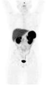
Indium 111 DTPA Octreotide (Octreoscan) has been the pillar of Somatostatin receptor (SSTRs) imaging in nuclear medicine for over three decades. The advent of PET/CT brought new analogs of somatostatin that have higher affinity and improved resolution due to their labeling to Gallium 68 for positron imaging. The most used analogs include DOTATATE, DOTATOC and DOTANOC. However, Gallium 68-1,4,7,10-tetraazacyclododecane-1,4,7,10-tetraacetic acid (DOTA)-octreotate (DOTATATE) is probably the most common non-FDG (fluoro-2-deoxy glucose) PET tracer alongside PSMA (prostate specific membrane antigen). In contrast to F18-labeled FDG, it does not require proximity to a cyclotron due to the availability of the Ga68 generator. DOTATATE is a somatostatin analog which allows whole body imaging of somatostatin receptors on cell surfaces. 68Ga-DOTA compounds provide the imaging standard for well-differentiated (Grade 1 and low grade 2) neuro-endocrine tumors (NETs) and is utilized in the staging and characterization and restaging of patients with NETs. 68Ga DOTATATE has a complementary role with 18F-FDG where tumors may exhibit varying degrees of differentiation. It furthermore has application as a prelude to therapy in selecting patients for peptide receptor radionuclide therapy using a theranostic approach. A sound knowledge of the normal biodistribution of the radiotracer is imperative for optimal patient outcome and to avoid potential false positives such as inflammation, normal pancreatic uncinate process uptake and osteoblastic activity. In this review, we will describe the normal appearances of the 68Ga DOTATATE and the potential pitfalls with the support of images to aid in improving interpretation of this crucial innovative tool in the management of individuals with tumors expressing SSTRs.
Keywords: Ga68-DOTATATE; PET/CT; neuro-endocrine tumors; normal biodistribution; pitfalls; positron emission tomography/computer tomography; variants.
Copyright © 2022 Malan and Vangu.
Conflict of interest statement
The authors declare that the research was conducted in the absence of any commercial or financial relationships that could be construed as a potential conflict of interest.
Figures


References
-
- Servente L, Bianco C, Gigiret V, Alonso O. Benign pathologies and variants with 68Ga-DOTATATE uptake in PET/CT studies. Rev Argent Radiol. (2017) 81:184–91. 10.1016/j.rard.2017.08.001 - DOI
Publication types
LinkOut - more resources
Full Text Sources
Miscellaneous

