Review
. 2024 Aug 28;34(35):2305040.
doi: 10.1002/adfm.202305040. Epub 2023 Oct 10.

Strategies for Making High-Performance Artificial Spider Silk Fibers

Affiliations
Review

Strategies for Making High-Performance Artificial Spider Silk Fibers

Benjamin Schmuck et al. Adv Funct Mater. .

Abstract

Artificial spider silk is an attractive material for many technical applications since it is a biobased fiber that can be produced under ambient conditions but still outcompetes synthetic fibers (e.g., Kevlar) in terms of toughness. Industrial use of this material requires bulk-scale production of recombinant spider silk proteins in heterologous host and replication of the pristine fiber's mechanical properties. High molecular weight spider silk proteins can be spun into fibers with impressive mechanical properties, but the production levels are too low to allow commercialization of the material. Small spider silk proteins, on the other hand, can be produced at yields that are compatible with industrial use, but the mechanical properties of such fibers need to be improved. Here, the literature on wet-spinning of artificial spider silk fibers is summarized and analyzed with a focus on mechanical performance. Furthermore, several strategies for how to improve the properties of such fibers, including optimized protein composition, smarter spinning setups, innovative protein engineering, chemical and physical crosslinking as well as the incorporation of nanomaterials in composite fibers, are outlined and discussed.

Keywords: biomimetic spinning; mechanical properties; protein fibers; rational designs; wet‐spinning.


Conflict of interest statement

The authors declare no conflict of interest.

Figures

Figure 1
Figure 1
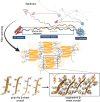
The different routes proposed in this review for improving the mechanical properties of biomimetic artificial spider silk fibers: a) To prepare mixtures of different silk proteins in order to mimic the composition of the natural spider spinning dope, b) post‐spin stretch of the fibers to increase the alignment of the protein chains and improve the intermolecular interactions, c) the use of protein engineering to increase the intermolecular contacts in the fibers, d) crosslinking the protein chains, and e) the design of a silk‐nanomaterial composite fiber. Protein structures in a) represent a recombinant mini‐spidroin (adapted from reference[ 36 ] under the creative commons attribution license) and the spider silk constituting element NMA1 from Trichonephila clavata.[ 46 ] The sequence for NMa1 was obtained from the National Center for Biotechnology Information (GFR3246.1) and the structural predictions were obtained using AlphaFold2 in ChimeraX.[ 315 , 316 ] The protein structures shown in c) were adapted under the terms of CC BY license.[ 36 ] Copyright 2022, The Authors, published by Wiley‐VCH.
Figure 2
Figure 2
Stretching a synthetic polymer or protein fiber, respectively, leads to increased orientational order and intermolecular interactions, which is why a post‐spin stretched fiber will improve in tensile strength and Young's modulus.
Figure 3
Figure 3
Scatter plot of the mean values of strength and strain at break of regenerated Bombyx mori silk and recombinant spider silk fibers. The mean values of the strength and the strain at break (dashed lines and big dots) are in general higher for fibers that were subjected to post‐spin stretch. Data was obtained from refs. [33, 34, 36, 37, 38, 39, 44, 54, 56, 64, 107, 108, 109, 110, 111, 112, 113, 114, 115, 116, 117, 118, 119, 120, 121, 122, 123, 124, 125, 126, 127, 128, 129, 130, 131] and is listed in File S1, Supporting Information.
Figure 4
Figure 4
Scatter plot of the average values of the strain at break and strength of the different artificial silk fibers (recombinant spider silk and regenerated Bombyx mori silk) post‐spin stretched in different solutions with a focus panel of the same graph. Dashed lines and big dots indicate the averages of all the values for fibers processed in the respective solutions. In this plot, methods in which post‐spin stretch was applied sequentially in different solutions were not considered. Data points obtained from refs. [33, 34, 36, 37, 38, 39, 44, 54, 56, 64, 107, 108, 109, 110, 111, 112, 113, 114, 115, 116, 117, 118, 119, 120, 121, 122, 123, 124, 125, 126, 127, 128, 129, 130, 131] are listed in File S1, Supporting Information.
Figure 5
Figure 5
Strength versus molecular weight of recombinant spidroins, using average values for stress at break obtained from the literature. Data was obtained from refs. [33, 34, 36, 37, 38, 39, 44, 54, 56, 64, 107, 108, 109, 110, 111, 112, 113, 114, 115, 116, 117, 118, 119, 120, 121, 122, 123, 124, 125, 126, 127, 128, 129, 130, 131].
Figure 6
Figure 6
Pearson correlation matrix of recombinant spidroin sequence properties and the resulting fiber mechanical properties. Data was extracted from Table 2.
Figure 7
Figure 7
Schematic representation of a strategy to increase the strength of artificial silk fibers by engineering the poly‐Ala segments of spidroins. Here, the poly‐Ala is replaced with peptides that have a much higher propensity to form β‐sheet crystals where they should adopt a steric zipper packing arrangement that increases the inter‐sheet interactions in the mature fiber. By doing so, the strength of the β‐sheet crystal could potentially be increased. NT domain PDB entry number: 4FBS; CT domain PDB entry number: 2MFZ. The steric zipper is represented by an amyloidogenic peptide from Transthyretin with the PDB entry number: 6C4O. The poly‐Ala zipper was obtained from the Zipper database. The protein structures were illustrated with ChimeraX and the final image was assembled with BioRender.com.[ 315 ]

References

    1. Gosline J. M., Guerette P. A., Ortlepp C. S., Savage K. N., J. Exp. Biol. 1999, 202, 3295. - PubMed
    1. Bourzac K., Nature 2015, 519, S4. - PubMed
    1. Pan L., Wang F., Cheng Y., Leow W. R., Zhang Y. W., Wang M., Cai P., Ji B., Li D., Chen X., Nat. Commun. 2020, 11, 1. - PMC - PubMed
    1. Geyer R., Jambeck J. R., Law K. L., Sci. Adv. 2017, 3, 3. - PMC - PubMed
    1. Manshoven S., Smeets A., Arnold M., Mortensen L. F., Eur. Top. Cent. Waste Mater. a Green Econ. 2021, 10.5281/zenodo.4475340. - DOI

LinkOut - more resources