. 2024 Oct 1;14(42):31227-31242.
doi: 10.1039/d4ra02969k. eCollection 2024 Sep 24.

Inhibition of cytotoxic self-assembly of HEWL through promoting fibrillation by new synthesized α-hydroxycarbamoylphosphinic acids

Affiliations

Inhibition of cytotoxic self-assembly of HEWL through promoting fibrillation by new synthesized α-hydroxycarbamoylphosphinic acids

Mohsen Mahdavimehr et al. RSC Adv. .

Abstract

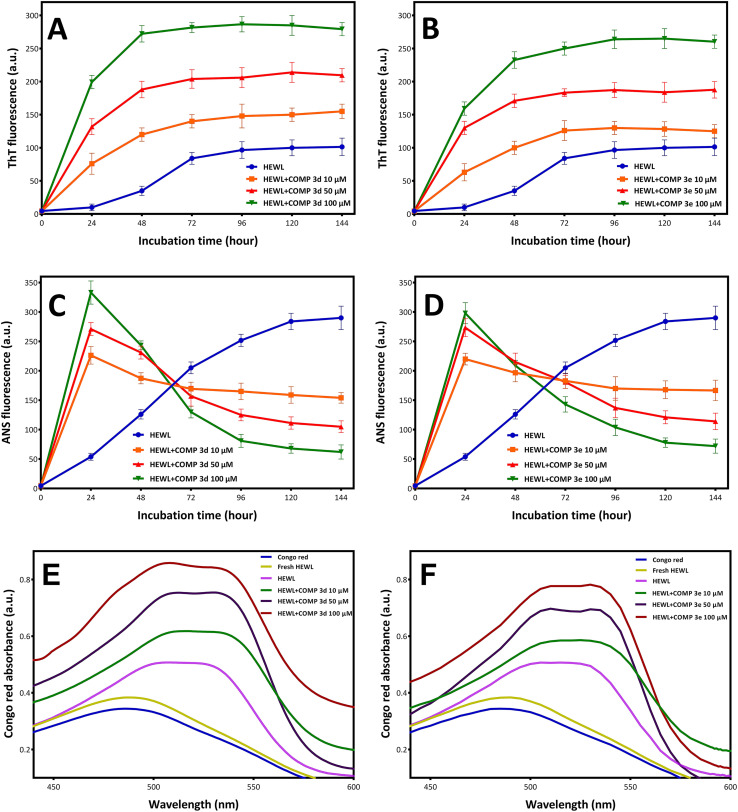
The main objective of the present study is to investigate the potency of new synthesized hydroxycarbamoyl phosphinic acid derivatives in modulating cytotoxic fibrillogenesis of hen egg white lysozyme (HEWL), as a common model in protein aggregation studies. Hydroxycarbamoyl phosphinic acid derivatives were prepared by the reaction of α-hydroxyalkylphosphinic acids with isocyanates (or isothiocyanates) in the presence of trimethylsilyl chloride (TMSCl). The designed process involves the condensation reaction leading to formation of new C sp2-P bond formation. The synthesis and purity of novel designed compounds were confirmed by NMR, LC-MS, and HPLC techniques. A range of experiments, including thioflavin T (ThT) and 8-anilino-1-naphthalenesulfonic acid (ANS) fluorescence assays, Congo red binding measurement, atomic force microscopy imaging, MTT-based cell viability and hemolysis assays were employed to investigate anti-amyloidogenic effects of tested compounds. The obtained results demonstrate that these compounds are able to significantly modulate the self-assembly process of HEWL via shortening of nucleation phase leading to the acceleration of fibrillation and appearance of very large and thick fibrils with decreased surface hydrophobicity and cytotoxicity. Based on ANS binding data, we suggest that increased exposure of hydrophobic patches of oligomeric species is the possible mechanism by which tested compounds promote self-assembly process of HEWL. Fluorescence anisotropy and molecular docking studies indicate the interaction of both synthesized compounds with HEWL, and more specifically with residues that are situated in the highly aggregation-prone β-domain region of protein. This study unveils the potential of hydroxyalkylphosphinic acids as modulators of amyloid fibrillation highlighting these compounds as a promising approach for targeting protein aggregates associated with neurodegenerative diseases.


Conflict of interest statement

The authors declare that they have no conflict of interest.

Figures

Scheme 1
Scheme 1. Synthesis of 1-hydroxyalkylphosphinic acids 1.
Scheme 2
Scheme 2. Synthesis of 1-hydroxcarbamoylphosphinic acids 3a.
Fig. 1
Fig. 1. Effect of compounds 3d and 3e on the HEWL amyloid fibrillation. Protein samples (100 μM) were incubated under amyloidogenic conditions without or with increasing concentrations of compound 3d and 3e for 6 days. (A and B) Kinetics of HEWL fibrillation indicated by increasing fluorescence intensity of ThT at specified concentrations of compounds 3d and 3e, respectively. (C and D) Changes in the surface hydrophobicity of HEWL indicated by increasing fluorescence intensity of ANS at specified concentrations of compounds 3d and 3e, respectively. (E and F) Changes in the Congo red binding absorption spectra of HEWL at specified concentrations of compounds 3d and 3e, respectively. Every test was conducted three times and the average was used.
Fig. 2
Fig. 2. Effect of increasing concentrations of compounds 3d and 3e on the surface hydrophobicity of HEWL aggregates incubated for 6 days. The incubated solutions were centrifuged at 21 000g for 30 min and changes in the surface hydrophobicity of supernatant (dark bars) and pellet (white bars) was measured by ANS fluorescence assay. *p < 0.05, **p < 0.01, significantly different from control samples.
Fig. 3
Fig. 3. AFM images of HEWL incubated under amyloidogenic conditions without or with increasing concentrations of compounds 3d and 3e for 3 and 6 days. The scale bar represents 100 nm.
Fig. 4
Fig. 4. Effect of increasing concentrations of compounds 3d and 3e on (A) viability of SH-SY5Y cells and (B) erythrocyte membrane integrity indicating no significant cytotoxicity. (C) Optical microscopy images of erythrocytes incubated with increasing concentrations of compounds 3d and 3e. Further details are provided in the Materials and methods section.
Fig. 5
Fig. 5. Effect of compounds 3d and 3e on the HEWL amyloid cytotoxicity evaluated by MTT assay. (A and B) Cytotoxicity evaluation of HEWL aggregates produced in the absence or presence of increasing concentrations of compounds 3d and 3e after 3 and 6 days, respectively. **p < 0.01, significantly different from control cells. #p < 0.05, ##p < 0.01, significantly different from cells exposed only to HEWL amyloid fibrils.
Fig. 6
Fig. 6. Cytotoxicity evaluation of (A) supernatant and (B) pellet of HEWL aggregates incubated for 6 days in the absence or presence of increasing concentrations of compounds 3d and 3e. **p < 0.01, significantly different from control cells. #p < 0.05, ##p < 0.01, significantly different from cells exposed only to HEWL amyloid fibrils.
Fig. 7
Fig. 7. Binding modes of compounds 3d and 3e to HEWL. Compounds 3d (A) and 3e (B) are interacting with HEWL active site. The compounds are depicted in red in stick model. Protein backbone of HEWL is shown in cartoon model. The secondary structure of HEWL is depicted as follows: β-strand: yellow; α-helix: purple; 3/10 helix: blue; random coil: white; turn: cyan. The N- and C-termini of HEWL are displayed as LYS1 and LEU129, respectively.
Fig. 8
Fig. 8. Binding site of compounds 3d and 3e within HEWL. HEWL residues surrounding compounds 3d (A) and 3e (B) are shown with numbers. Hydrogen bonds within 1 Å are represented as dashed green lines.
Scheme 3
Scheme 3. Schematic representation of proposed mechanism by which compounds 3d/3e modulate HEWL fibril formation leading to acceleration of assembly process of protein.

Similar articles

References

    1. Sadigh-Eteghad S. Sabermarouf B. Majdi A. Talebi M. Farhoudi M. Mahmoudi J. Amyloid-Beta: A Crucial Factor in Alzheimer's Disease. Med. Princ. Pract. 2015;24(1):1–10. doi: 10.1159/000369101. - DOI - PMC - PubMed
    1. Lücking C. B. Brice A. Alpha-Synuclein and Parkinson's Disease. Cell. Mol. Life Sci. 2000;57(13–14):1894–1908. doi: 10.1007/PL00000671. - DOI - PMC - PubMed
    1. Chen S. Ferrone F. A. Wetzel R. Huntington's Disease Age-of-Onset Linked to Polyglutamine Aggregation Nucleation. Proc. Natl. Acad. Sci. U. S. A. 2002;99(18):11884–11889. doi: 10.1073/pnas.182276099. - DOI - PMC - PubMed
    1. Sakaguchi S. Katamine S. Molecular Biology of Prion Diseases. J. Virol. 1999;49(2):193–204. doi: 10.2222/jsv.49.193. - DOI - PubMed
    1. Bonhommeau S. Talaga D. Hunel J. Cullin C. Lecomte S. Tip-Enhanced Raman Spectroscopy to Distinguish Toxic Oligomers from Aβ 1 - 42 Fibrils at the Nanometer Scale. Angew. Chem. 2017;129(7):1797–1800. doi: 10.1002/ange.201610399. - DOI - PubMed

LinkOut - more resources