Review
. 2024 Aug;86(3):370-382.
doi: 10.18999/nagjms.86.3.370.

Spontaneous regression of lumbar disc herniation: four cases report and review of the literature

Affiliations
Review

Spontaneous regression of lumbar disc herniation: four cases report and review of the literature

Yang Zheng et al. Nagoya J Med Sci. 2024 Aug.

Abstract

Spontaneous regression of lumbar disc herniation refers to shrinkage or disappearance of herniated nucleus pulposus without invasive surgical treatments. This phenomenon has been reported and is supported by improved clinical symptoms and radiographic after conservative treatment, but the underlying mechanism remains unclear. This article reports 4 cases of disc reabsorption and reviews the distribution of several clinical and radiographic factors of disc herniation reabsorption of total 46 patients, including the four from our study, gathered from 28 recent publications. Some of these factors are present with anomalous distributions. But some factors have similar deviations in patients with lumbar disc herniation. Therefore, more research is needed to explore the correlation between those factors and disc reabsorption.

Keywords: clinical factor; lumbar disc herniation; radiographic factor; reabsorption; spontaneous regression.


Figures

Fig. 1
Fig. 1
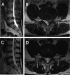
Case 1 Fig. 1A:MRI sagittal view revealed L4-L5 intervertebral disc herniation. Fig. 1B:MRI transverse view showed that the herniated disc was compressing the nerve root on the left side. Fig. 1C:MRI sagittal view revealed significant resorption of the L4-L5 disc herniation after 12 months. Fig. 1D:MRI transverse view showed resorption of the disc and release of the nerve root compression after 12 months. MRI: magnetic resonance imaging
Fig. 2
Fig. 2
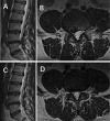
Case 2 Fig. 2A: MRI sagittal view revealed L4-L5 intervertebral disc herniation. Fig. 2B: MRI transverse view showed that the herniated disc was compressing the nerve root on the left side. Fig. 2C: MRI sagittal view revealed almost complete resorption of the L4-L5 disc fragment after 4 months. Fig. 2D:MRI transverse view showed almost resorption of the disc fragment and release of the nerve root compression after 4 months. MRI: magnetic resonance imaging
Fig. 3
Fig. 3
Case 3 Fig. 3A:MRI sagittal view revealed L4-L5 intervertebral disc herniation. Fig. 3B:MRI transverse view showed that the herniated disc was compressing the nerve root on the right side. Fig. 3C:MRI sagittal view revealed almost resorption of the L4-L5 disc fragment after 9 months. Fig. 3D:MRI transverse view showed almost resorption of the disc and significant release of the nerve root compression after 9 months. MRI: magnetic resonance imaging
Fig. 4
Fig. 4
Case 4 Fig. 4A, B:MRI transverse view revealed a right extreme lateral lumbar disc herniation at L4-L5 level (red arrow). Fig. 4C, D:MRI transverse view showed partial resorption of the protruding nucleus pulposus after 2 months (red arrow). MRI: magnetic resonance imaging
Fig. 5
Fig. 5
Visual representation of the Komori classification
Fig. 6
Fig. 6
Visual representation of the Pfirrmann grade
Fig. 7
Fig. 7
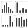
Distribution of age, disease duration, affected disc level, and MRI interval time MRI: magnetic resonance imaging

Similar articles

References

    1. Haro H, Ebata S, Inoue G, et al. Japanese Orthopaedic Association (JOA) clinical practice guidelines on the management of lumbar disc herniation, third edition - secondary publication. J Orthop Sci. 2022;27(1):31–78. doi:10.1016/j.jos.2021.07.028. - DOI - PubMed
    1. Hu C, Lin B, Li Z, Chen X, Gao K. Spontaneous regression of a large sequestered lumbar disc herniation: a case report and literature review. J Int Med Res. 2021;49(11):3000605211058987. doi:10.1177/03000605211058987. - DOI - PMC - PubMed
    1. Kobayashi K, Sato K, Ando T. Factors associated with disc degeneration based on Pfirrmann criteria after condoliase treatment for lumbar disc herniation. J Orthop Sci. 2023;28(5):976–983. doi:10.1016/j.jos.2022.08.001. - DOI - PubMed
    1. Modic MT, Steinberg PM, Ross JS, Masaryk TJ, Carter JR. Degenerative disk disease: assessment of changes in vertebral body marrow with MR imaging. Radiology. 1988;166(1):193–199. doi:10.1148/radiology.166.1.3336678. - DOI - PubMed
    1. Splendiani A, Puglielli E, De Amicis R, Barile A, Masciocchi C, Gallucci M. Spontaneous resolution of lumbar disk herniation: predictive signs for prognostic evaluation. Neuroradiology. 2004;46(11):916–922. doi:10.1007/s00234-004-1232-0. - DOI - PubMed

MeSH terms

LinkOut - more resources