

Cryoablation of osteoid osteomas: Is it a valid treatment option?
- PMID: 39355386
- PMCID: PMC11440277
- DOI: 10.4329/wjr.v16.i9.389
Cryoablation of osteoid osteomas: Is it a valid treatment option?
Abstract
Background: Osteoid osteoma is a benign bone tumor with characteristic clinical symptomatology. The selected method for its treatment is percutaneous radiofrequency ablation. However, percutaneous cryoablation is an alternative method with certain advantages.
Aim: To evaluate percutaneous computed tomography (CT)-guided cryoablation for the treatment of osteoid osteoma in young patients and adults.
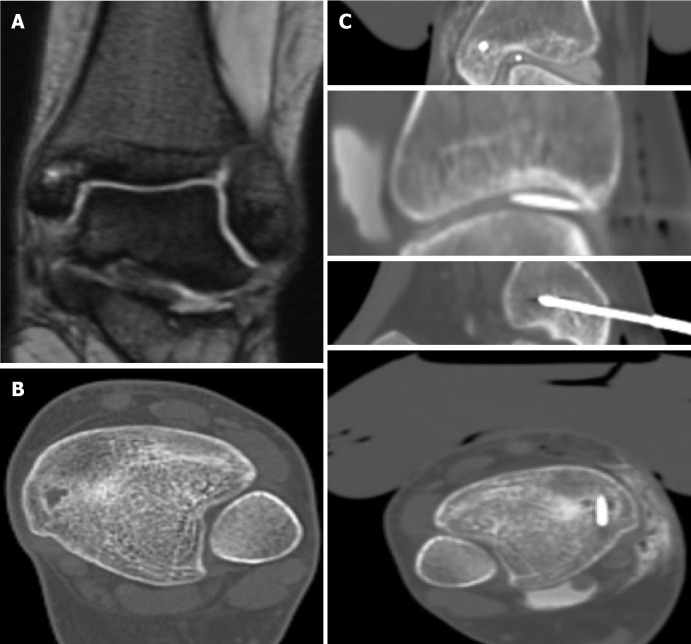
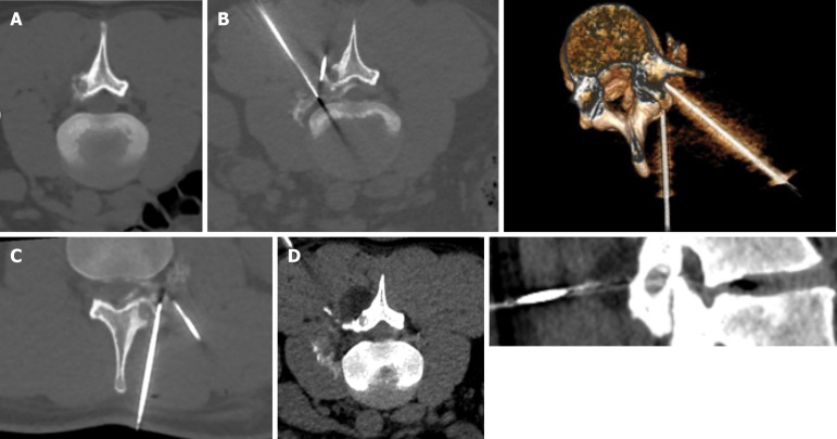
Methods: A total of 25 patients were treated with percutaneous CT- guided cryoablation for osteoid osteomas between October 2020 and March 2023 at a single institution. All patients were above 14-years-old (mean age, 24-years-old), and all procedures were performed under local anesthesia. Of the 25 patients, 8 were female and 17 were male. Tumor sites included the femur (n = 9), medial malleolus (n = 4), sacral ala (n = 4), facets (n = 4), humerus (n = 3), and tibia (n = 1). One cryoprobe was used in each procedure and, when possible, the lesion was covered by the ice-ball using an extraosseous position without penetrating the nidus. All necessary thermal protective techniques were used depending on the anatomical structure at risk.
Results: All patients treated had complete response (100% clinical success rate) starting on the day of the procedure. Technical success was achieved in all cases. Visual analog scale (VAS) scores at 1 year were 0, compared to a mean VAS score of 8.5 ± 1 (SD) before the procedure. No recurrences were reported at the 1-year follow-up and no complications were observed. In 11/25 cases, an extraosseous position of the cryoprobe was used with less procedural time achieving technical and clinical success and no complications with less patient discomfort. All patients were discharged from the hospital on the same day as the procedure.
Conclusion: Cryoablation of osteoid osteomas is an efficacious and safe procedure with durable clinical results. Its greatest advantage is that the procedure can be performed under local anesthesia using an extraosseous position of the cryoprobe when possible.
Keywords: Bone tumors; Computed tomography guidance; Cryoablation; Interventional radiology; Orthopedics; Osteoid osteoma.
©The Author(s) 2024. Published by Baishideng Publishing Group Inc. All rights reserved.
Conflict of interest statement
Conflict-of-interest statement: The authors declare that they have no conflicts of interest.
Figures


References
-
- Jaffe HL. "OSTEOID-OSTEOMA". Arch Surg. 1935;31:709.
-
- Tepelenis K, Skandalakis GP, Papathanakos G, Kefala MA, Kitsouli A, Barbouti A, Tepelenis N, Varvarousis D, Vlachos K, Kanavaros P, Kitsoulis P. Osteoid Osteoma: An Updated Review of Epidemiology, Pathogenesis, Clinical Presentation, Radiological Features, and Treatment Option. In Vivo. 2021;35:1929–1938. - PMC - PubMed
-
- Lee EH, Shafi M, Hui JH. Osteoid osteoma: a current review. J Pediatr Orthop. 2006;26:695–700. - PubMed
LinkOut - more resources
Full Text Sources
