Review
. 2022 Jan 17:1:826109.
doi: 10.3389/fnume.2021.826109. eCollection 2021.

F-18 FDG PET/CT Imaging in Normal Variants, Pitfalls and Artifacts in the Abdomen and Pelvis

Affiliations
Review

F-18 FDG PET/CT Imaging in Normal Variants, Pitfalls and Artifacts in the Abdomen and Pelvis

Mboyo D T Vangu et al. Front Nucl Med. .

Abstract

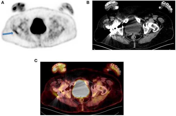
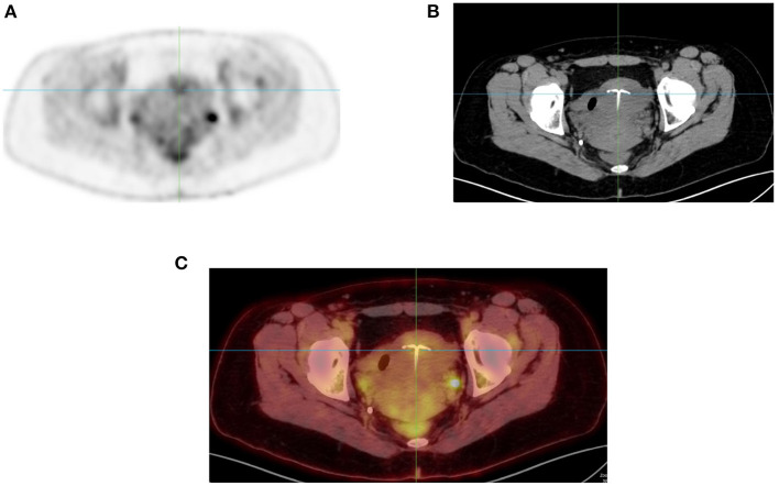
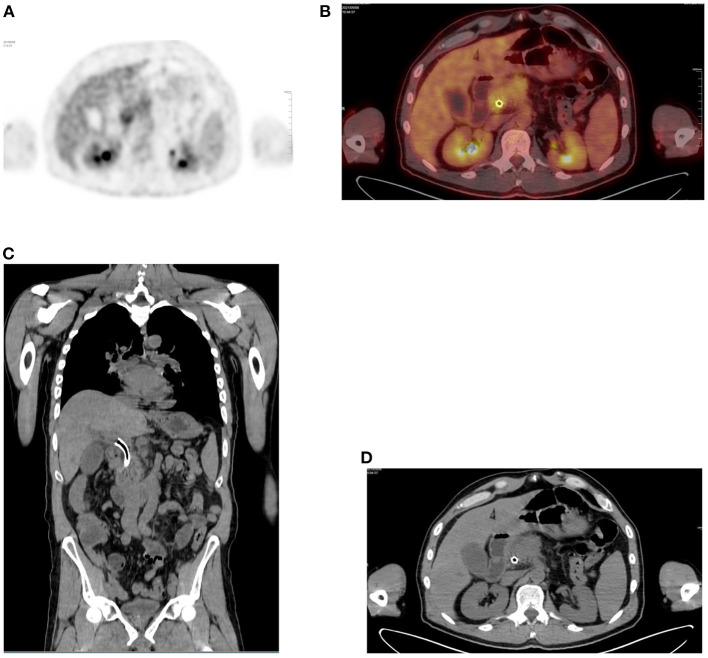
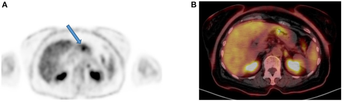
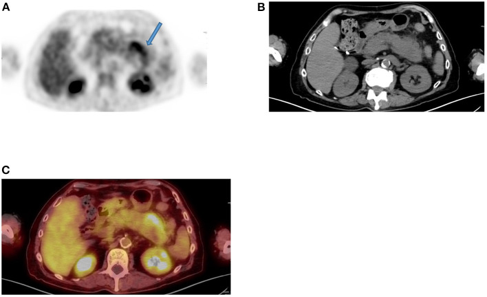
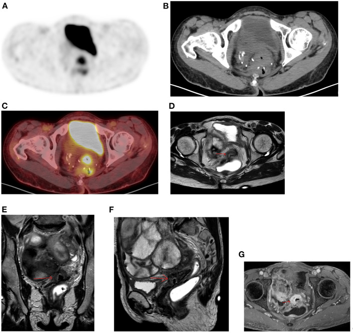
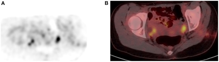
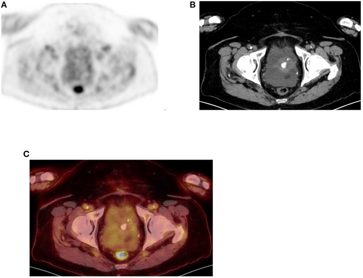
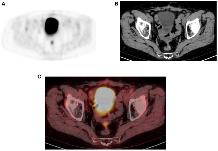
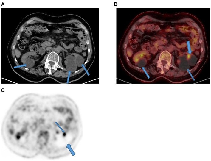
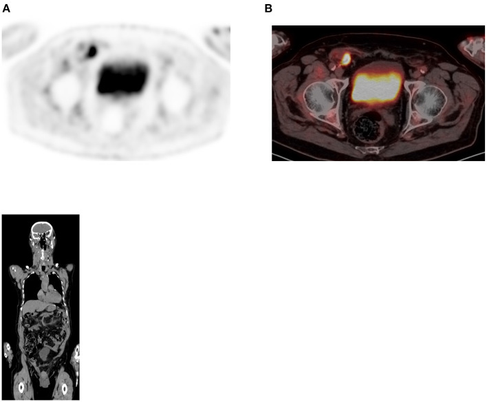
Since its introduction into clinical practice, multimodality imaging has revolutionized diagnostic imaging for both oncologic and non-oncologic pathologies. 18F-fluorodeoxyglucose (18F-FDG) PET/CT imaging which takes advantage of increased anaerobic glycolysis that occurs in tumor cells (Warburg effect) has gained significant clinical relevance in the management of most, if not all oncologic conditions. Because FDG is taken by both normal and abnormal tissues, PET/CT imaging may demonstrate several normal variants and imaging pitfalls. These may ultimately impact disease detection and diagnostic accuracy. Imaging specialists (nuclear medicine physicians and radiologists) must demonstrate a thorough understanding of normal and physiologic variants in the distribution of 18F-FDG; including potential imaging pitfalls and technical artifacts to minimize misinterpretation of images. The normal physiologic course of 18F-FDG results in a variable degree of uptake in the stomach, liver, spleen, small and large bowel. Urinary excretion results in renal, ureteric, and urinary bladder uptake. Technical artifacts can occur due to motion, truncation as well as the effects of contrast agents and metallic hardware. Using pictorial illustrations, this paper aims to describe the variants of physiologic 18F-FDG uptake that may mimic pathology as well as potential benign conditions that may result in misinterpretation of PET/CT images in common oncologic conditions of the abdomen and pelvis.

Keywords: FDG; PET/CT; abdomen; pelvis; pitfalls; variants.


Conflict of interest statement

The authors declare that the research was conducted in the absence of any commercial or financial relationships that could be construed as a potential conflict of interest.

Figures

Figure 1
Figure 1
Severe reflux. FDG PET sagittal MIP image demonstrates diffuse uptake in esophagus and much increased in the lower third, in a 65-year-old male patient referred for therapy evaluation and he is known with gastro-esophageal reflux.
Figure 2
Figure 2
FDG PET MIP image (A) showing uptake in large, mainly in the transverse colon while the small bowel is almost not seen. This is a common finding in most patients who undergo PET/CT imaging. Axial PET/CT image (B) enhanced the describe finding in the large bowel with less uptake in small bowel that may be identified in the corresponding anatomical CT (C).
Figure 3
Figure 3
Metformin and bowel uptake. FDG PET MIP image demonstrates diffuse increased bowel uptake, in a diabetic patient who stopped medication 24 h before the scheduled PET/CT study visualization of large bowel with impressive details of the small bowel. Possibly, the increased sensitivity of the digital PET machine may also play a role of the visualized small bowel.
Figure 4
Figure 4
Appendix uptake. FDG PET Axial image (A) showing linear increased uptake in right iliac fossa that corresponds to appendix as correlated in anatomical CT (B) and combined PET/CT (C) images of the same plane (see crosshair in each image).
Figure 5
Figure 5
A 50-year-old man with a prosthesis in his right hip join. A PET attenuation-corrected image (A) shows FDG uptake in the lateral aspect of the prosthesis (solid arrow) l) caused by an artifact induced by attenuation correction. Clearly, no corresponding tissue is seen on the CT of the same region, despite the streaks effects from the metal implant (B) and confirmed in the fused PET/CT axial image (C). When not sure on the reason of increased FDG uptake, interpreting physicians must assess the non-attenuation-corrected image of the same region to prevent misinterpretation.
Figure 6
Figure 6
Uptake in intrauterine device (IUD). FDG PET MIP image in axial plane (A) showing focal uptake (Arrow) in anterior uterus. Corresponding CT (B) and combined PET/CT (C) confirms the uptake in the IUD.
Figure 7
Figure 7
Focal uptake due to stent. (A) FDG PET Axial and (B) combined PET/CT with focal liver uptake in a 64-year-old male patient with pancreas adenocarcinoma and who underwent a placement of stent at CBD. Corresponding anatomical images alone, both coronal and axial planes (C,D) show the stent in place.
Figure 8
Figure 8
Diffuse gastric uptake. (A) FDG PET Axial and (B) combined PET/CT, showing diffuse uptake in inferior greater curvature of the stomach of a young patient who received a liver transplant. He continues to complain of abdominal pain following transplant procedure and all his inflammatory markers were raised. The visualized increased diffuse uptake was suggestive of gastritis and gastroscopy confirmed inflammation.
Figure 9
Figure 9
Focal gastric uptake. A 76-year-old female with newly diagnosed poorly differentiated gastric adenocarcinoma. (A) A focus FDG PET seen in Axial image (arrow) and (B) confirmed in the combined PET/CT. Focal uptake in gastric wall is typical for carcinoma and there was gastric outlet obstruction seen.
Figure 10
Figure 10
Inflammation uptake. FDG PET Axial image (A) demonstrates diffuse increased uptake in left central abdomen (arrow) that corresponds to bulky and edematous tail of pancreas (B,C), in an 82-year-old male patient with previous history of rectal cancer and suspected liver metastases. The finding is consistent with chronic inflammatory process due to pancreatitis.
Figure 11
Figure 11
Diffuse uptake in pelvis. FDG PET Axial image (A) demonstrates diffuse increased uptake in central pelvis posterior to bladder that may be consider normal. The corresponding CT (B) showed a soft tissue mass with central air at the suture lines in a 56-year-old female patient with previous history of rectal cancer and had a complete response to chemotherapy. She was referred for restaging due to weight loss and the combined PET/CT Axial (C) image suspected a metastatic lesion despite diffuse uptake. MRI of pelvis was performed, and the Axial, coronal, and sagittal planes (D–F) confirmed the suspicious and showed overt enhancement of the soft tissue mass (G) that is consistent with metastasis.
Figure 12
Figure 12
Omental cake. FDG PET Axial (A) image demonstrates diffuse moderate increased linear uptake in anterior abdomen in a “band like” patterns (arrows) that mimic large bowel uptake, in a patient with ovarian carcinoma who was referred for therapy response evaluation. Corresponding CT (B) and combined PET/CT (C) confirmed the features of “omental cake” that are suggestive of peritoneal metastasis.
Figure 13
Figure 13
Uptake in ovaries. FDG PET Axial (A) image showed focal increased uptake in pelvis corresponding to ovaries in combined PET/CT (B) image, in a female patient of childbearing age. The uptake is physiologic due to menstrual cycle.
Figure 14
Figure 14
Uterine uptake. FDG PET Axial (A) image showing diffuse moderate uptake in central lower pelvis that correspond to calcified uterine fibroid as confirmed in CT and combined PET/CT (B,C), in a 66-year-old female patient who was referred with non-alcoholic steatohepatitis (NASH) and ascites.
Figure 15
Figure 15
Physiologic pelvic focal uptake. FDG PET Axial (A) image showing focal increased uptake in posterior pelvic, in a 75-year-old female patient diagnosed with high grade serous carcinoma of the left ovary. She underwent oophorectomy and received chemotherapy. Her serum CA started to rise and was referred for restaging. The PET/CT showed active abdominal and pelvic adenopathy and in focal area of increased uptake that was localized in the vaginal fornix on CT (B) and combined PET/CT (C).
Figure 16
Figure 16
Focal kidney uptake. Axial CT (A) component of the PET/CT showed multiple bilateral Bosniak type 1 renal cysts that appear hypodense (arrows) as compared to thin normal renal tissue. Their magnitude in left kidney (thin arrows) showed compression and thinning of renal cortex with a focal FDG uptake (thick arrow) on corresponding combined PET/CT (B) that may mimic focal disease. Corresponding axial PET (C) demonstrates photon deficient areas due to renal cysts (thick arrow) with adjacent focal urinary uptake (thin arrow).
Figure 17
Figure 17
Uptake in hernia. FDG PET Axial (A) image showing focal increased uptake within right iliac fossa (RIF) in a 82 year old male patient with uncomplicated inguinal hernia as seen in combined PET/CT (B). Coronal plane of the CT confirmed the small bowel content of the right inguinal hernia.

Similar articles

Cited by

References

    1. Jian Y, Mohan D. Normal variants and pitfalls encountered in PET assessment of gynaecologic malignancies. PET Clin. (2018) 13:249–68. 10.1016/j.cpet.2017.11.009 - DOI - PMC - PubMed
    1. Corrigan AJG, Schleyer PJ, Cook GJ. Pitfalls and artifacts in the use of PET/CT in oncology imaging. Semin Nucl Med. (2015) 45:481–99. 10.1053/j.semnuclmed.2015.02.006 - DOI - PubMed
    1. Lin E, Alavi A. PET and PET/CT- A Clinical Guide. 3rd ed. New York, NY: Thieme; (2019).
    1. Jayaprakasam VS, Paroder V, Schöder H. Variants and pitfalls in PET/CT imaging of gastrointestinal cancers. Semin Nucl Med. (2021) 51:485–501. 10.1053/j.semnuclmed.2021.04.001 - DOI - PMC - PubMed
    1. Cronin CG, Scott J, Kambadakone A, Catalano OA, Sahani D, Blake MA, et al. . Utility of positron emission tomography / CT in the evaluation of small bowel pathology. Br J Radiol. (2012) 85:1211–21. 10.1259/bjr/64534573 - DOI - PMC - PubMed

LinkOut - more resources