

Case Reports
doi: 10.1016/j.hrcr.2024.06.013.
eCollection 2024 Sep.
Unipolar morphology-guided critical isthmus emphasis in a patient with scar-related ventricular tachycardia
Affiliations
- PMID: 39355827
- PMCID: PMC11440152
- DOI: 10.1016/j.hrcr.2024.06.013
Item in Clipboard
Case Reports
Unipolar morphology-guided critical isthmus emphasis in a patient with scar-related ventricular tachycardia
HeartRhythm Case Rep.
.
No abstract available
Keywords: Activation-recovery interval; High-pass filter; Reentry arrhythmias; Unipolar potential; Ventricular tachycardia.
Conflict of interest statement
There were no relationships with industry.
Figures

The targeted ventricular tachycardia (VT) and the endocardial mapping. A: The left panel shows the electrocardiogram during sinus rhythm; the right panel, VT. B: Activation map of the targeted VT. The white circled area indicates the location where the diastolic potentials were recorded during the tachycardia. C: Bipolar voltage map during sinus rhythm, with a cut-off for low voltage set at <1.5 mV. D: Unipolar voltage map during sinus rhythm, with a cut-off for low voltage set at <8.3 mV. E: Isochronal late activation map for revealing the deceleration zone, which is depicted by the pink circles. The yellow lines indicate localized functional lines of block detected by the isochronal activation map.
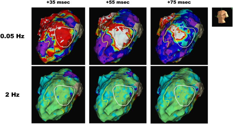
Unipolar activation maps with 2 high-pass filter settings. Figures where the duration from the onset to the maximum dV/dt point of the unipolar waveform was color-coded with reference to the S wave of lead I in surface electrocardiogram. The number of milliseconds indicates the blanking time from the S wave of lead I.

Unipolar potential morphologies. Maps identifying from the S wave in lead I to the maximum dV/dt point using the local unipolar morphologies. (1) and (2) indicate the critical isthmus point and another area, respectively, which are the same points irrespective of the filter setting. A: High-pass filter set at 0.05 Hz. B: High-pass filter set at 2 Hz.
References
-
- Tung R., Raiman M., Liao H., et al. Simultaneous endocardial and epicardial delineation of 3D reentrant ventricular tachycardia. J Am Coll Cardiol. 2020;75:884–897. - PubMed
-
- Higuchi K., Yavin H.D., Sroubek J., et al. How to use bipolar and unipolar electrograms for selecting successful ablation sites of ventricular premature contractions. Heart Rhythm. 2022;19:1067–1073. - PubMed
-
- Khan H., Bonvissuto M.R., Rosinski E., et al. Comparison of combined substrate-based mapping techniques to identify critical sites for ventricular tachycardia ablation. Heart Rhythm. 2023;20:808–814. - PubMed
-
- Yalin K., Golcuk E., Bilge A.K., et al. Combined analysis of unipolar and bipolar voltage mapping identifies recurrences after unmappable scar-related ventricular tachycardia ablation. Europace. 2015;17:1580–1586. - PubMed
-
- Yanagisawa S., Inden Y., Goto T., et al. Visualization of repolarization heterogeneity in Brugada syndrome: a quantitative analysis of unipolar electrogram T-wave. JACC Clin Electrophysiol. 2023;9:2401–2411. - PubMed
Publication types
LinkOut - more resources
Full Text Sources
