Review
. 2025 Jan;31(1):32-55.
doi: 10.3350/cmh.2024.0734. Epub 2024 Oct 2.

Sinusoidal communication in chronic liver disease

Affiliations
Review

Sinusoidal communication in chronic liver disease

Albert Gibert-Ramos et al. Clin Mol Hepatol. 2025 Jan.

Abstract

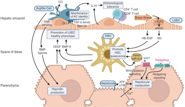
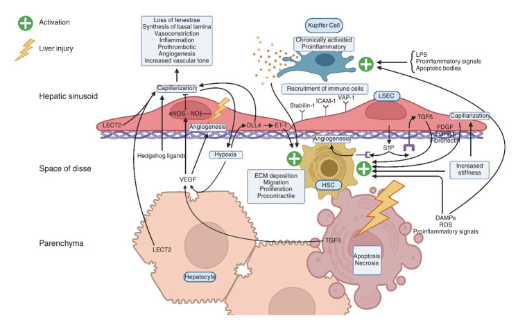
The liver sinusoid, mainly composed of liver sinusoidal endothelial cells, hepatic macrophages and hepatic stellate cells, shapes the hepatic vasculature and is key to maintaining liver homeostasis and function. During chronic liver disease (CLD), the function of sinusoidal cells is impaired, being directly involved in the progression of liver fibrosis, cirrhosis, and main clinical complications including portal hypertension and hepatocellular carcinoma. In addition to their roles in liver diseases pathobiology, sinusoidal cells' paracrine communication or cross-talk is being studied as a mechanism of disease but also as a remarkable target for treatment. The aim of this review is to gather current knowledge of intercellular signalling in the hepatic sinusoid during the progression of liver disease. We summarise studies developed in pre-clinical models of CLD, especially emphasizing those pathways characterized in human-based clinically relevant models. Finally, we describe pharmacological treatments targeting sinusoidal communication as promising options to treat CLD and its clinical complications.

Keywords: Chronic liver disease; Hepatic sinusoid; Hepatic stellate cells; Kupffer cells; Liver sinusoidal endothelial cells.


Conflict of interest statement

Conflicts of Interest

Dr. Gracia-Sancho reports grants from Gilead Sciences, Novo Nordisk, GAT Therapeutics and Oncomatryx. In addition, he was offered lecture fees by Cell Pharma and BrudyLab, outside the submitted work. The rest of the authors declare no conflicts of interest.

Figures

Figure 1.
Figure 1.
Liver sinusoidal crosstalk in health. In physiological conditions, the hepatic cells interact with each other, maintaining liver homeostasis. The bone morphogenetic protein 9 (BMP-9) released by HSCs and the vascular endothelial growth factor (VEGF) secreted by HSCs and hepatocytes promote the maintenance of LSECs healthy phenotype. LSECs contribute to the differentiation and phenotype maintenance of KCs through delta-like protein 4 (DLL4) and the secretion of transforming growth factor-β (TGF-β) family ligands. Healthy LSECs also promote HSCs quiescence through different factors, such as nitric oxide (NO) and the secretion of the heparin binding epidermal growth factor-like growth factor (HB-EGF). An increase in mechanical shear stress in LSECs induces the expression of Krüppel-like factor 2 (KLF2), a vasoprotective transcription factor that modulates the endothelial nitric oxide synthase (eNOS) pathway, further increasing NO synthesis. HSCs can also promote their quiescence by autocrinally secreting microvesicles that contain the transcription factor Twist1 or the microRNA-214 (miR-214), and as a result of haemoglobin degradation, KCs also produce vasoprotective mediators, such as carbon monoxide (CO). Communication in the liver sinusoids also coordinates the liver metabolic and synthetic functions. Together with the oxygen gradient, the Wnt/β-catenin and the Hedgehog signalling pathways are suggested to regulate hepatocyte liver zonation. Hepatocytes can also modulate their metabolism autocrinally via the intracellular calcium (Ca2+) signalling system or the release of extracellular nucleotides, mainly ATP and UTP, to the sinusoidal space. Hepatocyte synthetic functions can be regulated by non-parenchymal cells. For instance, LSECs sense changes in iron levels and secrete signals, such as bone morphogenetic proteins (BMP) ligands, that induce hepcidin production, and KCs can also modulate hepcidin transcription. Finally, interleukin 10 (IL-10) secretion by LSECs and KCs and LSEC antigen presentation to naïve CD4+ and CD8+ T cells are key to confer immunological tolerance to the organ.
Figure 2.
Figure 2.
Crosstalk in liver sinusoids upon liver injury. Following liver injury, hepatocytes release damage-associated molecular patterns (DAMPs), reactive oxygen species (ROS), and proinflammatory signals that activate Kupffer cells (KCs). Activated KCs secrete various proinflammatory factors that orchestrate the immune response to resolve the liver injury. In parallel, liver damage induces the capillarisation of liver sinusoidal endothelial cells (LSECs), which are responsible for activating hepatic stellate cells (HSCs) through the secretion of different paracrine signals, including platelet-derived growth factor (PDGF), transforming growth factor-beta (TGF-β), fibroblast growth factor receptor 1 (FGFR1), and fibronectin. At the initial stages of liver damage, the long non-coding RNA Airn maintains LSECs in a differentiated state through the activation of the KLF2-eNOS-sGC pathway, inhibiting the process of capillarization. This allows for the secretion of Wnt2a and hepatocyte growth factor (HGF), which maintain HSCs in a quiescent state and signal hepatocytes to regenerate. Moreover, the process of autophagy within the liver sinusoids protects hepatocytes from apoptosis by removing damaged mitochondria and protein aggregates, and by mitigating oxidative stress. Autophagy in KCs also prevents the activation of other immune cells and HSCs, thus protecting against fibrogenesis and chronic inflammation.
Figure 3.
Figure 3.
Liver sinusoidal crosstalk in chronic liver disease. Chronic liver disease (CLD) induces persistent hepatocyte damage, leading to the release of apoptotic bodies, damage-associated molecular patterns (DAMPs), reactive oxygen species (ROS), and proinflammatory extracellular vesicles. These factors collectively activate the inflammatory response. Kupffer cells (KCs) are particularly chronically activated by these factors, as well as by gut-derived compounds such as lipopolysaccharides (LPS). Hepatocyte damage also promotes the secretion of transforming growth factor-beta (TFGβ), which in turn induces liver sinusoidal endothelial cell (LSECs) angiogenesis and tube formation while activating the endothelial nitric oxide synthase (eNOS) pathway. However, due to endothelial damage, inflammation, and oxidative stress, nitric oxide (NO) inhibition of capillarisation is arrested. Moreover, in the context of CLD, there is an increase in LECT2 in hepatocytes and endothelial cells, which promotes LSECs capillarization. This process is further influenced by Hedgehog ligands released by various sinusoidal cell types in response to cellular damage, NO signaling inhibition, and Delta-like 4 (DLL4) activation, all of which are key drivers of LSECs capillarization. Capillarized LSECs contribute to liver inflammation by recruiting immune cells via Stabilin-1, intercellular adhesion molecule 1 (ICAM-1), and vascular adhesion protein-1 (VAP-1) surface receptors. The capillarisation of LSECs also leads to hepatic stellate cell (HSCs) activation. This activation is caused by decreased NO signaling and the secretion of sphingosine-1-phosphate (S1P) contained within exosomes, which also autocrinally induce endothelial TGFβ secretion, a critical activator of HSC. Activated HSCs synthesize excessive extracellular matrix (ECM) components, resulting in ECM accumulation and increased liver stiffness, which further promotes HSC activation and LSEC capillarization. Additionally, the loss of fenestrae during LSEC capillarisation and the architectural distortion of the liver due to fibrosis lead to hypoxia. Hypoxia not only induces rapid endothelial growth through vascular endothelial growth factor (VEGF) but also further activates HSC via the DLL4-endothelial differentiation gene-1 (ET-1) pathway.
Figure 4.
Figure 4.
Liver sinusoidal intercellular communication in healthy and cirrhotic human livers. (A) Number of ligand-receptor pairs predicted in cirrhotic vs healthy livers. (B) Difference in ligand-receptor pairs by cell type. Red=increased in cirrhotic vs. healthy; blue=decreased in cirrhotic vs. healthy. (C) Specific ligand-receptor pathways predicted in livers described in (A). Pathways in white are overrepresented in healthy livers, while pathways in black are overrepresented in cirrhotic livers; *P<0.05. Epithelia (including hepatocytes), endothelial cells (including LSECs), mesenchyma (including HSCs) and macrophages (including KCs). Reanalysis of scRNA-seq data from Ramachandran et al. [123] (2019) Nature.

Similar articles

Cited by

References

    1. Boyer TD, Wright TL, Manns MP. Zakim and Boyer’s hepatology e-book: a textbook of liver disease. Philadelphia: Elsevier Health Sciences; 2011. p. 1330.
    1. Augustin HG, Koh GY. Organotypic vasculature: from descriptive heterogeneity to functional pathophysiology. Science. 2017;357:eaal2379. - PubMed
    1. Marrone G, Shah VH, Gracia-Sancho J. Sinusoidal communication in liver fibrosis and regeneration. J Hepatol. 2016;65:608–617. - PMC - PubMed
    1. Gracia-Sancho J, Caparrós E, Fernández-Iglesias A, Francés R. Role of liver sinusoidal endothelial cells in liver diseases. Nat Rev Gastroenterol Hepatol. 2021;18:411–431. - PubMed
    1. DeLeve LD, Wang X, Hu L, McCuskey MK, McCuskey RS. Rat liver sinusoidal endothelial cell phenotype is maintained by paracrine and autocrine regulation. Am J Physiol Gastrointest Liver Physiol. 2004;287:G757–G763. - PubMed