Review
. 2024 Nov;32(11):405-416.
doi: 10.1007/s12471-024-01904-5. Epub 2024 Oct 2.

Photon-counting detector computed tomography in cardiac imaging

Affiliations
Review

Photon-counting detector computed tomography in cardiac imaging

Simran P Sharma et al. Neth Heart J. 2024 Nov.

Abstract

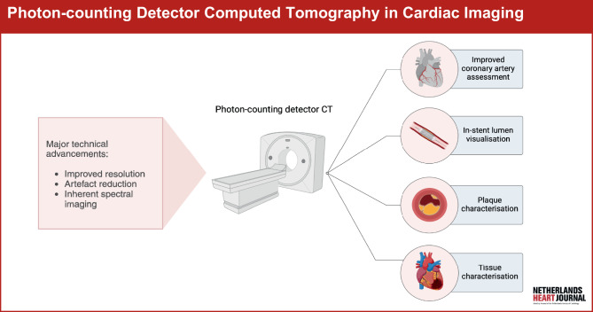
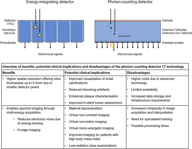
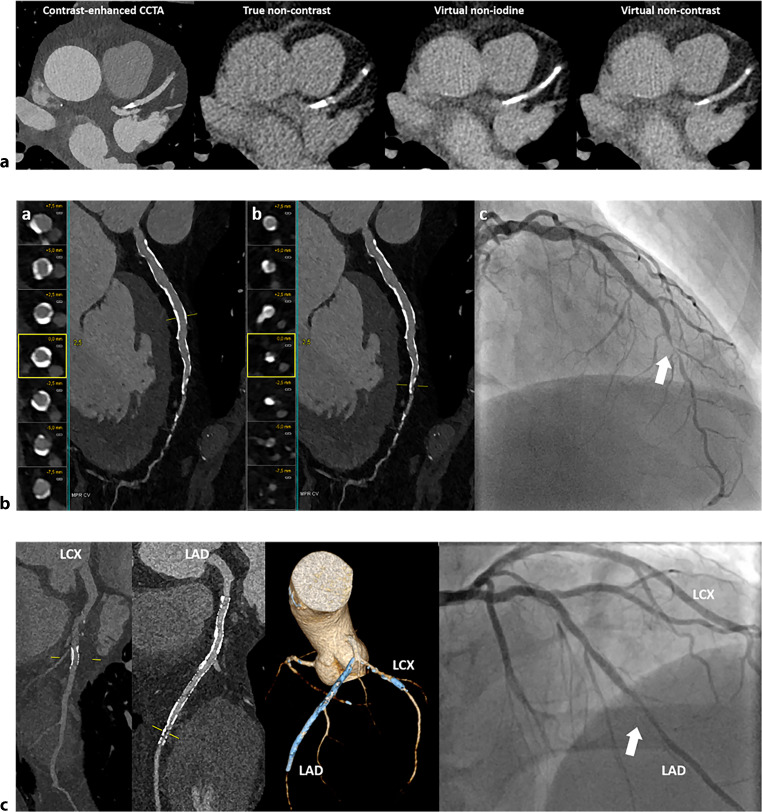
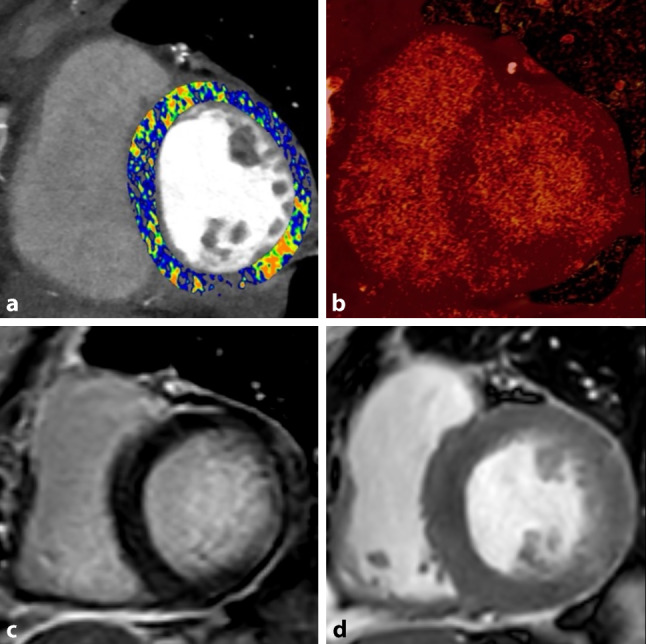
Photon-counting detector computed tomography (PCD-CT) has emerged as a revolutionary technology in CT imaging. PCD-CT offers significant advancements over conventional energy-integrating detector CT, including increased spatial resolution, artefact reduction and inherent spectral imaging capabilities. In cardiac imaging, PCD-CT can offer a more accurate assessment of coronary artery disease, plaque characterisation and the in-stent lumen. Additionally, it might improve the visualisation of myocardial fibrosis through qualitative late enhancement imaging and quantitative extracellular volume measurements. The use of PCD-CT in cardiac imaging holds significant potential, positioning itself as a valuable modality that could serve as a one-stop-shop by integrating both angiography and tissue characterisation into a single examination. Despite its potential, large-scale clinical trials, standardisation of protocols and cost-effectiveness considerations are required for its broader integration into clinical practice. This narrative review provides an overview of the current literature on PCD-CT regarding the possibilities and limitations of cardiac imaging.

Keywords: Angiography; Cardiac imaging techniques; Coronary artery disease; Tomography, X‑ray computed.


Conflict of interest statement

M.W. Smulders: Speakers fee ESC highlights ’22 and ’23—Daiichi Sankyo Europe. Academic Grant by Maastricht UMC+. R.P.J. Budde: Institutional support to EMC by Siemens and Heartflow. Speakers fee Bayer. A. Hirsch: Editor for Netherlands Heart Journal. Received a research grant and consultancy fees from GE Healthcare and speaker fees from GE Healthcare and Bayer. He is also a member of the medical advisory board of Medis Medical Imaging Systems and was MRI corelab supervisor of Cardialysis BV until 2022. C. Mihl: Speakers bureau Bayer Healthcare. Academic Grant by Maastricht UMC+. MJ.D.K. Lemmens and S.P. Sharma declare that they have no competing interests.

Figures

Fig. 1
Fig. 1
Infographic: Photon-counting detector computed tomography in cardiac imaging
Fig. 2
Fig. 2
Overview of energy-integrating versus photon-counting detector. The energy-integrating detector uses a scintillator (e.g. gadolinium oxysulfide) to absorb X‑rays and convert them into visible light, which is then detected and converted into electrical signals by photodiodes. The photon-counting detector (PCD) uses a Cadmium Telluride/Cadmium Zinc Telluride or Silicon detector to directly convert incoming X‑rays into a charge cloud of electrons, which are collected by pixelated anodes. The electronic signals’ varying colours indicate the different energy levels of the photons, highlighting PCD’s energy-resolving capability
Fig. 3
Fig. 3
Advanced coronary artery disease assessment using photon-counting detector CT (PCD-CT). a Calcium artery calcium scoring using PCD-CT on contrast-enhanced, true non-contrast, virtual non-iodine and virtual non-contrast images. The calcified plaque in the left anterior descending (LAD) artery appears more pronounced on the virtual non-iodine image and less pronounced on the virtual non-contrast image, relative to the true non-contrast image. b Ultra-high resolution imaging of coronary arteries in a 66-year-old man with stable angina and multiple risk factors. (a) Multiplanar reformatted (MPR) images of the LAD, depicting extensive calcification of the vessel wall. MPR and cross-sectional views reveal no significant stenosis in the proximal and mid-LAD. (b) MPR images of the LAD with cross-sectional views illustrate significant lumen tapering in the transition from the mid to distal LAD. (c) Invasive coronary angiography confirms the presence of significant stenosis in the mid/distal LAD. c Stent-imaging using ultra-high resolution PCD-CT. Coronary CT angiography of a 67-year-old male patient with typical chest pain was performed using PCD-CT in ultra-high resolution mode. The patient has a history of stent placement in the LAD and left circumflex (LCX) artery. Multiplanar reconstruction images display a patent stent in the LCX. However, in the LAD, there is evidence of in-stent restenosis located distally within the stent. The 3D volume rendering clearly depicts the stents in both the LAD and LCX. Corresponding invasive coronary angiography confirms the presence of in-stent restenosis in the LAD and the patent stent in the LCX
Fig. 4
Fig. 4
Photon-counting detector CT imaging protocol as a one-stop-shop for both coronary artery assessment and late enhancement imaging. The duration of a regular coronary CT angiography (CCTA) protocol is approximately 10 min, an additional late enhancement (LE) CT acquisition extends the total duration to approximately 15–20 min. Acquisition times may differ between centres. A haematocrit assessment is only necessary for the optional extracellular volume (ECV) assessment acquired 5–10 min after contrast administration. A native scan is optional as it can be reconstructed as a virtual non-iodine image
Fig. 5
Fig. 5
Short-axis photon-counting detector CT (PCD-CT, a and b) and cardiovascular magnetic resonance images (c and d) of a 59-year-old female recently diagnosed with myocardial infarction. Image a shows a PCD-CT extracellular volume (ECV) map of the left ventricle with focal increased ECV in the basal inferolateral segment. Image b shows a calculated iodine map with retainment of iodine in an ischaemic pattern, corresponding with an area of late gadolinium enhancement on cardiovascular magnetic resonance (Image c). Image d shows a short-axis cine image (after gadolinium administration) in which increased signal intensity and regional wall motion abnormalities were observed in the basal inferolateral segment, indicative of myocardial infarction

References

    1. Willemink MJ, Persson M, Pourmorteza A, Pelc NJ, Fleischmann D. Photon-counting CT: technical principles and clinical prospects. Radiology. 2018;289:293–312. - PubMed
    1. van der Bie J, van Straten M, Booij R, et al. Photon-counting CT: review of initial clinical results. Eur J Radiol. 2023;163:110829. - PubMed
    1. Douek PC, Boccalini S, Oei EHG, et al. Clinical applications of photon-counting CT: a review of pioneer studies and a glimpse into the future. Radiology. 2023;309:e222432. - PMC - PubMed
    1. Flohr T, Schmidt B. Technical basics and clinical benefits of photon-counting CT. Invest Radiol. 2023;58:441–50. - PMC - PubMed
    1. Nakamura Y, Higaki T, Kondo S, Kawashita I, Takahashi I, Awai K. An introduction to photon-counting detector CT (PCD CT) for radiologists. Jpn J Radiol. 2023;41:266–82. - PMC - PubMed

LinkOut - more resources