. 2024 Dec 20;5(4):103353.
doi: 10.1016/j.xpro.2024.103353. Epub 2024 Oct 1.

Protocol for culturing patient-derived organoids of cervical cancer

Affiliations

Protocol for culturing patient-derived organoids of cervical cancer

Rui Wang et al. STAR Protoc. .

Abstract

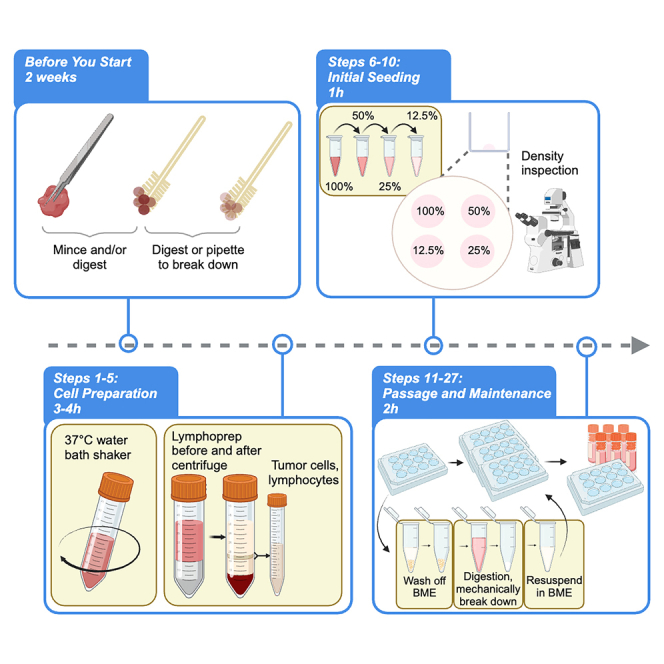
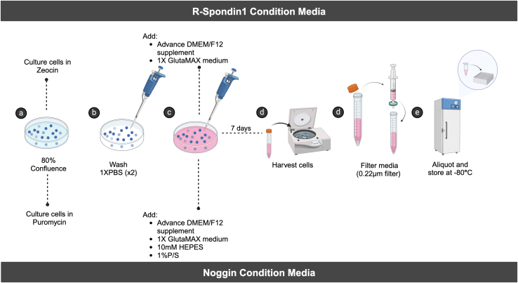
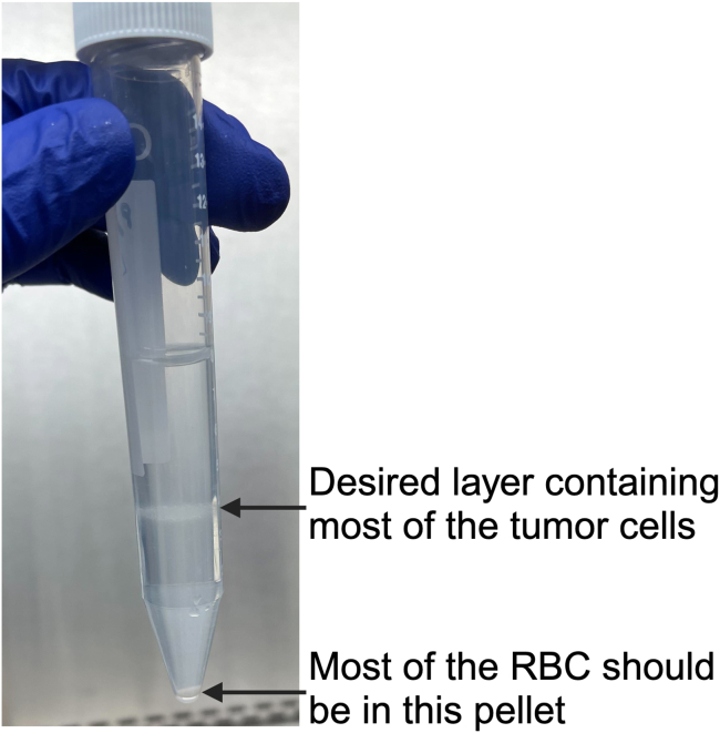
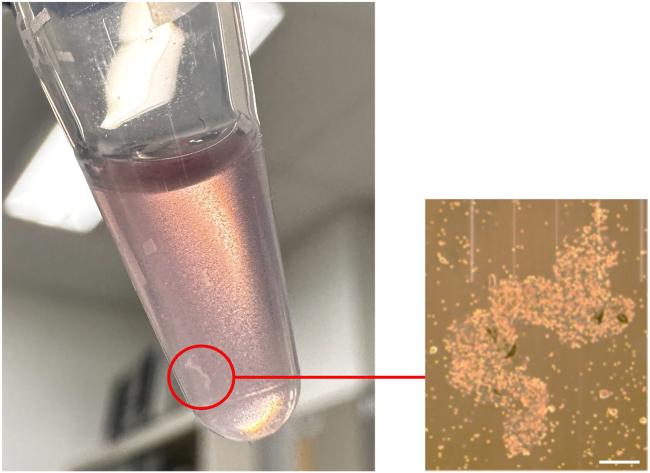
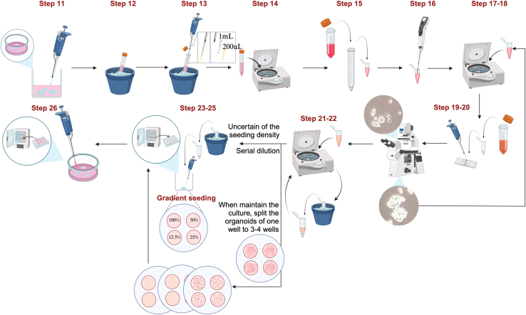
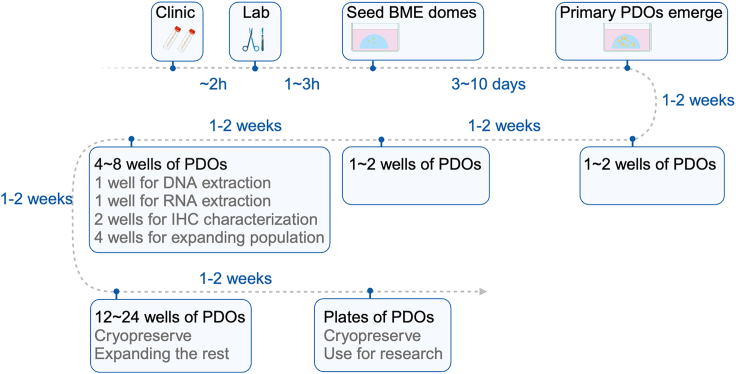
Herein, we present a protocol for culturing patient-derived organoids (PDOs) of cervical cancer that includes workflows for tumor biopsy/resection tissue and cytobrush-sampled cells. We describe steps for PDO culture initiation, including rinsing, gentle dissociation, Lymphoprep separation, and cell assessment, as well as seeding cells from surgical and cytobrush tissue digestion. We then provide guidance on PDO maintenance and passage and techniques for producing conditioned medium. Overall, this protocol serves as a valuable guide for establishing and maintaining cervical cancer PDOs. For complete details on the use and execution of this protocol, please refer to Colbert et al.1.

Keywords: cancer; genomics; metabolism; organoids.


Conflict of interest statement

Declaration of interests The authors declare no competing interests.

Figures

None
Graphical abstract
Figure 1
Figure 1
Schematic of the detailed protocol for preparing the conditioned media from Noggin- and R-spondin1–expressing 293T cells Created with BioRender.com.
Figure 2
Figure 2
Representative photomicrographs of cell types that can be seen in cytobrush samples The red arrows indicate RBCs, the white arrow indicates a tumor cell, the white asterisks indicate superficial squamous epithelial cells, and the area within the yellow dashed line represents debris. Scale bar = 100 μm.
Figure 3
Figure 3
Representative images of cytobrush samples The image in (A) shows fewer RBCs than does the image in (B). Most of the RBCs were seen on top of the cytobrushes. The medium in the right tube turned deep red owing to many RBCs released from the cytobrushes, whereas the left medium was pale pink.
Figure 4
Figure 4
Desired separation of cells and RBCs in Lymphoprep solution after centrifugation
Figure 5
Figure 5
Representative images of overdigested cells in TrypLE The enlarged microscopic image (right) shows a cell clump trapped by leaking DNA. Scale bar = 250 μm.
Figure 6
Figure 6
Schematic of the detailed protocol for maintenance and passage of PDOs with troubleshooting strategies for specific challenges Created with BioRender.com.
Figure 7
Figure 7
The general timeline for successful PDO line establishment In this timeline, we assume that the BME droplets are seeded in a 12- or 24-well plate at one 20- to 30-μL droplet per well. Created with BioRender.com.
Figure 8
Figure 8
Representative photomicrographs and characterization of successfully growing PDOs (A–C) Bright-field images of live culture PDOs derived from cervical adenocarcinoma (A) and cervical squamous cell carcinoma (B and C). (D and H) Hematoxylin and eosin (HE) staining of formalin-fixed, paraffin-embedded PDOs derived from cervical adenocarcinoma (D) and cervical squamous cell carcinoma (H). (E) Periodic acid-Schiff (PAS) staining of PDOs derived from cervical adenocarcinoma. (F and G) Immunofluorescent staining of PDOs derived from cervical adenocarcinoma for PAX8 and Ki67. (I and J) Immunofluorescent staining of PDOs derived from cervical squamous cell carcinoma for P63 and Ki67. Scale bar = 100 μm.
Figure 9
Figure 9
Representative image of undissolved BME in medium after being broken down into pieces and centrifuged

References

    1. Colbert L.E., El Alam M.B., Wang R., Karpinets T., Lo D., Lynn E.J., Harris T.A., Elnaggar J.H., Yoshida-Court K., Tomasic K., et al. Tumor-resident Lactobacillus iners confer chemoradiation resistance through lactate-induced metabolic rewiring. Cancer Cell. 2023;41:1945–1962.e11. doi: 10.1016/j.ccell.2023.09.012. - DOI - PMC - PubMed
    1. Heijmans J., van Lidth de Jeude J.F., Koo B.K., Rosekrans S.L., Wielenga M.C.B., van de Wetering M., Ferrante M., Lee A.S., Onderwater J.J.M., Paton J.C., et al. ER stress causes rapid loss of intestinal epithelial stemness through activation of the unfolded protein response. Cell Rep. 2013;3:1128–1139. doi: 10.1016/j.celrep.2013.02.031. - DOI - PubMed
    1. Lohmussaar K., Oka R., Espejo Valle-Inclan J., Smits M.H.H., Wardak H., Korving J., Begthel H., Proost N., van de Ven M., Kranenburg O.W., et al. Patient-derived organoids model cervical tissue dynamics and viral oncogenesis in cervical cancer. Cell Stem Cell. 2021;28:1380–1396.e6. doi: 10.1016/j.stem.2021.03.012. - DOI - PubMed
    1. Trillsch F., Czogalla B., Kraus F., Burges A., Mahner S., Kessler M. Protocol to optimize the biobanking of ovarian cancer organoids by accommodating patient-specific differences in stemness potential. STAR Protoc. 2023;4 doi: 10.1016/j.xpro.2023.102484. - DOI - PMC - PubMed
    1. Driehuis E., Gracanin A., Vries R.G.J., Clevers H., Boj S.F. Establishment of pancreatic organoids from normal tissue and tumors. STAR Protoc. 2020;1 doi: 10.1016/j.xpro.2020.100192. - DOI - PMC - PubMed

Publication types

Substances