. 2024 Oct 4;10(40):eadp5491.
doi: 10.1126/sciadv.adp5491. Epub 2024 Oct 2.

Substrate recognition principles for the PP2A-B55 protein phosphatase

Affiliations

Substrate recognition principles for the PP2A-B55 protein phosphatase

Thomas Kruse et al. Sci Adv. .

Abstract

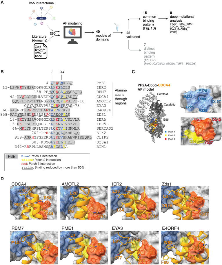
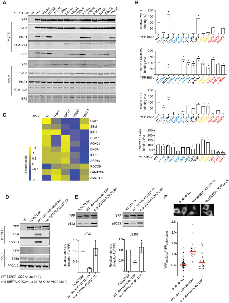
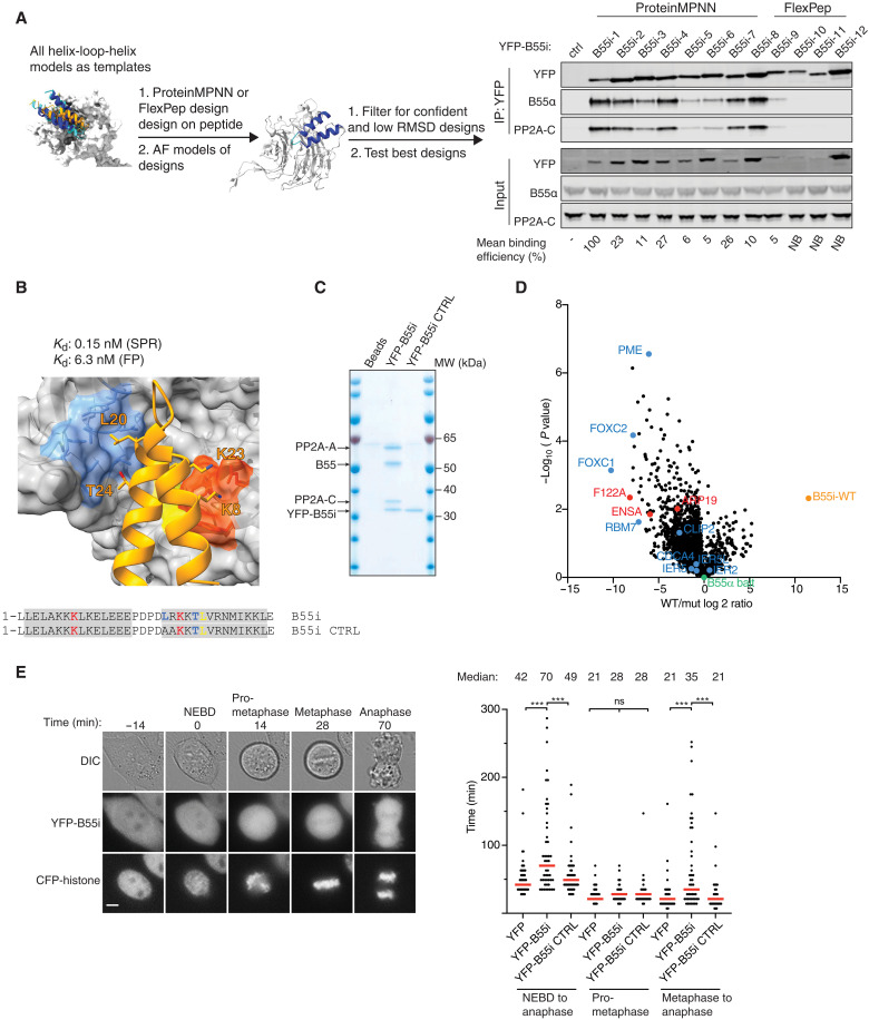
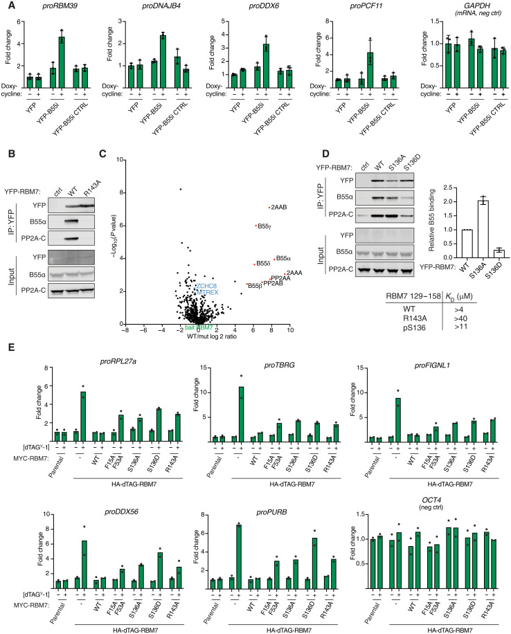
The PP2A-B55 phosphatase regulates a plethora of signaling pathways throughout eukaryotes. How PP2A-B55 selects its substrates presents a severe knowledge gap. By integrating AlphaFold modeling with comprehensive high-resolution mutational scanning, we show that α helices in substrates bind B55 through an evolutionary conserved mechanism. Despite a large diversity in sequence and composition, these α helices share key amino acid determinants that engage discrete hydrophobic and electrostatic patches. Using deep learning protein design, we generate a specific and potent competitive peptide inhibitor of PP2A-B55 substrate interactions. With this inhibitor, we uncover that PP2A-B55 regulates the nuclear exosome targeting (NEXT) complex by binding to an α-helical recruitment module in the RNA binding protein 7 (RBM7), a component of the NEXT complex. Collectively, our findings provide a framework for the understanding and interrogation of PP2A-B55 function in health and disease.


Figures

Fig. 1.
Fig. 1.. Helical motifs engage a conserved binding pocket on PP2A-B55.
(A) Schematic of pipeline to identify PP2A-B55 binding elements. Generated with BioRender. (B) Alignment of validated instances with helices in gray and residues contacting patch 1 in blue, patch 2 in yellow, and patch 3 in red. The first eight proteins on the list were the proteins that were scanned by single alanine mutagenesis, and residues found to reduce binding by at least 50% upon mutation to alanine are in italic. (C) Model of the PP2A-B55-CDCA4 complex with the different patches in B55 indicated in different colors. (D) AF2 models of the indicated proteins and their interaction with B55. Core residues forming interactions in the different patches are presented as sticks. AF, AlphaFold.
Fig. 2.
Fig. 2.. Helical motifs can act as substrate specifying elements.
(A) Indicated B55 mutants were purified and binding to IER2, PME1, CDCA4, and FAM122A determined and quantified. (B) Quantifications of relative binding of indicated proteins to B55 of two or four biological replicates and the averages are shown. (C) Heatmap illustrating the changes in binding pattern of the indicated proteins to the different B55 variants as determined by MS. The scale is log 2. n = 4 technical replicates. (D) CDCA4 WT or mutant SERTA domain was fused to FOXO3 2A, and binding to PP2A-B55 was monitored by Western blot. (E) As in (D) but immunoprecipitations (IPs) probed with FOXO3 phospho-specific antibodies as indicated. n = 3 biological replicates. Error bars are mean with SD. (F) Subcellular localization of FOXO3 fusion proteins by live-cell microscopy. Shown is a representative experiment of three independent experiments. Each dot represents a cell analyzed. Median is indicated with red line. Scale bar, 10 μM.
Fig. 3.
Fig. 3.. Generation of a specific B55 inhibitor.
(A) Design pipeline for generating a specific and tight binder of B55 and test of the top designs by immunopurification. (B) Model of the B55i bound to B55 and measured Kd and Ki indicated above. (C) Coomassie stained gel of B55i and B55i CTRL purified from HeLa cells. MW, molecular weight. (D) B55 was affinity purified after incubation with either B55i or B55i CTRL peptides, and samples were analyzed by MS. (E) Left: Mitotic duration in cells expressing B55i measured by time-lapse microscopy. Scale bar, 5 μM. Right: Quantification of (E). Shown are pooled data from three independent experiments. Each circle represents the timing of a single cell. Red line indicates the median time. A Mann-Whitney U test was applied. ns, not significant. ***P < 0.001.
Fig. 4.
Fig. 4.. PP2A-B55 regulates NEXT complex function through binding RBM7.
(A) Stable HeLa cell lines expressing the indicated constructs and RNA levels of indicated NEXT substrates measured by reverse transcription quantitative polymerase chain reaction (RT-qPCR). Quantifications of data from three biological replicates. Error bars show mean and SD. (B) IP of RBM7 constructs and monitoring binding to B55. (C) MS comparison of RBM7 WT and R143A with NEXT components highlighted in blue. (D) Binding of RBM7 S136A and S136D to PP2A-B55 and table of affinities measured of RBM7 peptides measured by SPR. Quantifications of data from three biological replicates. Error bars show mean and SD. (E) Endogenous RBM7 was tagged with dTAG, allowing the rapid removal of RBM7; cells were complemented with the indicated RBM7 variants; and the indicated RNAs were quantified by RT-qPCR. Quantifications of data from two biological replicates and the average are shown.

Update of

References

    1. Hunter T., Why nature chose phosphate to modify proteins. Philos. Trans. R. Soc. Lond. B Biol. Sci. 367, 2513–2516 (2012). - PMC - PubMed
    1. Jin J., Pawson T., Modular evolution of phosphorylation-based signalling systems. Philos. Trans. R. Soc. Lond. B Biol. Sci. 367, 2540–2555 (2012). - PMC - PubMed
    1. Johnson J. L., Yaron T. M., Huntsman E. M., Kerelsky A., Song J., Regev A., Lin T. Y., Liberatore K., Cizin D. M., Cohen B. M., Vasan N., Ma Y., Krismer K., Robles J. T., van de Kooij B., van Vlimmeren A. E., Andree-Busch N., Kaufer N. F., Dorovkov M. V., Ryazanov A. G., Takagi Y., Kastenhuber E. R., Goncalves M. D., Hopkins B. D., Elemento O., Taatjes D. J., Maucuer A., Yamashita A., Degterev A., Uduman M., Lu J., Landry S. D., Zhang B., Cossentino I., Linding R., Blenis J., Hornbeck P. V., Turk B. E., Yaffe M. B., Cantley L. C., An atlas of substrate specificities for the human serine/threonine kinome. Nature 613, 759–766 (2023). - PMC - PubMed
    1. Ubersax J. A., Ferrell J. E. Jr., Mechanisms of specificity in protein phosphorylation. Nat. Rev. Mol. Cell Biol. 8, 530–541 (2007). - PubMed
    1. Garvanska D. H., Nilsson J., Specificity determinants of phosphoprotein phosphatases controlling kinetochore functions. Essays Biochem. 64, 325–336 (2020). - PubMed

LinkOut - more resources