. 2024 Oct 4;10(40):eadm9801.
doi: 10.1126/sciadv.adm9801. Epub 2024 Oct 2.

Single-molecule tracking reveals dynamic regulation of ribosomal scanning

Affiliations

Single-molecule tracking reveals dynamic regulation of ribosomal scanning

Hea Jin Hong et al. Sci Adv. .

Abstract

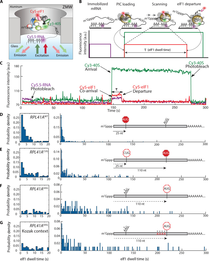
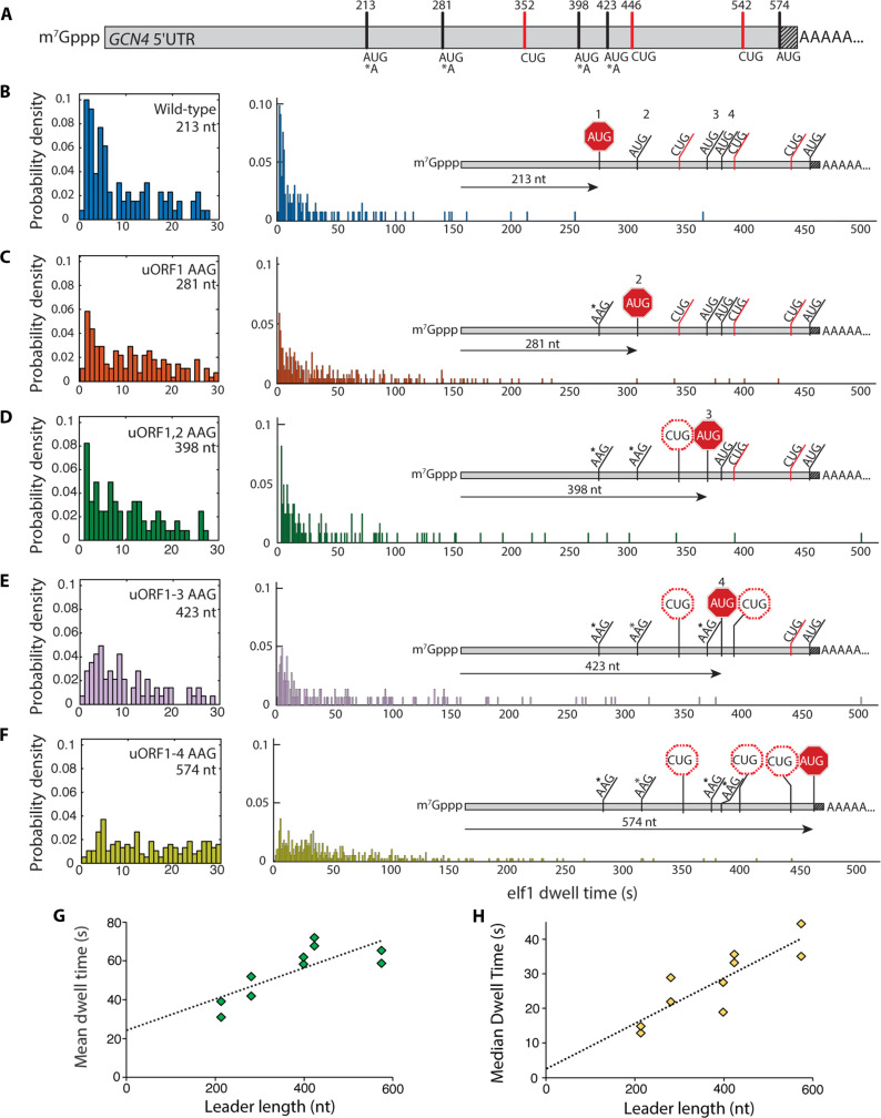
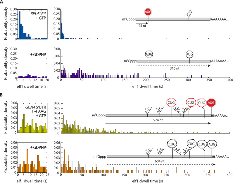
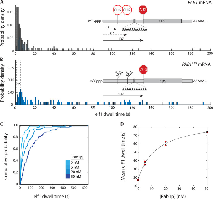
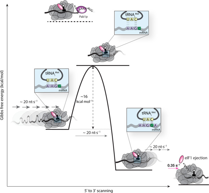
How eukaryotic ribosomes traverse messenger RNA (mRNA) leader sequences to search for protein-synthesis start sites remains one of the most mysterious aspects of translation and its regulation. While the search process is conventionally described by a linear "scanning" model, its exquisitely dynamic nature has restricted detailed mechanistic study. Here, we observed single Saccharomyces cerevisiae ribosomal scanning complexes in real time, finding that they scan diverse mRNA leaders at a rate of 10 to 20 nt s-1. We show that specific binding of a protein to its mRNA leader sequence substantially arrests scanning. Conversely, impairing scanning-complex guanosine 5'-triphosphate hydrolysis results in native start-site bypass. Our results illustrate an mRNA-centric, kinetically controlled regulatory model where the ribosomal pre-initiation complex amplifies a nuanced energetic landscape to regulate scanning and start-site selection fidelity.


Figures

Fig. 1.
Fig. 1.. Direct observation of scanning in real time.
(A) Experimental setup for single-molecule fluorescence scanning assays in zero-mode waveguides. (B) Idealized single-molecule fluorescence trace for scanning signal, indicating eIF1 dwell time, τ. (C) Representative trace with scanning signal observed on the wild-type RPL41A mRNA. (D to G) Representative eIF1 dwell-time distributions on wild-type RPL41A mRNA (n = 193 molecules), RPL41AAAG (n = 143), RPL41AAAG/Kozak (n = 94), and RPL41ACUG (n = 254) mRNAs, respectively. The distribution in the 0 to 20 s time domain is expanded to the left of the full-timescale distribution. Distributions from replicate experiments were statistically indistinguishable. Inset schematics indicate distances (in nucleotides) between the +1 nt and the initiation codon.
Fig. 2.
Fig. 2.. Scanning dynamics on the GCN4 leader.
(A) Schematic of the GCN4 leader indicating positions of canonical upstream ORF AUG codons, near-cognate CUG triplets, and the GCN4 main-ORF AUG codon. The shaded portion indicates 30 nt of the GCN4 coding sequence included in the transcript. (B to F) eIF1 dwell-time distributions for the wild-type GCN4 leader sequence and the four sequential upstream ORF AUG-to-AAG variants. Left panels expand the short-time component of the distributions. Insets indicate relative linear distance traversed by the PIC for each leader sequence. For (B) to (F), the number of molecules in each distribution was 136, 115, 165, 98, and 160, respectively. (G and H) Mean and median eIF1 dwell times plotted against the cap to AUG leader length in nucleotides. Trend lines are from linear regression to all datapoints.
Fig. 3.
Fig. 3.. Regulation of scanning by GTP hydrolysis.
(A and B) eIF1 dwell-time distributions for scanning on (A) RPL41Awt (n = 193 in the presence of GTP; n = 146 in the presence of GDPNP) and (B) the GCN4 quadruple uORF AUG-to-AAG variant (n = 378 in the presence of GTP; n = 172 in the presence of GDPNP), contrasted between PICs formed with GTP versus GDPNP in the eIF2 TC (top versus bottom panel, respectively).
Fig. 4.
Fig. 4.. Regulation of scanning by S. cerevisiae poly(A)-binding protein.
(A) eIF1 dwell-time distribution for the PAB1 mRNA (n = 125 molecules). Inset: Relative locations of upstream CUG triplets and PAB1 main-ORF AUG codon, and location of internal oligo(A) 11-mer. (B) eIF1 dwell-time distribution for the PAB1AAG mRNA, in which both upstream CUG triplets are substituted by AAG (n = 95). (C) eIF1 dwell-time cumulative distribution functions for scanning on the PAB1 mRNA in the presence of varying concentrations of the poly(A)-binding protein, Pab1p (5 nM, n = 110; 20 nM, n = 103; 50 nM, n = 112). (D) Dependence of mean eIF1 dwell time on PAB1 mRNA on the concentration of Pab1p, with hyperbolic fit (Kd 13 nM; 95% confidence intervals 1.1, 25.6 nM). Replicate data points at 0 and 50 nM Pab1p overlap. The time added to scanning at saturating Pab1p concentration is 72 s (95% confidence intervals: 64 to 79 s).
Fig. 5.
Fig. 5.. Energetics of scanning.
The measured scanning rate of ~20 nt s−1 implies an energy barrier of ~16 kcal mol−1, on average, for PIC movement to the next nucleotide at each scanning step. The magnitude of this barrier corresponds to breakage of the hydrogen bonds in base-paired tRNA and mRNA; this base pairing by itself has an overall stability in the range of −3 kcal mol−1, with an enthalpic component of around −15 kcal mol−1. On the left of the schematic, one possible interaction during scanning includes five hydrogen bonds formed between a near-cognate mRNA triplet with a noncognate tRNA. Thermal fluctuation that disengages the base pair is expected to traverse an activation barrier equal to or exceeding the enthalpy of the hydrogen-bond interactions, as measured by our assay. Steric hindrance from Pablp binding to an oligo(A) site abutting the PIC increases this activation barrier, resulting in blocked scanning. eIF1 ejection stabilizes the PIC to the point where no further forward motion is possible.

References

    1. Aitken C. E., Lorsch J. R., A mechanistic overview of translation initiation in eukaryotes. Nat. Struct. Mol. Biol. 19, 568–576 (2012). - PubMed
    1. Kozak M., How do eucaryotic ribosomes select initiation regions in messenger RNA? Cell 15, 1109–1123 (1978). - PubMed
    1. Peters J. M., Vangeloff A. D., Landick R., Bacterial transcription terminators: The RNA 3′-end chronicles. J. Mol. Biol. 412, 793–813 (2011). - PMC - PubMed
    1. Richardson J. P., Rho-dependent termination and ATPases in transcript termination. Biochim. Biophys. Acta 1577, 251–260 (2002). - PubMed
    1. Berthelot K., Muldoon M., Rajkowitsch L., Hughes J., McCarthy J. E. G., Dynamics and processivity of 40S ribosome scanning on mRNA in yeast. Mol. Microbiol. 51, 987–1001 (2004). - PubMed

Publication types

MeSH terms

Substances