. 2024 Oct 4;10(40):eadp0466.
doi: 10.1126/sciadv.adp0466. Epub 2024 Oct 2.

Pachytene piRNAs control discrete meiotic events during spermatogenesis and restrict gene expression in space and time

Affiliations

Pachytene piRNAs control discrete meiotic events during spermatogenesis and restrict gene expression in space and time

Jacob Ortega et al. Sci Adv. .

Abstract

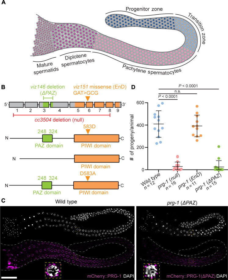
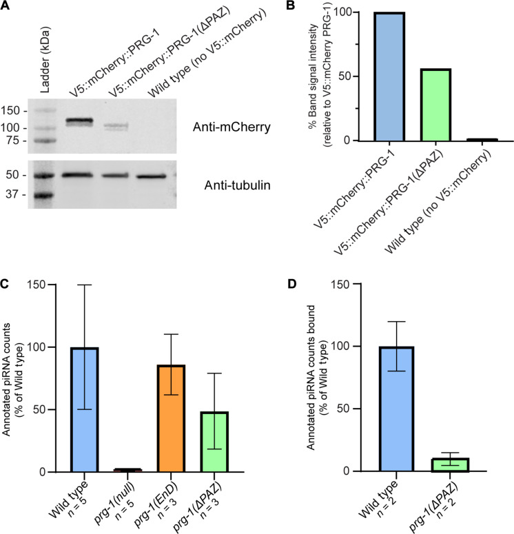
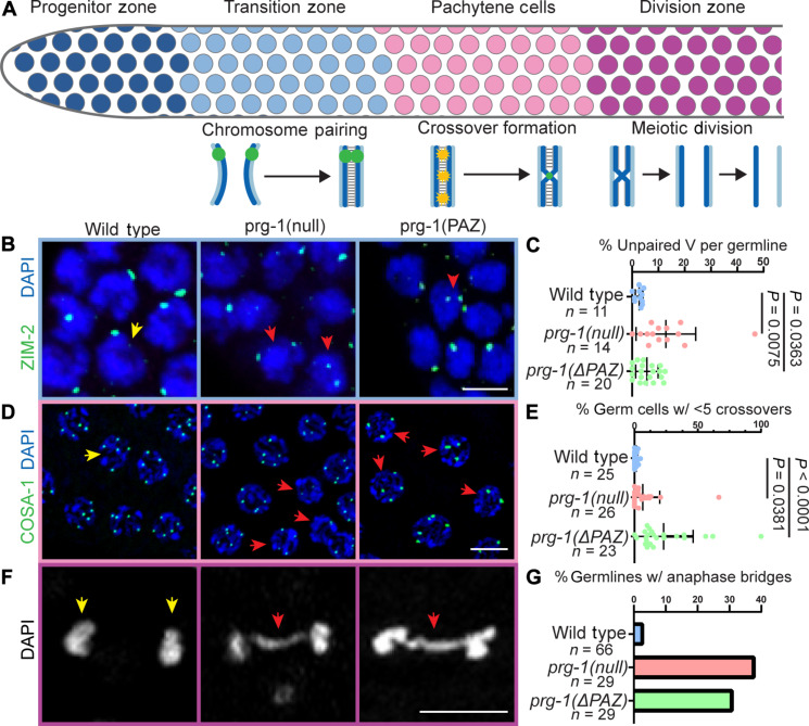
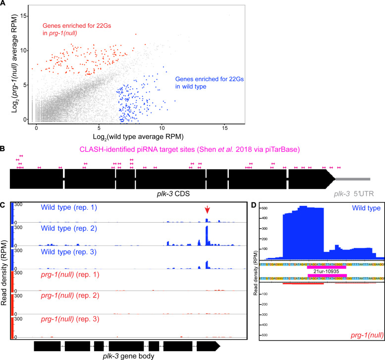
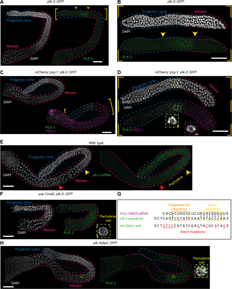
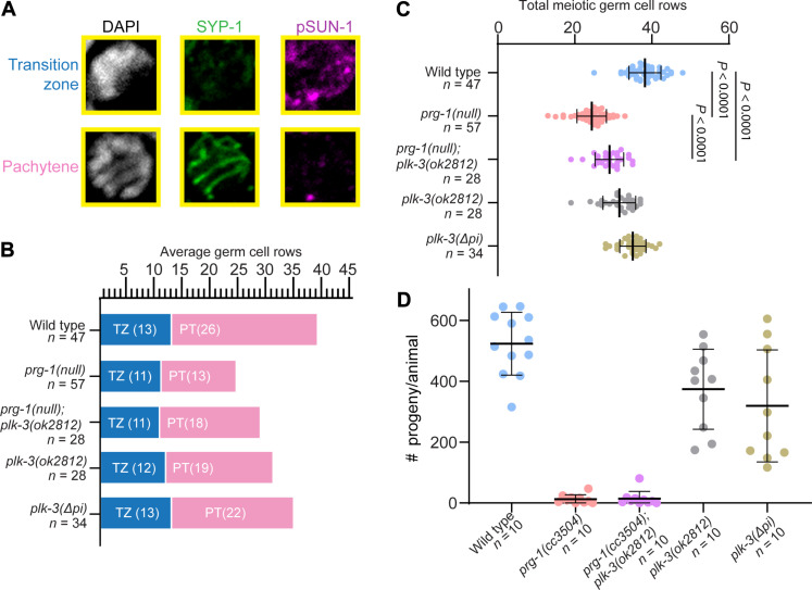
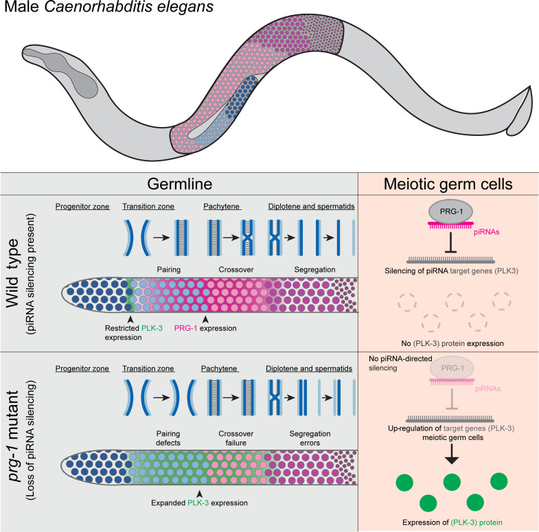
Pachytene piRNAs, a Piwi-interacting RNA subclass in mammals, are hypothesized to regulate non-transposon sequences during spermatogenesis. Caenorhabditis elegans piRNAs, the 21URNAs, are implicated in regulating coding sequences; the messenger RNA targets and biological processes they control during spermatogenesis are largely unknown. We demonstrate that loss of 21URNAs compromises homolog pairing and makes it permissive for nonhomologous synapsis resulting in defects in crossover formation and chromosome segregation during spermatogenesis. We identify Polo-like kinase 3 (PLK-3), among others, as a 21URNA target. 21URNA activity restricts PLK-3 protein to proliferative cells, and expansion of PLK-3 in pachytene overlaps with the meiotic defects. Removal of plk-3 results in quantitative genetic suppression of the meiotic defects. One discrete 21URNA inhibits PLK-3 expression in late pachytene cells. Together, these results suggest that the 21URNAs function as pachytene piRNAs during C. elegans spermatogenesis. We identify their targets and meiotic events and highlight the remarkable intricacy of this multi-effector mechanism during spermatogenesis.


Figures

Fig. 1.
Fig. 1.. Loss of the PRG-1 PAZ domain regulates male fertility while retaining germ granule association.
(A) Schematic of a male C. elegans germline displaying the spatiotemporal arrangement of germ cell development. Cells of the progenitor zone (oriented left in the schematic) are located at the most distal end of the germline and enter meiosis in the TZ. Progression through meiosis I continues in the pachytene and diplotene stages before meiotic divisions giving rise to the mature spermatids in the most proximal portion of the germline. (B) prg-1 gene structure with corresponding protein structure. PAZ-domain (green) and PIWI-domain (orange) functions were ablated with targeted CRISPR mutations (Materials and Methods) creating ΔPAZ and RNase-dead (RD) alleles. (C) Representative 4′,6-diamidino-2-phenylindole (DAPI)–stained (white) adult male germlines displaying expression of wild-type PRG-1 and PRG-1(ΔPAZ) (magenta) and subcellular localization around the nucleus in pachytene cells (inset, yellow boxes). (D) Quantitative analysis of male fertility in prg-1 mutant alleles. Statistical significance was calculated by Student’s t test; mutant groups were compared to wild type. Scale bar, 50 μm. Each experiment was conducted at least in triplicate and over at least 25 to 30 germlines analyzed each time.
Fig. 2.
Fig. 2.. Loss of the PRG-1 PAZ domain reduces PRG-1 expression and disrupts piRNA loading.
(A) Western blot data demonstrating reduction of PRG-1 expression after PAZ-domain deletion relative to a tagged control strain. (B) Quantification of band signal intensity from Western blot after normalization to tubulin loading control. Signal intensity is given as a percentage relative to tagged control strain signal. (C) Quantification of piRNA counts from sequenced samples as a percentage of average piRNA reads from wild-type samples, normalized to total mapping reads. (D) Quantification of piRNA counts from RNA IP experiments as a percentage of average piRNA reads from wild-type samples, normalized to total mapping reads.
Fig. 3.
Fig. 3.. Multiple meiotic processes are defective in prg-1 mutants.
(A) Schematic of meiotic progression and meiotic events by germ cell stage. Homolog pairing occurs in the TZ, followed by crossover formation in pachytene stage. Meiotic divisions occur in the division zone following diplotene stage. (B) Immunolocalization of ZIM-2 (green) in DAPI-stained nuclei (blue) of TZ cells. Two foci in a nucleus indicates unpaired homologs. One focus in a nucleus indicates paired homologs. Red arrowheads indicate nuclei with unpaired homologs. (C) Quantification of the percentage of germ cells with unpaired homologs per germline analyzed. (D) Localization of GFP::COSA-1 (green) in late pachytene nuclei stained with DAPI (blue). Red arrowheads indicate nuclei with fewer than five crossovers. (E) Quantification of the percentage of germ cells with less than five crossovers per nucleus per germline analyzed. (F) Representative DAPI (DNA, white) images of meiotically dividing germ cells from the division zone. Red arrowheads indicate anaphase bridges. (G) Quantification of percentage of germlines displaying meiotically dividing cells with anaphase bridges. Statistical significance was calculated by Student’s t test, mutant groups were compared to wild type. Scale bars, 5 μm. Each experiment was conducted at least in triplicate and over at least 25 to 30 germlines analyzed each time and at least 500 germ cells each time.
Fig. 4.
Fig. 4.. The piRNA pathway targets genes involved in meiosis and germ cell development, including plk-3, a member of the PLK family.
(A) Scatterplot of mapped 22G reads per C. elegans gene in matched wild-type versus prg-1 mutant samples. Highlighted genes met the 2log (2) cutoff for enrichment of 22G reads in either wild-type (blue) or prg-1 mutant (red). (B) Schematic displaying CLASH-identified piRNA binding sites within the plk-3 coding sequence. (C) Histogram for 22G reads mapped to plk-3 in wild-type (blue) or prg-1 mutant (red) replicates. Red arrowhead denotes cluster of 22G reads that coincided with 21ur-10935 binding site. (D) Magnification of histogram including all wild-type (top, blue) or prg-1 mutant (bottom, red) 22G reads mapping to plk-3 around the location of the 21ur-10935 binding site (magenta).
Fig. 5.
Fig. 5.. PLK-3 expression is restricted to the distal germline by piRNA function.
(A and B) Wild-type adult male germline expressing PLK-3::GFP (green) stained with DAPI (white). (A) PLK-3::GFP is expressed in the distal regions of the germline in the areas noted by the yellow arrowheads. (B) Zoomed in region of the distal germline from (A), displaying PLK-3::GFP expression between the progenitor zone and meiotic regions of the germline. (C and D) Wild-type adult male germline expressing PLK-3::GFP (green) and V5::mCherry::PRG-1 (magenta) stained with DAPI (white). (D) Zoomed in distal germline from (C), showing that PLK-3 expression is spatially expressed before and almost mutually exclusive with PRG-1 expression at the end of the progenitor zone, but PRG-1 expression is highest in the meiotic region. Inset 1 (yellow, dashed lines) shows a single TZ nucleus with visible PLK-3 perinuclear granules, whereas inset 2 (red, dashed lines) shows a pachytene nucleus with PRG-1 perinuclear granules. (E) Wild-type adult male germline stained with DAPI (white) and plk-3 HCR-FISH, showing expression of plk-3 mRNA throughout the distal regions with the mRNA expression significantly reduced in mid/late pachytene (red arrowhead). (F) A prg-1(null) adult male germline expressing PLK-3::GFP (green) stained with DAPI (white). Inset (yellow, dashed lines) displays single pachytene nucleus in the meiotic region expression PLK-3::GFP. (G) Schematic displaying the plk-3 coding sequence where 21ur-10935’s target sequence maps. Below, plk-3(Δpi) designates CRISPR-edited sequence with specific base changes noted (red, underlined). Predicted base-pairing within seed sequence (yellow box) and supplemental sequence (orange box) is noted. (H) Germline with PLK-3(Δpi)::GFP (green) stained with DAPI (white). Inset (yellow, dashed lines) displays pachytene nucleus in the meiotic region expressing PLK-3(Δpi)::GFP. Scale bars, 50 μm. Each experiment was conducted at least in triplicate and over at least 25 to 30 germlines analyzed each time.
Fig. 6.
Fig. 6.. Loss of plk-3 partially rescues prg-1 meiotic progression defects.
(A) Representative images of TZ and pachytene germ cells stained with DAPI (white), SYP-1 (green), and pSUN-1 (magenta). (B) Graphical representation of average TZ and pachytene cell rows per genotype across germlines analyzed. (C) Quantification of the average total meiotic germ cell rows. Numbers graphed are averages across germlines analyzed. Statistical significance between groups was calculated by one-way analysis of variance (ANOVA) with Bonferroni correction; comparisons not shown on graph: Wild type versus plk-3(ok2812), P < 0.0001; wild type versus plk-3(Δpi), P = 0.0039; prg-1(null);plk-3(ok2812) versus plk-3(Δpi), P < 0.0001; prg-1(null) versus plk-3(ok2812), P < 0.0001; prg-1(null) versus plk-3(Δpi), P < 0.0001; plk-3(ok2812) versus plk-3(Δpi), P = 0.0050. (D) Brood sizes for corresponding genotypes from (B) and (C). Each experiment was conducted at least in triplicate and over at least 25 to 30 germlines analyzed each time.
Fig. 7.
Fig. 7.. Pachytene piRNAs control spatiotemporal gene expression in the male germline.
Schematic model for regulation of spatiotemporal gene expression in the male germline by the piRNA pathway. In wild-type animals, PRG-1 is expressed most highly beginning in the TZ through the pachytene stage. This expression corresponds with essential meiotic processes such as pairing of homologous chromosomes and crossover formation. Conversely, PLK-3 is only expressed between the progenitor zone and the early TZ. In PRG-1 mutants, the loss of piRNAs leads to meiotic defects and desilencing of PLK-3 during meiotic stages. Together, our data suggest that loss of piRNA silencing such as PLK-3 during meiosis underlies observed meiotic defects.

References

    1. Bartel D. P., Metazoan microRNAs. Cell 173, 20–51 (2018). - PMC - PubMed
    1. Czech B., Munafo M., Ciabrelli F., Eastwood E. L., Fabry M. H., Kneuss E., Hannon G. J., piRNA-guided genome defense: From biogenesis to silencing. Annu. Rev. Genet. 52, 131–157 (2018). - PMC - PubMed
    1. Okamura K., Lai E. C., Endogenous small interfering RNAs in animals. Nat. Rev. Mol. Cell Biol. 9, 673–678 (2008). - PMC - PubMed
    1. Aravin A., Gaidatzis D., Pfeffer S., Lagos-Quintana M., Landgraf P., Iovino N., Morris P., Brownstein M. J., Kuramochi-Miyagawa S., Nakano T., Chien M., Russo J. J., Ju J., Sheridan R., Sander C., Zavolan M., Tuschl T., A novel class of small RNAs bind to MILI protein in mouse testes. Nature 442, 203–207 (2006). - PubMed
    1. Vagin V. V., Sigova A., Li C., Seitz H., Gvozdev V., Zamore P. D., A distinct small RNA pathway silences selfish genetic elements in the germline. Science 313, 320–324 (2006). - PubMed

MeSH terms

Substances