

Influence of the digital file format on radiographic diagnostic in dentistry: a scoping review
- PMID: 39356906
- PMCID: PMC11441824
- DOI: 10.1590/1807-3107bor-2024.vol38.0100
Influence of the digital file format on radiographic diagnostic in dentistry: a scoping review
Abstract
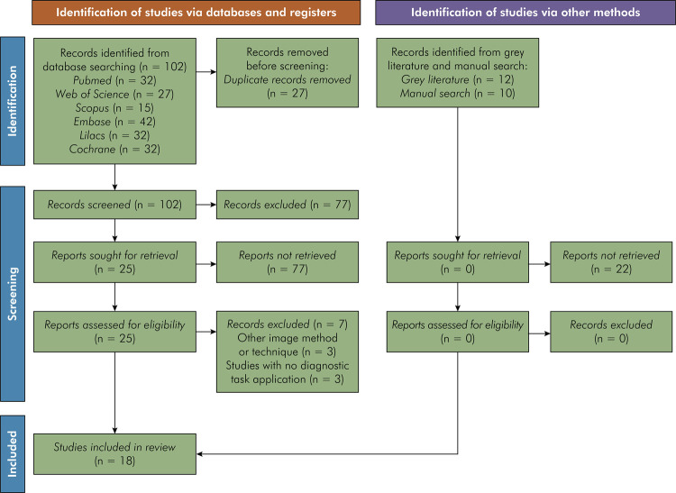
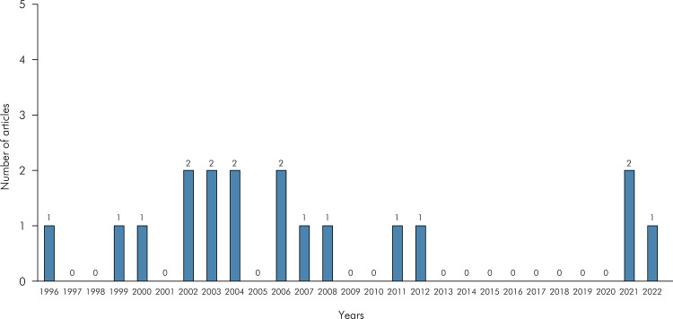
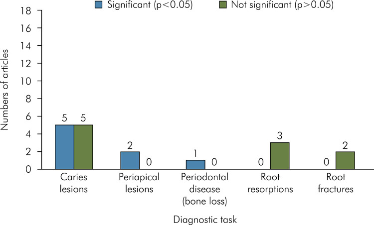
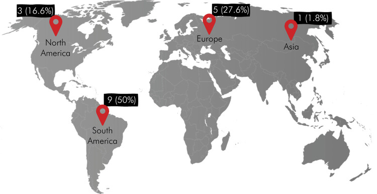
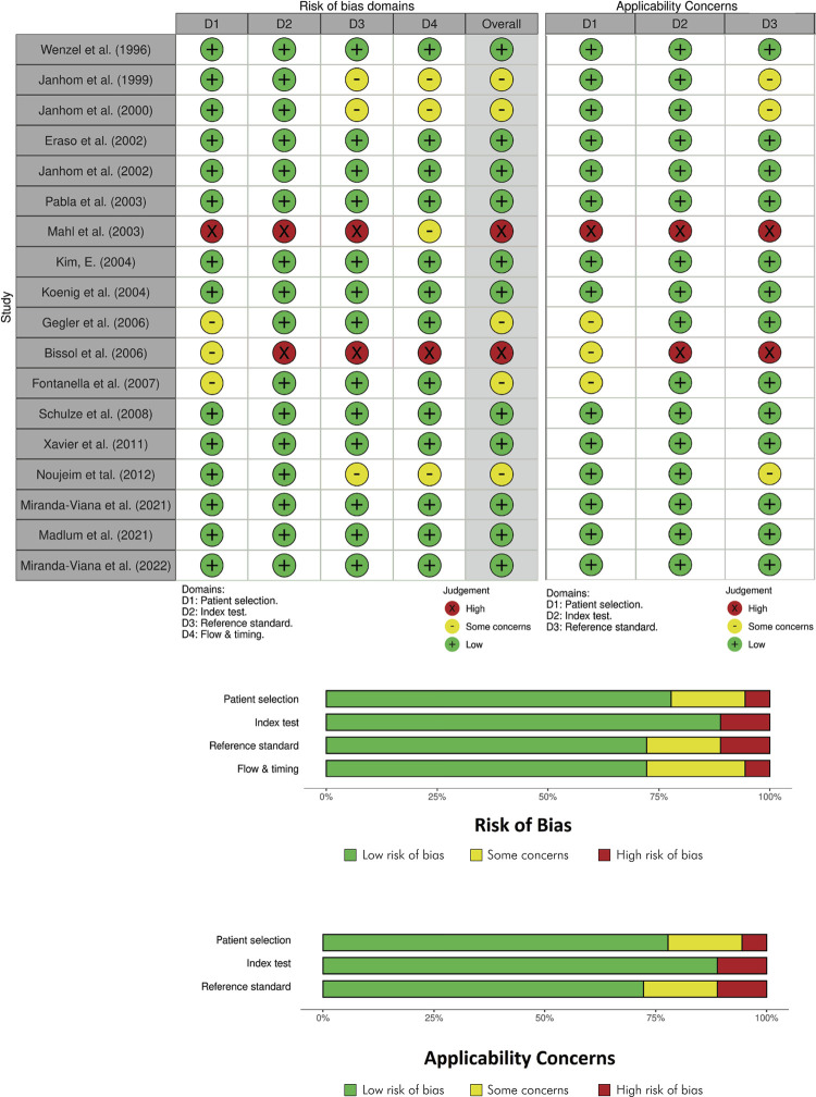
Given today's higher demand for online transmission of radiographic images, clinicians and regulatory agencies should be given the evidence they need to guide them in choosing the best image file format to be adopted. To this end, the present scoping review aims to explore, map, and evaluate the literature, with the object of reporting the influence of image file formats on dental diagnostic tasks by assessing intraoral radiographic images. This scoping review complies with PRISMA-ScR. It was customized to assess the risk of bias of the included studies, and was registered on the Open Science Framework platform. The data extraction protocol was developed based on the PCC acronym. An electronic search was conducted in six databases (Pubmed, Web of Science, Scopus, Embase, Lilacs, Cochrane) in December 2023. Original articles were screened, having observational, diagnostic accuracy, and consisting of in vivo or ex vivo laboratory studies investigating the influence of file formats on different diagnostic tasks in dentistry. Eighteen studies, published between the years 1996 and 2022, were included. The following data were extracted from the selected articles: article title, authors' citation, publication date, country, diagnostic task, image file formats tested, compression level, and main conclusion. The most widely investigated diagnostic task was caries lesions (n = 10), led by root resorptions (n = 3), root fractures (n = 2), periapical lesions (n = 2), and periodontal disease (n = 1). The most commonly used radiographic techniques were periapical (n = 12) and bitewing (n = 6). The most frequently investigated image file formats were JPEG (all studies) and TIFF (n = 10 studies). BMP, PNG, and JPEG2000 were also included in 7, 3 and 3 studies, respectively. No studies included the DICOM file format. In regard to the subjective assessment of the several dental diagnostic tasks, the studies mostly showed that the influence of the file formats was not significant (n = 10/55.5%). As for the quality assessment of the included papers, more than 70% of the studies featured a low risk of bias. Current evidence on image file formats and dental radiographic diagnosis is reliable. Any image file format can be used without impairing diagnostic accuracy.
Conflict of interest statement
Figures
References
Publication types
MeSH terms
LinkOut - more resources
Full Text Sources
Research Materials
