. 2024 Nov:57:101060.
doi: 10.1016/j.neo.2024.101060. Epub 2024 Oct 1.

Distinction of papillary and adamantinomatous craniopharyngioma: Clinical features, surgical nuances and hypothalamic outcomes

Affiliations

Distinction of papillary and adamantinomatous craniopharyngioma: Clinical features, surgical nuances and hypothalamic outcomes

Le Yang et al. Neoplasia. 2024 Nov.

Abstract

Objective: Understanding the differences of suprasellar papillary and adamantinomatous craniopharyngiomas (PCPs/ACPs) is pivotal for target therapy, surgical strategy or postoperative management. Here, the clinical features, surgical nuances and postoperative hypothalamic outcomes of PCPs were systematically recapitulated.

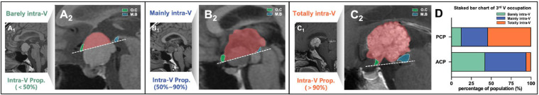
Methods: 24 PCPs and 52 ACPs underwent initial surgery were retrospectively reviewed. Clinical data, quantified third ventricle (3rd V) occupation and optic chiasm distortion were compared, as well as intra-operative findings, operating notes and prognosis. Moreover, analysis of tumor/3rd V relationship and hypothalamic outcomes were also performed.

Results: Tumors were more likely to occupies the 3rd V cavity in PCPs. Chiasm distortion of "compressed forward" was the most common pattern (45.8 %) in PCPs, whereas "stretched forward" pattern accounted the highest (42.5 %) in ACPs. Besides, round-shaped with less calcification, duct-like recess, solid consistency, rare subdiaphragmatic invasion, visible lower stalk and improved postoperative visual outcome were more frequently observed in PCPs. The basal membranes of the tumor epithelium and the reactive gliosis were separated by a layer of collagen fibers in most PCPs, which differs from ACPs in the morphological examination of tumor/3rd V floor interface. In daytime sleepiness and memory difficulty, the PCPs showed significantly better outcomes than the ACPs groups, and PCPs suffered less postoperative weight gain (p < 0.05) than ACPs among adult-onset cases.

Conclusion: PCPs are different from ACPs regards the clinical features, operative techniques and outcomes. If necessary, PCPs are suggested more amenable to total removal since its less invasiveness to the 3rd V floor and better hypothalamic outcomes.

Keywords: Clinical characteristics; Hypothalamic outcomes; Papillary craniopharyngioma; Surgical technique; Third ventricle floor; Tumor topography.


Conflict of interest statement

Declaration of competing interest The authors declare that they have no known competing financial interests or personal relationships that could have appeared to influence the work reported in this paper.

Figures

Fig 1
Fig. 1
3rd V occupation of T-CPs based on intra-ventricular proportion of tumor mass. Intra-ventricular occupation of tumor mass is calculated as the proportion of the tumor above the reference dotted line that from ventral O.C (green) to ventral M.B (blue) in midsagittal MRI. 3rd V occupation is categorized in 3 types: Barely intra-ventricular type (intra-ventricular proportion<50 %, A1-A2); Mainly intra-ventricular type (intra-ventricular proportion within 50-90 %, B1-B2) and Totally intra-ventricular type (intra-ventricular proportion>90 %, C1-C2). (D) Stacked bar chart of 3rd V occupation among T-type PCP and ACP group. 3rd V, third ventricle; Intra-V Prop., intra-ventricular proportion; M.B, mamillary body; O.C, optic chiasm.
Fig 2
Fig. 2
Five main patterns of optic chiasm distortion caused by T-CPs identified by preoperative midsagittal MRI and corresponding intra-operative view. (A1) Non-distorted or normal positioned O.C: Normal morphology in MRI and verified with surgical view (A2); (B1) Compressed downward O.C: Locates at the antero-inferior margin of the tumor due to its downward compressive force. A distorted O.C that bulged down could be seen under endoscopic view (B2); (C1) Compressed forward O.C: Displaced forward by the tumor and compressed against the tuberculum sellae. The O.C being pushed forward is visible after remove the tuberculum sellae (C2); (D1) Stretched forward O.C: Stretched along the anterior margin of the tumor. The O.C is bended forward by the suprasellar tumor under endoscopic view (D2); (E1) Stretched upward O.C: Stretched along the superior surface of the tumor. The O.C is bended upward and barely be seen since the tumor mass occupied the suprasellar cistern (E2). (F) Stacked bar chart of O.C distortion among T-type PCP and ACP group. O.C, optic chiasm; P.G, pituitary gland; TU, tumor.
Fig 3
Fig. 3
Illustrative cases of PCP with radiological findings and intra-operative observation. (A-C) Pre-operative CT and MRI of a “Totally intra-V” type PCP. The O.C remained with normal morphology and tumor occupies the 3rd V chamber without calcification; (D) Under EEA, normal positioned O.C is confirmed. Note the chiasm-tuberculum distance is narrow and the tumor mass is hidden above the 3rd V floor; (E) The tumor is dissected precisely from the infundibular recess; (F) Intraventricular tumor is easily stripped away from the 3rd V wall through a cleavage pial plane (white dotted line); (G) Choroid plexus (pentagram) and each foramen of Monro (asterisk) are visible when tumor is removed en bloc; (H) Overall view after tumor removed totally; (I-J) MRI obtained 4 months after surgery. 3rd V, third ventricle; O.C, optic chiasm; O.N, optic nerve; P.S, pituitary stalk; P.G, pituitary gland; TU, tumor.
Fig 4
Fig. 4
Illustrative cases of ACP with radiological findings and intra-operative observation. (A-C) Pre-operative CT and MRI of a “Mainly intra-V” type ACP. MRI indicates O.C as “stretched forward” pattern and CT scan reveals the calcification; (D) Under EEA, O.C that bended forward by the suprasellar tumor is confirmed. (E) Precisely handling of the perforating arteries that supplying the suprasellar part of tumor; (F) Blunt dissecting the suprasellar part from the O.C along the plane in between (white dotted line); (G) Intraventricular tumor is easily stripped away from the 3rd V wall through a cleavage pial plane (white dotted line); (H) Overall view after tumor removed totally; (I-J) MRI obtained 2 weeks after surgery. 3rd V, third ventricle; M.B, mamillary body; O.C, optic chiasm; O.N, optic nerve; P.S, pituitary stalk; P.G, pituitary gland; P.CoA, posterior communicating artery; TU, tumor.
Fig 5
Fig. 5
Histological observation in tumor–ventricle floor interface and post-operative weight gain in PCPs/ACPs. Immunofluorescent staining shows a layer of gliosis (GFAP positive) with various thickness between the tumor (Pan-CK positive) and the neural tissue of the third ventricle floor in PCP (A1–A3) and ACP (B1–B3). PCP in (C1–C3) shows that the basal membranes of tumor epithelium (Pan-CK positive) and reactive gliosis were separated by a layer of fibers (collagen positive, white arrow). ACP in (D1–D3) indicates a finger-like structure in which the tumor forms a finger-like bulge into the third ventricle floor, with the absence of the pia mater (Laminin β1 positive) at some areas (yellow arrowhead) and the development of gliosis between the tumor and the neural tissue of the third ventricle floor. Scale bar =200 μm. (E–F) The trend and comparison of obesity rate from the pre-operative period to the last follow-up. (G) shows the changes of BMI in groups. The BMI in pre-operative period is considered as the baseline value, △BMI in (H) indicates the difference between the peak value and the baseline (*p < 0.05). △BMI in (I) indicates the difference between the latest value (documented in the last follow-up) and the baseline (**p < 0.01). 3VF, third ventricle floor; Tu, tumor; mo, months; pre-op, pre-operative period; F-U, follow-up.

Similar articles

References

    1. Yamini B, Narayanan M. Craniopharyngiomas: an update. Expert. Rev. Anticancer Ther. 2006;6(Suppl 9):S85–S92. doi: 10.1586/14737140.6.9s.S85. - DOI - PubMed
    1. Prieto R, Barrios L, Pascual JM. Papillary craniopharyngioma: a type of tumor primarily impairing the hypothalamus - a comprehensive anatomo-clinical characterization of 350 well-described cases. Neuroendocrinology. 2022;112:941–965. doi: 10.1159/000521652. - DOI - PubMed
    1. Brastianos PK, Twohy E, Geyer S, et al. BRAF-MEK inhibition in newly diagnosed papillary craniopharyngiomas. N. Engl. J. Med. 2023;389:118–126. doi: 10.1056/NEJMoa2213329. - DOI - PMC - PubMed
    1. Brastianos PK, Taylor-Weiner A, Manley PE, et al. Exome sequencing identifies BRAF mutations in papillary craniopharyngiomas. Nat. Genet. 2014;46:161–165. doi: 10.1038/ng.2868. - DOI - PMC - PubMed
    1. Sartoretti-Schefer S, Wichmann W, Aguzzi A, Valavanis A. MR differentiation of adamantinous and squamous-papillary craniopharyngiomas. AJNR Am. J. Neuroradiol. 1997;18:77–87. - PMC - PubMed

Publication types