

doi: 10.52054/FVVO.16.3.041.
European Society for Gynaecological Endoscopy (ESGE) Good Practice Recommendations on surgical techniques for removal of fibroids: part 1 abdominal (laparoscopic and open) myomectomy
- PMID: 39357857
- PMCID: PMC11569431
- DOI: 10.52054/FVVO.16.3.041
Item in Clipboard
European Society for Gynaecological Endoscopy (ESGE) Good Practice Recommendations on surgical techniques for removal of fibroids: part 1 abdominal (laparoscopic and open) myomectomy
Facts Views Vis Obgyn.
2024 Sep.
Abstract
Uterine fibroids are the most common benign tumours of the female reproductive tract and can cause a range of symptoms including abnormal uterine bleeding, pain, pressure symptoms and subfertility. Surgery may be required for some symptomatic fibroids via abdominal or transvaginal routes. The European Society for Gynaecological Endoscopy Uterine Fibroids Working Group developed recommendations based on the best available evidence and expert opinion for the surgical treatment of uterine fibroids. In this first part of the recommendations, abdominal approaches to surgical treatment of fibroids including laparoscopic, robot- assisted and open myomectomy are described.
Figures
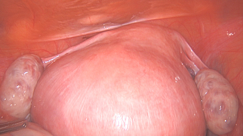
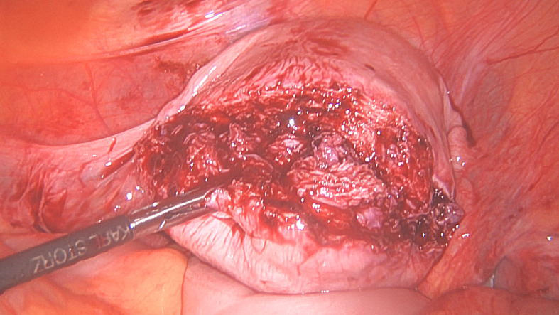
Surgical steps for laparoscopic removal of a large posterior wall fibroid. Figure 1A. A 9 cm fibroid protruding outwards on the posterior wall described as intramural by MRI.
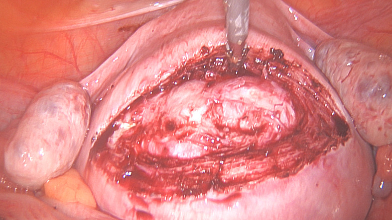
The incision over the fibroid to reach the fibroid before extending it to the desired length.
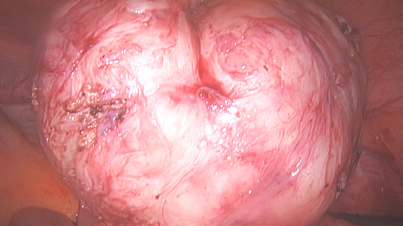
Removed fibroid.
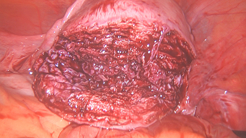
Uterine myometrial defect after fibroid removal.
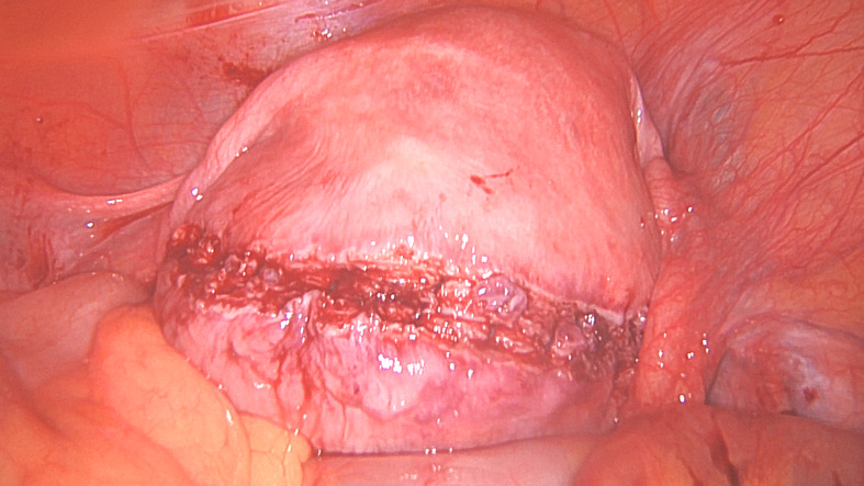
The first layer myometrial repair completed.
Second layer myometrial repair completed.
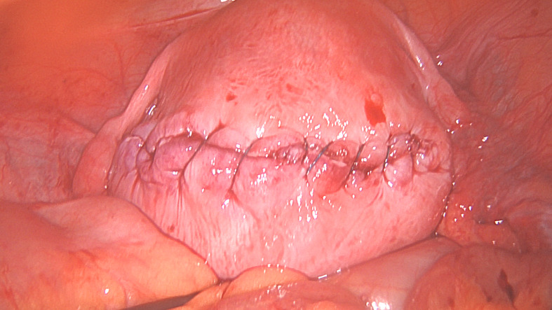
Serosal closure.
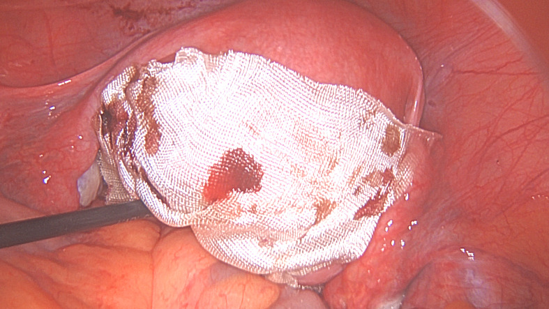
Second layer myometrial repair completed. G. Serosal closure. H. Incision and exposed barbed suture covered with a sheet of oxidised regenerated cellulose. (Pictures courtesy of Professor E. Saridogan)
References
-
- Acién P, Quereda F. Abdominal myomectomy: results of a simple operative technique. Fertil Steril. 1996;65:41–51. - PubMed
-
- Alessandri F, Remorgida V, Venturini PL, et al. Unidirectional barbed suture versus continuous suture with intracorporeal knots in laparoscopic myomectomy: a randomized study. J Minim Invasive Gynecol. 2010;17:725–729. - PubMed
LinkOut - more resources
Full Text Sources