. 2024 Nov 20;44(47):e1761232024.
doi: 10.1523/JNEUROSCI.1761-23.2024.

Growth Hormone Receptor in Lateral Hypothalamic Neurons Is Required for Increased Food-Seeking Behavior during Food Restriction in Male Mice

Affiliations

Growth Hormone Receptor in Lateral Hypothalamic Neurons Is Required for Increased Food-Seeking Behavior during Food Restriction in Male Mice

Mariana R Tavares et al. J Neurosci. .

Abstract

Growth hormone (GH) action in the brain regulates neuroendocrine axes, energy and glucose homeostasis, and several neurological functions. The lateral hypothalamic area (LHA) contains numerous neurons that respond to a systemic GH injection by expressing the phosphorylated STAT5, a GH receptor (GHR) signaling marker. However, the potential role of GHR signaling in the LHA is unknown. In this study, we demonstrated that ∼70% of orexin- and leptin receptor (LepR)-expressing neurons in the LHA are responsive to GH. Male mice carrying inactivation of the Ghr gene in the LHA were generated via bilateral injections of an adeno-associated virus. In ad libitum-fed mice, GHR ablation in LHA neurons did not significantly change energy and glucose homeostasis. Subsequently, mice were subjected to 5 d of 40% food restriction. Food restriction decreased body weight, energy expenditure, and carbohydrate oxidation. These effects were similarly observed in control and LHAΔGHR mice. While food-deprived control mice progressively increased ambulatory/exploratory activity and food-seeking behavior, LHAΔGHR mice did not show hyperactivity induced by food restriction. GHR ablation in the LHA reduced the percentage of orexin neurons expressing c-Fos during food restriction. Additionally, an acute GH injection increased the expression of c-Fos in LHAORX neurons. Inactivation of Ghr in LepR-expressing cells did not prevent hyperactivity in food-deprived mice, whereas whole-brain Ghr knock-out mice showed reduced ambulatory activity during food restriction. Our findings indicate that GHR signaling in the LHA regulates the activity of orexin neurons and is necessary to increase food-seeking behavior in food-deprived male mice.

Keywords: GH; arousal; food-seeking; hunger; hypocretin; orexin.


Conflict of interest statement

The authors declare no competing financial interests.

Figures
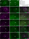
Figure 1.
Figure 1.
LepR and ORX neurons in the LHA are highly responsive to GH. A, B, Distribution of pSTAT5 immunoreactive neurons in the LHA of C57BL/6J mice after an injection of porcine GH (pGH; A) or saline (B). C, The percentage of neurons responsive to GH in the LHA (n = 3–4 mice for each reaction). D–H, Epifluorescence photomicrographs showing the expression of different neuronal markers (magenta) and pSTAT5 (green) in the LHA of mice that received an intraperitoneal pGH injection. Double-labeled neurons may appear as white. Abbreviations: DMH, dorsomedial nucleus of the hypothalamus; fx, fornix. Scale bar, 100 µm.
Figure 2.
Figure 2.
Ablation of GHR in the LHA using bilateral stereotaxic injections of AAV-Cre-GFP. A, A representative mouse showing the injection site through the expression of GFP after a bilateral infusion of AAV-Cre-GFP into the LHA. B–D, Distribution of GH-induced pSTAT5 in the hypothalamus of GHRflox/flox mice that received a bilateral injection of AAV-Cre-GFP (B) or AAV-GFP (C) in the LHA. Notice the absence of pSTAT5 in the LHA of AAV-Cre injected (LHAΔGHR) mice compared with the AAV-GFP (control) injection. The bar graphs show the quantification of the number of pSTAT5+ cells in the LHA of control (n = 4) and LHAΔGHR (n = 6) mice. Abbreviations: 3V, third ventricle; ARH, arcuate nucleus of the hypothalamus; fx, fornix; VMH, ventromedial nucleus of the hypothalamus. Scale bar, 500 µm. Differences between groups were analyzed by unpaired two-tailed Student’s t test. ***p < 0.001 significant difference between groups.
Figure 3.
Figure 3.
GHR ablation in the LHA does not affect energy and glucose homeostasis in ad libitum-fed mice. A–F, Body weight (n = 16–17/group), changes in body weight after surgery (4 weeks), fat mass (n = 4/group), lean body mass, daily food intake (n = 11–13/group), and glucose and insulin tolerance tests (n = 6–8/group) of control and LHAΔGHR mice. Possible differences between groups were analyzed by unpaired two-tailed Student’s t test or two-way repeated–measure ANOVA (glucose and insulin tolerance tests).
Figure 4.
Figure 4.
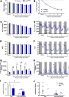
GHR in the LHA is required for the increased activity and food-seeking behavior of food-deprived mice. A–H, Body weight (n = 9–10/group), weight loss, VO2 (n = 7–9/group), RER, and ambulatory activity (n = 13–14/group) of control and LHAΔGHR mice in the basal (fed) state and during 5 d of food restriction (40% of basal intake). The recording time on the fifth day of food restriction ended ∼3–4 h before completing 24 h, leading to a lower cumulative ambulatory activity count than the previous days. The other metabolic parameters were not affected. The arrows indicate when the animals received the food (2 h before lights off). The representative figures are the average of all mice in each group. Gray and white backgrounds represent dark and light cycles, respectively. I, Latency to find food in the buried food-seeking test in ad libitum-fed mice and after 4 d of food restriction (n = 5–8/group). J, Plasma GH levels in ad libitum-fed mice and after 4 d of food restriction (n = 5–18/group). Differences between groups were analyzed by two-way repeated–measure ANOVA. *p < 0.05; **p < 0.01 significant differences between groups.
Figure 5.
Figure 5.
GHR ablation in the LHA reduces the percentage of ORX neurons expressing c-Fos in food-deprived mice. A–C, The percentage of ORX neurons expressing c-Fos, the percentage of double-labeled neurons in relation to the total number of c-Fos+ neurons, and representative photomicrographs of control (n = 10) and LHAΔGHR (n = 9) mice after 5 d of food restriction. c-Fos protein is observed through black nuclear staining, whereas ORX neurons present cytoplasmic brownish staining. D–E, ARH neurons present intense c-Fos expression in food-deprived control (n = 10) and LHAΔGHR (n = 9) mice. F, Representative photomicrograph demonstrating that c-Fos expression in the ARH of food-deprived mice is mainly restricted to AgRP neurons (discreet cytoplasmic brownish staining). Abbreviations: 3V, third ventricle; ARH, arcuate nucleus of the hypothalamus. Scale bar: C, 50 µm; D, 50 µm; F, 25 µm. Differences between groups were analyzed by unpaired two-tailed Student’s t test. *p < 0.05; **p < 0.01 significant difference between groups.
Figure 6.
Figure 6.
GH injection increases the expression of c-Fos in LHAORX neurons. A–E, The percentage of ORX neurons expressing c-Fos and representative photomicrographs of C57BL/6J mice that received an acute injection of saline (A, B; n = 5) or GH (C, D; n = 6). c-Fos protein is observed through black nuclear staining, whereas ORX neurons present cytoplasmic brownish staining. Arrows indicate double-labeled neurons. B and D represent higher-magnification photomicrographs of the selected areas in A and C, respectively. Abbreviation: fx, fornix. Scale bar, 100 µm. Differences between groups were analyzed by unpaired two-tailed Student’s t test. *p < 0.05 significant difference between groups.
Figure 7.
Figure 7.
GHR expression in non-LepR neurons is necessary to increase activity and food-seeking during food restriction. A, Epifluorescence photomicrograph showing that ORX neurons (green) and LepR-expressing cells (magenta) represent distinct neuronal populations in the LHA. B, Representative photomicrograph showing that GH-injected BrainΔGHR mice exhibit only a few pSTAT5 cells in the LHA. C, Representative photomicrograph showing that GH-injected LepRΔGHR mice present numerous pSTAT5 cells in the LHA (green staining), although most LepR neurons (magenta) were no longer responsive to GH. Abbreviations: 3V, third ventricle; fx, fornix; VMH, ventromedial nucleus of the hypothalamus. Scale bar, 100 µm. D–E, Ambulatory activity of control (n = 8), BrainΔGHR (n = 5), and LepRΔGHR (n = 11) mice in the basal (fed) state and during 5 d of food restriction (40% of basal intake). The arrows indicate when the animals received the food (2 h before lights off). The representative figure is the average of all mice in each group. Gray and white backgrounds represent dark and light cycles, respectively. F, Latency to find food in the buried food-seeking test in ad libitum-fed mice and after 4 d of food restriction (n = 6–8/group). Differences between groups were analyzed by two-way repeated–measure ANOVA. *p < 0.05; **p < 0.01 significant differences between groups.

References

    1. Alvarez-Crespo M, Martinez-Sanchez N, Ruiz-Pino F, Garcia-Lavandeira M, Alvarez CV, Tena-Sempere M, Nogueiras R, Dieguez C, Lopez M (2013) The orexigenic effect of orexin-A revisited: dependence of an intact growth hormone axis. Endocrinology 154:3589–3598. 10.1210/en.2013-1251 - DOI - PubMed
    1. Arrigoni E, Chee MJS, Fuller PM (2019) To eat or to sleep: that is a lateral hypothalamic question. Neuropharmacology 154:34–49. 10.1016/j.neuropharm.2018.11.017 - DOI - PubMed
    1. Barrile F, et al. (2023) Ghrelin’s orexigenic action in the lateral hypothalamic area involves indirect recruitment of orexin neurons and arcuate nucleus activation. Psychoneuroendocrinology 156:106333. 10.1016/j.psyneuen.2023.106333 - DOI - PMC - PubMed
    1. Bohlooly YM, et al. (2005) Growth hormone overexpression in the central nervous system results in hyperphagia-induced obesity associated with insulin resistance and dyslipidemia. Diabetes 54:51–62. 10.2337/diabetes.54.1.51 - DOI - PubMed
    1. Burton KA, Kabigting EB, Clifton DK, Steiner RA (1992) Growth hormone receptor messenger ribonucleic acid distribution in the adult male rat brain and its colocalization in hypothalamic somatostatin neurons. Endocrinology 131:958–963. 10.1210/endo.131.2.1353444 - DOI - PubMed

LinkOut - more resources