

COVID-19 related cognitive, structural and functional brain changes among Italian adolescents and young adults: a multimodal longitudinal case-control study
- PMID: 39358346
- PMCID: PMC11447249
- DOI: 10.1038/s41398-024-03108-2
COVID-19 related cognitive, structural and functional brain changes among Italian adolescents and young adults: a multimodal longitudinal case-control study
Abstract
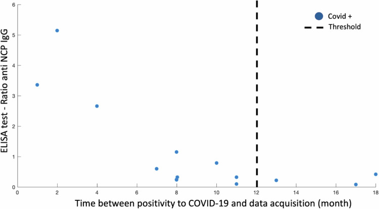
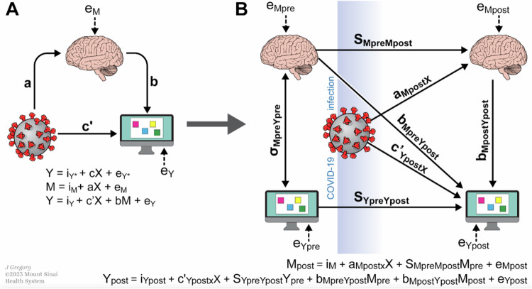
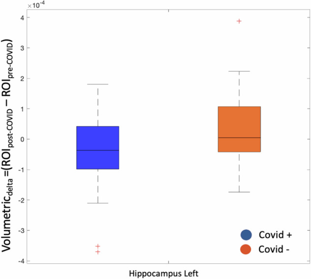
Coronavirus disease 2019 (COVID-19) has been associated with brain functional, structural, and cognitive changes that persist months after infection. Most studies of the neurologic outcomes related to COVID-19 focus on severe infection and aging populations. Here, we investigated the neural activities underlying COVID-19 related outcomes in a case-control study of mildly infected youth enrolled in a longitudinal study in Lombardy, Italy, a global hotspot of COVID-19. All participants (13 cases, 27 controls, mean age 24 years) completed resting-state functional (fMRI), structural MRI, cognitive assessments (CANTAB spatial working memory) at baseline (pre-COVID) and follow-up (post-COVID). Using graph theory eigenvector centrality (EC) and data-driven statistical methods, we examined differences in ECdelta (i.e., the difference in EC values pre- and post-COVID-19) and Volumetricdelta (i.e., the difference in cortical volume of cortical and subcortical areas pre- and post-COVID) between COVID-19 cases and controls. We found that ECdelta significantly between COVID-19 and healthy participants in five brain regions; right intracalcarine cortex, right lingual gyrus, left hippocampus, left amygdala, left frontal orbital cortex. The left hippocampus showed a significant decrease in Volumetricdelta between groups (p = 0.041). The reduced ECdelta in the left amygdala associated with COVID-19 status mediated the association between COVID-19 and disrupted spatial working memory. Our results show persistent structural, functional and cognitive brain changes in key brain areas associated with olfaction and cognition. These results may guide treatment efforts to assess the longevity, reversibility and impact of the observed brain and cognitive changes following COVID-19.
© 2024. The Author(s).
Conflict of interest statement
The authors declare no competing interests.
Figures

Update of
-
Covid-19 related cognitive, structural and functional brain changes among Italian adolescents and young adults: a multimodal longitudinal case-control study.medRxiv [Preprint]. 2023 Jul 23:2023.07.19.23292909. doi: 10.1101/2023.07.19.23292909. medRxiv. 2023. Update in: Transl Psychiatry. 2024 Oct 2;14(1):402. doi: 10.1038/s41398-024-03108-2. PMID: 37503251 Free PMC article. Updated. Preprint.
Similar articles
-
Covid-19 related cognitive, structural and functional brain changes among Italian adolescents and young adults: a multimodal longitudinal case-control study.medRxiv [Preprint]. 2023 Jul 23:2023.07.19.23292909. doi: 10.1101/2023.07.19.23292909. medRxiv. 2023. Update in: Transl Psychiatry. 2024 Oct 2;14(1):402. doi: 10.1038/s41398-024-03108-2. PMID: 37503251 Free PMC article. Updated. Preprint.
-
Cognitive impact and brain structural changes in long COVID patients: a cross-sectional MRI study two years post infection in a cohort from Argentina.BMC Neurol. 2024 Nov 18;24(1):450. doi: 10.1186/s12883-024-03959-8. BMC Neurol. 2024. PMID: 39558250 Free PMC article.
-
Brain and cognitive changes in patients with long COVID compared with infection-recovered control subjects.Brain. 2024 Oct 3;147(10):3611-3623. doi: 10.1093/brain/awae101. Brain. 2024. PMID: 38562097
-
Eigenvector Centrality Mapping Reveals Volatility of Functional Brain Dynamics in Anti-NMDA Receptor Encephalitis.Biol Psychiatry Cogn Neurosci Neuroimaging. 2024 Nov;9(11):1222-1229. doi: 10.1016/j.bpsc.2024.07.021. Epub 2024 Jul 27. Biol Psychiatry Cogn Neurosci Neuroimaging. 2024. PMID: 39074556
-
Graph theory analysis of resting-state functional magnetic resonance imaging in essential tremor.Hum Brain Mapp. 2019 Nov 1;40(16):4686-4702. doi: 10.1002/hbm.24730. Epub 2019 Jul 22. Hum Brain Mapp. 2019. PMID: 31332912 Free PMC article.
Cited by
-
Functional connectivity of default mode network in non-hospitalized patients with post-COVID cognitive complaints.Front Neurosci. 2025 Apr 10;19:1576393. doi: 10.3389/fnins.2025.1576393. eCollection 2025. Front Neurosci. 2025. PMID: 40276574 Free PMC article.
-
Multimodal MRI analysis of COVID-19 effects on pediatric brain.Sci Rep. 2025 Apr 5;15(1):11691. doi: 10.1038/s41598-025-96191-4. Sci Rep. 2025. PMID: 40188214 Free PMC article.
-
Short-Term Restriction of Physical and Social Activities Effects on Brain Structure and Connectivity.Brain Sci. 2024 Dec 25;15(1):7. doi: 10.3390/brainsci15010007. Brain Sci. 2024. PMID: 39851375 Free PMC article.
-
Structural and functional neuroimaging of hippocampus to study adult neurogenesis in long COVID-19 patients with neuropsychiatric symptoms: a scoping review.PeerJ. 2025 Jun 27;13:e19575. doi: 10.7717/peerj.19575. eCollection 2025. PeerJ. 2025. PMID: 40589858 Free PMC article.
-
On the merits and potential of advanced neuroimaging techniques in COVID-19: A scoping review.Neuroimage Clin. 2024;42:103589. doi: 10.1016/j.nicl.2024.103589. Epub 2024 Mar 6. Neuroimage Clin. 2024. PMID: 38461701 Free PMC article.
References
MeSH terms
Grants and funding
LinkOut - more resources
Full Text Sources
Medical
